Abstract
The ethyl acetate extract of pomegranate (Punica granatum) fruits was fractionated by chromatographic techniques to afford the ellagitannin punicalagin. The substance was found to be active against methicillin-resistant Staphylococcus aureus strains and was identified by HPLC/UV and 1HNMR. The antibacterial assays that guided the isolation of the tannin were conducted using the disc diffusion method. Minimum inhibitory concentration (MIC) was determined by the dilution method according to NCCLS (National Committee for Clinical Laboratory Standards) procedure.
KEYWORDS:
INTRODUCTION
As part of our effort to identify the substances responsible for the pharmacological activities attributed to plants utilized in India in popular medicine, we have studied the epicarp of pomegranate fruits to identify the components with antimicrobial activity. Punica granatum Linn. (Punicaceae) is a shrub or small tree native to Asia (CitationJafri et al., 2000) where its several parts have been used as an astringent, a haemostatic, a remedy for diabetes, an anthelmintic against tapeworms, and for diarrhea and dysentery (CitationDas et al., 1999). In India fruits are known as “romã” and are used for the treatment of throat infections, coughs, and fever. There are several commercial phytopreparations in India containing extracts from pomegranate. For the validation of such products, it is necessary to define chemical markers, substances that when present in the preparations attest their quality (CitationGunther, 1996). Although many reports on the antimicrobial activity of pomegranate exist in the literature, none of them relate such activity with its chemical composition. We describe here for the first time the isolation and identification of the tannin responsible for the activity against a bacterium of medical importance (Staphylococcus aureus). Interest in plants with antimicrobial properties has been revived due to current problems associated with the use of antibiotics. With the increasing prevalence of methicillin-resistant Staphylococcus aureus (MRSA) strains as pathogens in both hospitals and the community, the investigation of plant extracts active against this organism provides an example of prospecting for new compounds that may be effective against infections currently difficult to treat (CitationCowan, 1999).
MATERIALS AND METHODS
General Experimental Procedures
All reagents were of analytical grade. The NMR spectrum of the compound was taken on a Varian-Gemini 200 (1H: 200 MHZ spectrometer). Column chromatography was performed using Sephadex LH-20 (Pharmacia) and XAD-16 resin (Sigma). Thin-layer chromatography was performed on cellulose plates (Merck), HPLC/UV on a Shimadzu instrument equipped with a diode array detector and RP-18 column (5 µm, 20 × 5 mm, Merck) and preparative HPLC on a Shimadzu instrument equipped with UV detector and RP-18 column (10 µm, 25 × 2 cm). Antibacterial assays were performed on Mueller Hinton agar medium.
Extraction and Isolation of the Constituents
Fresh fruit pericarp (240 gm) was exhaustively extracted with EtOH. The dried ethanolic extract was suspended in water and successively partitioned with hexane, chloroform, ethyl acetate, and butanol. The most active fraction on bioassay (ethyl acetate) was chromatographed on an XAD-16 column using a water:methanol gradient. The active fraction eluted from the column with H2O:MeOH (1:1) was submitted to chromatography on a Sephadex LH-20 column using a gradient H2O:MeOH, and the active fraction was purified on a preparative HPLC column to afford the active compound punicalagin and the inactive ones, ellagic acid and punicalin.
Bacterial Strains
Agra Methicillin–resistant Staphyloccus aurens prevalent clone of MRSA strains were obtained from hospitalized patients in two Agra hospitals (India Hospitals) and identified at the Institute of Microbiology. Furthermore, as a comparison parameter, 10 MRSA strains from other clones were tested, as well as 8 MSSA (methicillin-sensitive S. aureus) and 2 reference strains (S. aureus ATCC 29213 [MSSA] and ATCC 33591 [MRSA]).
Assay for Antibacterial Testing
Disc-diffusion method (Citationsantos et al., 1999)
Petriplates containing 20 mL of Mueller Hinton agar medium were seeded with a 24-hour-old culture of the bacterial strains. The extracts, fractions, and pure compounds were tested in concentration of 25 or 50 mg mL−1, applying 10 µL of each sample to sterile filter paper discs (5 mm in diameter) and placed on the surface of the medium. The inoculum size was adjusted so as to deliver a final inoculum of approximately 108 colony-forming units (CFU)/mL (CitationBate-Smith, 1972). Incubation was conducted at 37°C for 24 hours. The assessment of antibacterial activity was based on the measurement of diameter of the inhibition zone formed around the disc.
Dilution Method
The minimum inhibitory concentration (MIC) was determined by dilution according to National Committee for Clinical Laboratory Standards (NCCLS) (CitationHatano, 1988). Mueller Hinton agar is prepared and sterilized in the usual fashion by autoclaving. Before solidification, 20 mL of agar medium is added to each of the Petri dishes containing the samples, and the Petri dishes are swirled carefully until the agar begins to set. Final concentrations from 250 to 0,97 µg mL−1 were used for each plant sample. The bacterial inoculum size was adjusted so as to deliver a final inoculum of approximately 104 colony-forming units (CFU/mL8) and was added to the medium using a Steers replicator.
RESULTS AND DISCUSSION
The active fraction when analyzed by TLC on cellulose plates (H2O:CH3COOH, 4:1) initially showed the presence of two orange-colored spots with NaNO2 reagent, turning purple after some minutes. This is a characteristic reaction for ellagitannins (CitationBates-Smith, 1972). However, these two substances show up as four when analyzed by analytical HPLC/UV (). Each one of the four peaks was obtained by preparative HPLC (H2O:CH3CN, 9:1) and after liophilization was reanalyzed by analytical HPLC/UV. shows the presence of two substances for each peak. This is in fact a characteristic phenomenon of hydrolysable tannins with an unsubstituted anomeric hydroxyl group (CitationDoig et al., 1990).
FIGURE 1 Characteristic HPLC chromatograms and UV spectra of the anomers punicalin and punicalagin (silicagel RP-18, KH2PO4 0.01 mol L−1 + H3PO4 0.01 mol L−1 + CH3CN - 4:42). 1A- Analytical chromatograms of the mixture. 1B - UV spectra of each compound. 1C-Analytical chromatogram after the preparative HPLC separation.
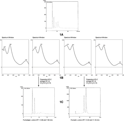
The substances were identified as punicalin α1 and punicalin β2, punicalagin α3 and punicalagin β4, the major ellagitannins from the pomegranate (CitationBurapadaja and Bunchoo, 1995). shows the UV spectra of these substances (λmax = 218, 260, and 379 nm). These values are characteristic of a gallagyl cromophore (Doig et al., 1990) conferring to the compounds a bright yellow color. Acid hydrolysis of punicalin afforded gallagyldilactone (identified by HPLC/UV, RT 3.2 minutes) and glucose (identified by co-chromatography on silica gel plates). Acid hydrolysis of punicalagin afforded gallagyldilactone, ellagic acid (HPLC/UV, 3.2 min and 4.9 minutes), and glucose.
FIGURE 2 Ellagitannins from the pericarp of the Punica granatum.
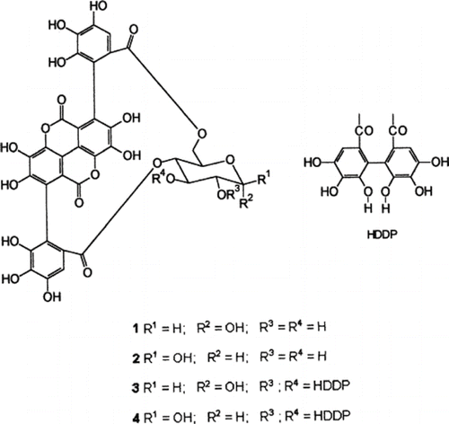
Punicalagin (3 and 4) yellow amorphous powder (320 mg), [α]D 20 –123.43 (c = 1.28, MeOH); 1H-NMR (CD3OD) δ 2.15 (1H, m, H-5), 3.15–3.45 (1H, m, H-4), 3.98–4.17 (1H, m, H-6), 6.42, 6.55, 6.65, 6.82 (each H, s, aromatic-H; CitationBurapadaja and Bunchoo, 1995).
The antibacterial activity for punicalagin (250 µg - disc diffusion method Fig. 4) afforded a clear inhibition zone of 20 mm for all bacteria tested. The minimum inhibitory concentration was established as 61.5 µg mL−1. It is noteworthy that CitationBurapadaja and Bunchoo (1995) in a phytochemical study of Terminalia citrina, isolated punicalagin and assayed it against several bacterial colonies. For a methicillin-resistant S. aureus colony, they found a MIC value of 768 µg mL−1.
CONCLUSIONS
Our results show that the ellagitannin punicalagin is the substance responsible for the antimicrobial activity of the pomegranate. Furthermore, this compound presented an activity of 10-fold higher than the one found by CitationBurapadaja and Bunchoo (1995). For the standardization of phytopharmaceuticals it could be the chemical marker of choice.
ACKNOWLEDGEMENTS
The authors thank Dr. S. K. Gupta, Department of Chemistry, Bipin Bihari College, Jhansi, and Dr. Ashok Kumar, Department of Chemistry, St John's College, Agra, India for their assistance.
LITERATURE CITED
- Bate-Smith , E.C. 1972 . Phytochemistry , 11 : 1153
- Burapadaja , S. and Bunchoo , A. 1995 . Planta Med. , 61 : 365
- Cowan , M.M. 1999 . Clin. Microbiol. Rev. , 12 : 564
- Das , A.K. , Mandal , S.C , Banerjee , S.K , Sinha , S. , Das , J. , Saha , B.P. and Pal , M.J. 1999 . J. Ethnopharmacol. , 68 : 205
- Doig , A.J. , Williams , D.H. , Oelrichs , P.B. and Baczynskyj , L. 1990. J . Chem. Soc . Perkin Trans. , 1 : 2317
- Gunther , B. and Wagner , H. 1996 . Phytomedicine , 3 : 59
- Hatano , T. , Yoshida , T. , Shingu , T. and Okuda , T. 1988 . Chem. Pharm. Bull. , 36 : 2925
- Jafri , M.A. , Aslam , M. , Javed , K. and Singh , S. 2000 . Article Title . J. Ethnopharmacol , 70 : 309
- Santos , K.R.N. , Teixeira , L.M. , Leal , G.S. , Fonseca , L.S and Filho , P.P.G. 1999 . J. Med. Microbiol. , 48 : 17
- Tanaka , T. , Nonaka , G.I. and Nishioka , I. 1986 . Chem. Pharm. Bull. , 34 : 650