
ABSTRACT
In this article a detailed review of the fungal diseases that can affect pear fruit is provided. Each section comprises a complete description of the fungi responsible for the diseases and the symptoms they produce on the pears. The majority of pear fungal diseases, including blue mold, grey mold, bitter rot, black spot, brown rot, bull’s eye rot, Phytophthora rot, pink mold rot, powdery mildew, Rhizopus rot, scab, side rot, sooty blotch, and flyspeck, are presented. Sources of infection are comprehensively described in relation to harvesting or post-harvesting handling and intervention practices, respectively. As these diseases are dependent on a number of environmental parameters, the optimal temperature ranges for disease development are described and the causal agents are identified. Previous studies on the diagnostic indications are reviewed while information about the optimal environmental parameters of temperature, pressure, pH, water activity, and oxygen level for the fungal growth is comprehensively summarized.
Introduction
The growth of filamentous fungi in highly perishable fruits including pears is an important quality problem that may lead to significant economic losses. Considerable losses occur mostly in the post-harvest phase where Penicillium expansum, Botrytis cinerea, and Mucor piriformis are the three most common causative factors of pear rot and loss. Other etiological factors include Phialophora malorum, Alternaria spp., Cladosporium herbarum, and Neofabrea spp. (Sutton et al., Citation2014). These species, which are normally present in air and surrounding environments, such as soil and water, tend to infect wounds and senescent fruits (Mari et al., Citation2003).
Fungi are ubiquitous organisms and can grow in most moist environments found in pear storage plants, establishing themselves in ceilings, floors, walls, and drains if these are not properly sanitized. This article presents a comprehensive review of the fungi responsible for pear infections. Their source of origin, susceptibility, and disease severity, as well as the impact of the environmental conditions on their growth, is described.
Fungal diseases
At least 13 fungal diseases have been reported for pears of different cultivars worldwide. A summary of these diseases, the fungal species responsible, and the pear cultivars for which they have been reported are presented in . A more detailed description of these diseases is presented hereunder.
Table 1. Overview of fungal diseases for pears.
Blue mold
Blue mold, known also as soft rot or wet rot, has been reported as one of the most important post-harvest diseases in pears, which causes destructive rot in all of the producer countries, namely the U.S., U.K., Poland, Italy, Israel, India, and Australia (Snowdon, Citation1990). Under modern storage conditions, losses due to this rot should be less than 1%, but if storage problems occur, or conditions are less than optimal, losses can be 50% or more (Monroe, Citation2009).
Blue mold originates primarily from infection of wounds, such as punctures, bruises, and limb rubs, on the fruit. The first symptoms are soft watery brown spots, which undergo rapid enlargement at temperatures between 20 °C and 25 °C (Snowdon, Citation1990). The lesions have a very sharp margin between diseased and healthy tissues and decayed tissue can be readily separated from the healthy one, leaving it like a “bowl” (Pierson et al., Citation1971; Shim et al., Citation2002). Blue or blue-green spore masses may appear on the decayed area starting at the infection site. Decayed fruit has an earthy musty odor. The presence of blue-green spore masses at the decayed area and the associated musty odor are the positive diagnostic indications of blue mold (Jones and Aldwinckle, Citation1990; Shim et al., Citation2002). Blue mold occurs on all of the cultivars of pears around the world.
Three forms of the disease can occur: check rot, stem and neck rot, and pinhole rot (Monroe, Citation2009; Pierson et al., Citation1971). Check rot develops on the check of the fruit and it occurs on all cultivars of pears. It is the form of the disease most frequently found in the market. Stem and neck rot is a form of the disease that develops from stem infections in fleshy-stemmed varieties, such as Anjou and Comice. The fungus grows down through the stem, which remains firm but darkens. It spreads through vascular tissues into the neck of the fruit where the flesh becomes soft and watery (Pierson et al., Citation1971). Pinhole rot is a form of the disease that occurs mainly on Winter Nelis, a variety with large prominent lenticels. It first appears as numerous minute spots of decay scattered over the surface of the fruit, with infection apparently taking place at the lenticles. As the disease progresses the spots increase in size, finally coalescing to decompose the entire fruit (Pierson et al., Citation1971).
The causal organism of blue mold is Penicillium expansum Link (an example of a contaminated pear fruit is given in ). Several other Penicillium species, including P. solitum, P. commune, P. verrucosum, P. chrysogenum, and P.regulosum, have also been reported to cause decay on pears. P. expansum can grow at temperatures as low as –3 °C and conidia can germinate at 0 °C (WSU, Citation2015a). Penicillium expansum Link is the most important fungal pathogen of pear fruit, which is responsible for substantial global economic losses (Yu et al., Citation2012).
Grey mold
Grey mold is an important storage disease of pears, especially varieties that are stored for long periods (Pierson et al., Citation1971). Grey mold is considered as the second most important infection in pears after blue mold rot (Jones and Aldwinckle, Citation1990; Ogawa and English, Citation1991). Losses as high as 20% to 60% due to grey mold are not uncommon after an extended period of storage, particularly in fruit that was not treated with fungicides prior to storage. This is because grey mold has the ability to spread from decayed to healthy fruit by fruit-to-fruit contact during storage and this process is enhanced by high relative humidity (WSU, Citation2015a). The disease develops more rapidly in cold storage temperatures unlike other postharvest decay, with the exception of mucor rot (Jones and Aldwinckle, Citation1990).
Grey mold originates primarily from infection of wounds, such as punctures and bruises, which are created at harvest and during postharvest handling. Stem-end grey mold is common on ‘d’Anjou’ pears (Xiao, Citation2006). This rot is often difficult to identify because its symptoms vary with fruit age and cultivar (Pierson et al., Citation1971). The rot tends to start as a small brown spot with diffuse margins. As the rot progresses, older portions of the decay darken, whereas the edges remain lighter (Monroe, Citation2009). The decayed area is spongy and diseased tissue is not separable from the healthy tissue, which is different from blue mold (WSU, Citation2015a). Under high relative humidity conditions, fluffy white to grey mycelium and greyish spore masses may appear on the decayed area (Xiao, Citation2006). The internal decayed flesh appears light brown to brown at the margin and on advanced decayed fruit, sclerotia may form on the lesion surface (Xiao, Citation2006). Generally, grey mold does not have a distinct odor, but in advanced stages it may have a pleasant fermented odor that distinguishes it from other storage rots (WSU, Citation2015a; Pierson et al., Citation1971).
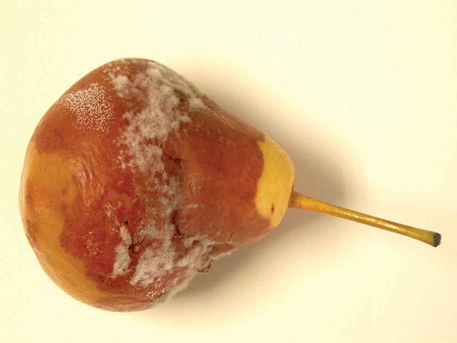
The causal agent of grey mold is Botrytis cinerea Pers.—teleomorph Botryotinia fuckeliana (de Bary) Whetzel; it is widely distributed in nature and thrives on decaying plant matter (Pierson et al., Citation1971) (an example on pear contamination is given in ). Conidia of the fungus are dispersed mainly by air currents and water splash. At harvest, pear fruit is detached from the trees and fresh wounds at the stem are exposed to potential contamination by B. cinerea (WSU, Citation2015a). Outdoors, Botrytis overwinters in the soil in the form of a mycelium on plant debris, and as black, hard, flat or irregular sclerotia in the soil and plant debris, or mixed with seed. The fungus requires free moisture for germination and cool (15 to 25 °C) weather with little wind for optimal infection, growth, sporulation and spore release. Botrytis is also active at lower temperatures (0 to 10 °C). Infection rarely occurs at temperatures above 25 °C and once it occurs, the fungus grows over a range of 0 to 35 °C (University of Illinois, Citation2000).
Figure 1. Pear fruit affected by blue mold.
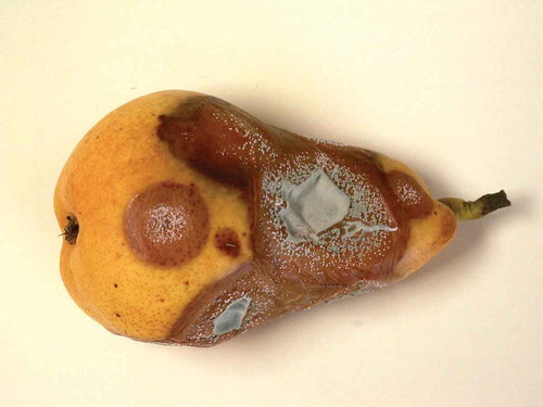
Figure 2. Pear fruit affected by grey mold.
B. cinerea is able to move through the stem of the fruit to reach the flesh after a period of time in cold storage and then cause decay. It has been shown that the incidence of grey mold is correlated with spore levels on the fruit surface. B. cinerea has been reported to invade floral parts of pear fruit during bloom, causing calyx-end rot during storage (WSU, Citation2015a).
Bitter rot
Bitter rot, caused by the fungus Colletotrichum gloesporioides (Penz.)—teleomorph Glomerella cingulata (Stonem.), is primarily an orchard disease. Immature pears may be infected on the tree, but more commonly the disease attacks fruits that are starting to ripen (Jones and Aldwinckle, Citation1990; Pierson et al., Citation1971). Bitter rot is one of the few diseases that affects uninjured fruits (Palmer and Riesselman, Citation1972). It is most likely to become a problem in areas with hot, humid summers (Monroe, Citation2009). The disease is of particular importance in the south-eastern U.S. and occasional epidemics have been reported in New Zealand, Israel, India, and in Poland (Snowdon, Citation1990). Symptoms may vary and depend on whether the infection could initiate by spores from a erithecial type, which produces ascospores and conidia, or by the conidial type, which produces only conidia. Fruit infection can occur as early as the time of bloom where it appears as minute, grey-brown flecks, which usually do not develop further until the fruit begins to ripen (University of Illinois, Citation2005a). Nevertheless, bitter rot symptoms are usually found only on the fruit, even if the pathogen may also infect stems to cause cankers (Monroe, Citation2009; Schubert, Citation1983).
Lesions originating from conidial type infections remain circular and become sunken as they enlarge. Lesions then become 1/2 to 1 inch in diameter and acervuli are produced in concentric circles around the point of infection. When dry, conidia in mass appear crystalline, but under moist, humid conditions the spores mass appears creamy and salmon to pink (University of Illinois, Citation2005a; Monroe, Citation2009).
Lesions initiated by perithecial types are usually not sunken and are often darker brown than those initiated by conidial types. Acervuli are widely scattered over the surface and perithecia are found in dark brown to black clusters scattered over the surface of the fruit. Conidial masses associated with the perithecial types tend to initially be orange to salmon, but then turn dark brown to black in older lesions (University of Illinois, Citation2005a). In cross section, the lesions appear V-shaped (Monroe, Citation2009).
The optimal temperature for infection is approximately 25 °C and epidemics are associated with wet growing seasons. Some fruits may rot completely on the tree, forming shriveled mummies, which survive the winter and together with cankers act as a source of spores the following year; some others might drop (Jones and Aldwinckle, Citation1990; Snowdon, Citation1990). Infected fruit that has not yet developed symptoms may have a latent infection, which develops later during the storage period (Monroe, Citation2009).
Black spot
Black spot, also known as Fabraea leaf spot, pear leaf blight, leaf spot, leaf scald, fruit spot, and quince leaf spot (Jones and Aldwinckle, Citation1990), is caused by the fungus Fabraea maculata Atk., previously also known as Diplocarpon mespili (Sor.) Sutton—anamorph Entomosporium maculatum Lev., or E. mespili (DC.) Sacc (Jones and Aldwinckle, Citation1990). The disease usually appears late in the growing season but can occasionally develop in late May and early June. Fabraea leaf spot attacks leaves, fruit, and twigs of pear (Douglas, Citation2003). The disease does not develop or spread during transit or storage and usually the spots are not followed by rot of any kind (Pierson et al., Citation1971). Abundant leaf spotting causes serious defoliation, thus the fruits are impaired not only by the spots but also by a lack of nourishment due to defoliation (Pierson et al., Citation1971). Heavily infected leaves are often yellow and drop prematurely. Severe defoliation can substantially reduce tree vigor and yield, especially if trees are defoliated several years in a row (Douglas, Citation2003).
Leaf spot can be found on petioles, leaves, shoots, and fruits. Initial lesions on leaves are tiny, round, purplish-black spots, which quickly enlarge to 1/8 to 1/4 inch in diameter and usually have a blackish-brown center. Fruit lesions are larger than those on leaves and cause the fruit to crack and drop. Lesions may be observed as small inconspicuous, purplish-black spots (Van der Zwet and Briggs, Citation2004).
Brown rot
The main causative fungi of brown rot are Monilinia fructicola, Monilinia fructigena, and Monilinia laxa. This disease is widespread in Europe, Asia, and South America, causing serious losses in apple and pear fruits (Ogawa and English, Citation1991; Snowdon, Citation1990). Monilinia laxa and M. fructigena are two fungi that are very closely related and indistinguishable to the naked eye. M. laxa more commonly causes blossom wilt on pears and stone fruit (The Royal Horticultural Society, Citation2015).
M. fructicola (Wint.) Honey is the cause of American brown rot. It is established throughout North America, New Zealand, and Australia. M. laxa (Aderh. & Ruhl.) Honey and M. laxa f. sp. mali cause European brown rot. M. laxa is found particularly on stone fruits in Europe, China, Japan, South Africa, North America, and South America, whereas M. laxa f. sp. mali is present only in Europe. However, M. fructigena is the most common species on apple and pear in Europe and it commonly causes fruit rot on both apples and pears (Jones and Aldwinckle, Citation1990).
The first indication of fruit infection is the development of small circular brown spots, spreading out from wounds, especially those made by birds (The Royal Horticultural Society, Citation2015; Snowdon, Citation1990). Under humid conditions, tufts of white mold break through the skin, often forming concentric circles around the point of infection. In the presence of light there is copious production of grey-brown spores (conidia). The fungus can spread into adjacent healthy fruit, but even in its advanced stage, the rot tends to remain firm and dry.
Fruits infected early in the growing season undergo rotting on the tree, turning into shriveled mummies, which may remain attached or fall to the ground. The fungus thereby survives the winter and spores are disseminated throughout the orchard (Snowdon, Citation1990).
Bull’s eye rot
Bull’s eye rot, previously known as “Gloeosporium rot” (Snowdon, Citation1990), is caused by Neofabraea spp. and is a common postharvest disease in the Pacific Northwest of the U.S. as well as in Europe and in other growing areas (WSU, Citation2015a; Henriquez et al., Citation2004, Citation2008). Fruit can be infected in the orchard at any time during the growing season, and spores or incipient infections remain latent until development of symptoms after several months of postharvest storage (Henriquez et al., Citation2004; Jones and Aldwinckle, Citation1990). Neofabraea perennans Kienholz (also known as Pezicula perennans Kienholz) and N. alba E.J. Guthrie Verkley (also known as Pezicula alba E.J. Guthrie) are the principal species associated with bull’s eye rot of pear in the Pacific Northwest (WSU, Citation2015a). These pathogens cause cankers on branches or develop saprophitically on trees. Conidia of N. perennans and N. alba are produced throughout the year on pear trees, but the highest sporulation occurs at the end of summer and during fall (Henriquez et al., Citation2008). These conidia are splash-dispersed to fruit. Infection can occur any time after petal-fall; however, fruit susceptibility increases as the growing season progresses (Henriquez et al., Citation2008).
Bull’s-eye rot spots have light brown centers and a dark brown border (Pscheidt and Ocamb, Citation2014). Decayed tissue is firm and cream-colored spore masses in the aged decayed area may appear. Rot does not spread from one fruit to another and it does not show up in the orchard until after months of storage (Pscheidt and Ocamb, Citation2014).
Phytophtora rot
Phytophthora root rot is not usually a major concern on pear trees (Gubler et al., Citation2007). The disease usually attacks the root and the trunks of apple and pear trees (Rivard, Citation2007). Susceptibility and disease severity depend on which species of the pathogen are present, the age and the health of the tree, the rootstock, water management, and climatic conditions that occur after the tree is infected (Gubler et al., Citation2007). The disease commonly infects relatively young trees and occurs in patches of one to five trees throughout the orchard block. Phytophthora is more likely to kill young trees, as their root and crown areas are smaller than those of mature trees (Monroe, Citation2009).
Infection occurs on windfalls or on low-hanging fruits that touch the ground or are splattered with infested soil. The rot develops as firm, brown to black lesions that may cover the fruit surface after several days (Pierson et al., Citation1971). Older infections can cause the whole fruit to rot on the tree. In some years, the disease may also spread to fruit pedicels and, to a lesser extend, into first-year wood causing dieback (Pscheidt and Ocamb, Citation2014). Discoloration of diseased flesh varies with pear fruit variety, ranging from clear and watersoaked in ‘Clairgeau’, brown with watersoaked margin in ‘Anjou’ to brown in ‘Kieffer’. In most cultivars the flesh is light brown, with the vascular bundles and strands more deeply discolored (Pierson et al., Citation1971).
Phytophtora is a cosmopolitan genus comprised of 67 described species and cultivars but pear rot is caused mainly by Phytophtora cactorum (Lebert & Cohn). However, P. citricola Sawada, P. cambivora (Petri) Buisman, and unidentified nonpapillate species have also been occasionally isolated from infected fruits and contaminated irrigation water (Yamak et al., Citation2002). These are all soil-borne fungi, many of which are common inhabitants of most orchard soils. Some species that are not common inhabitants may be introduced to the orchard on contaminated planting stock or through movement of contaminated soil (Ellis, Citation2008).
Infections progress slowly in cold storage but develop fairly rapidly at ripening temperatures. Irrigation water is frequently contaminated with the pathogen and the disease is prevalent in orchards using over-the-canopy irrigation or when the sprinkler angles of under-the-canopy systems are sufficiently high to moisten fruit (Harris and Dennis, Citation1980; Yamak et al., Citation2002).
Most Phytophthora species can survive for an extended period in moist soil, but they die fairly rapidly in dry soil. The optimal temperature range for disease development is 15 to 22.8 °C. Although root zones are routinely saturated from rainfall during the winter months, Phytophthora root rot is not a problem in the winter because temperatures are well below those necessary for infection. Additionally, soil temperatures above the optimum suppress the growth and development of the fungus, and soil temperatures over 32 °C significantly reduce fungal survival (Gubler et al., Citation2007).
Pink mold rot
Pink mold rot is caused by Cephalothecium roseum Cda. Early stages of the disease appear as small, irregularly shaped spots of about 1/8 inch in diameter, with brown margins and light centers (Pierson et al., Citation1971). White fungus threads may develop on the surface of the lesions, and under moist conditions they may also be accompanied by pink masses of spores. A second stage consisting of chocolate-brown, irregularly shaped, sunken lesions from 1/2 up to 2 inches across, may develop under favorable conditions. Depressed, lighter colored, circular spots may be scattered over the surface of rotted areas. The late stage of the rot is less likely to exhibit the white fungus threads and pink spore masses as compared to the first stage (WSU, Citation2015b; Pierson et al., Citation1971).
At any stage the rotted areas are rather firm and dry, never watery and the affected tissues have a bitter taste (WSU, Citation2015b). Superficially, pink mold resembles fish eye and bull’s eye rots. Spots of pink mold rot can generally be distinguished by their irregular shape and by a white to pinkish growth of mold. Below 10 °C, pink mold develops very slowly and is not likely to start at new places; at 0 °C it is checked almost entirely. Infection rarely spreads from one fruit to another unless the fruits are held at fairly high temperatures for some time after harvest (WSU, Citation2015b).
Powdery mildew
Powdery mildew is caused by Podosphaera leucotricha (Ell. & Ev.)—anamorph Oidium farinosum Cooke, a fungus that invades buds, blossoms, leaves, twigs, and fruits of susceptible varieties (Larsen and Pokharel, Citation2011). This disease has been reported as seriously affecting pears of the Flemish, Bartlett, Anjou, and Idaho cultivars (Pierson et al., Citation1971). Infected leaves, shoots, and fruit are covered by a light grey or white powdery coating of spores and mycelium. The leaves become curled, crinkled, and stunted. Infections on the young fruit surface damage the fruit skin cells and result in scarring of the fruit surface known as russet. The fungus spreads from older leaves to young, succulent leaves and shoots. Extensive infection on young trees can severely damage them (Larsen and Pokharel, Citation2011).
Most powdery mildews produce fungal threads that grow only on the plant surface and never invade the internal plant tissues. The fungi feed by sending haustoria, or root-like structures, into the epidermal cells of the plant. Being obliged parasites, powdery mildew fungi require living plant tissue to grow. Powdery mildew survives from one season to the next in infected buds or as fruiting bodies called “chasmothecia” that overwinter on leaf litter and on the bark of branches and stems. In spring, newly developing leaves become diseased as they emerge from infected buds (Larsen and Pokharel, Citation2011).
Powdery mildew infections occur when the relative humidity is greater than 90% and the temperature is between 10 to 25 °C. The optimal temperature range for the fungus is 18.9 to 22.2 °C. The high relative humidity that often occurs before and after wetting periods is conducive to powdery mildew development (Hartman and Eshenaur, Citation2002).
Rhizopus rot
Rhizopus soft rot occurs occasionally on pears that have been injured or are overripe (Pierson et al., Citation1971). This disease mainly occurs during sale, transports, market shelf, and storage (Pitt and Hocking, Citation2009; Snowdon, Citation1990).
Soft rot starts in a single fruit, which then becomes surrounded by a coarse, loose “nest” of mycelium. Growth spreads rapidly, involving several fruits adjacent to the originally infected one, and sometimes all of the fruit in a box, in only 2–3 days (Pitt and Hocking, Citation2009).
After harvest, Rhizopus is omnipresent as a saprophyte and sometimes as a weak parasite on stored organs of plants. When the epidermal cells are collapsed, the fungus emerges through the wounds and produces aerial sporangiophores, sporangia, stolons, and rhizoids, the latter, which are capable of piercing the softened epidermis.
The rot spots are brown and the affected flesh is soft and watery with a sour smell. A dense growth of the coarse grey fungus often covers the rotted areas (Pierson et al., Citation1971). Infections develop from spores or by contact, the latter method causing most of the losses in fruit boxes.
Rhizopus is a mold that grows at temperatures between 5 and 30 °C with an optimum at around 25 °C (El Arbi et al., Citation2014; Schipper and Stalpers, Citation1984). In a humid environment, this plant pathogen grows rapidly and produces a black and loose mycelium with white aerial fruiting structures that later become black. The establishment of the infection takes 2 to 6 h (El Arbi et al., Citation2014).
R. stolonifer is a cosmopolitan fungus located mainly in warehouses on the conditioning material and on stored fruits. In postharvest, the best way to control this disease is by rapid cooling of fruits and their conservation between 0 and 5°C (El Arbi et al., Citation2014; Harris and Dennis, Citation1980; Jones and Aldwinckle, Citation1990).
Scab
Pear scab, a fungal disease closely related to apple scab, is neither as common nor as economically important on pears as apple scab is on apples. Nevertheless, it can cause economic damage by marring the appearance of the fruit. Pear scab causes lesions on leaves, shoots, and fruit, and unlike apple scab, it infects twigs, where it can overwinter (Ames and Born, Citation2012).
Pear scab is a disease of European and Asian pear but also occurs in most other pear producing regions of the world, including North and South America, Australia, New Zealand, South Africa, Japan, and Israel (Spotts and Castagnoli, Citation2010). The pathogen can attack all known European pear varieties and cause serious disease in ‘Anjou’, ‘Bosc’, ‘Bartlett’, ‘Comice’, ‘Winter Nelis’, and ‘Forelle’ (Jones and Aldwinckle, Citation1990). Pear fruit is susceptible primarily early in the growing season, but pinpoint scab may occur throughout the growing season (Gubler et al., Citation2007).
This disease is caused by the fungus Venturia pirina Aderhold (Spotts and Cervantes, Citation1991), also known as Venturia nashicola Tanaka & Yamamoto—anamorph Fusicladium pyrorum (Lib.) Fuckel (Jones and Aldwinckle, Citation1990). On leaves and fruits, the disease appears as olive green to dark brown to black, velvety circular spots (lesions) that consist of sporulating mycelia growing under the epidermis. Although it causes tree damage, pear scab’s main impact is the loss of fruit quality (Spotts and Castagnoli, Citation2010; Won et al., Citation2014). When very young fruits are attacked, they drop from the tree or become either misshapen or badly scabbed. Later infections may be more numerous, but the spots are generally smaller. As the lesions age on fruit, the velvety look disappears and infected areas become cracked and corky (Spotts and Castagnoli, Citation2010).
Fruit lesions in the orchard are about 1/8 inch in diameter. Late-season fruit infections cause lesions that may not become visible until fruit have been in storage. These lesions are much smaller. Scab lesions on leaves are about 3/16 to 3/8 inch in diameter. Whether on leaves or on fruit, lesion margins are not smooth and sharp but irregular and diffuse (Spotts and Castagnoli, Citation2010).
Infection occurs most rapidly at temperatures between 12.8 and 23.9 °C. Leaves or fruit must remain wet continuously for 9 h for infection to occur (Monroe, Citation2009).
Side rot
Side rot of pear, caused by Phialophora malorum McColloch, known also as Sporotrichum malorum Kidd & Beaumont and S. carpogenum Ruehle (Jones and Aldwinckle, Citation1990), is an important disease of long-term stored pears, rarely observed before 3 months of storage and appearing more frequently after pears have been stored for 4–5 months at –1 °C (Sugar and Spotts, Citation1992). This disease, known earlier as “Sporotrichum rot,” produces roughly circular, slightly sunken, brown spots with distinct margins. The fungus appears to enter fruit most commonly through punctures and lenticels (Ogawa and English, Citation1991). The rotted tissues are wet and slimy under the intact skin, but are dry and spongy if the skin has been broken. The decayed tissues separate readily from the healthy tissues, leaving a saucer-shaped cavity usually less than 1/4 inch deep. If there is no visible mycelium, side rot is indistinguishable from the decay caused by Cladosporium herbarum (Pers.) Link, without isolating the pathogen from a lesion (Jones and Aldwinckle, Citation1990). Bosc pear is very susceptible (Pierson et al., Citation1971).
Sooty blotch and flyspeck
Sooty blotch and flyspeck are two of the most common diseases of apple and pear that often occur on the fruit at the same time (University of Illinois, Citation2005b). Both diseases are favored by extended periods of warm, humid weather (McManus, Citation1997). Sooty blotch is a disease complex caused by the fungi Peltaster fructicola, Geastrumia polystigmatis, and Leptodontium elatius. Flyspeck is caused by the fungus Zygophiala jamaicensis (University of Illinois, Citation2005b).
Since the fungi causing sooty blotch and flyspeck grow on the surface of the fruit, losses are primarily through downgrading fruit quality (University of Illinois, Citation2005b) and both of the diseases are favored by similar environmental and horticultural conditions (Wilcox, Citation1994). The fungi that causes both diseases overwinter on twigs of various wild woody plants, especially wild blackberry and raspberry canes. The fungi that causes both diseases require free water on the fruit surface to infect (Ellis, Citation2008).
Sooty blotch appears as sooty or cloudy blotches on the surface of the fruit. The blotches are usually 1/4 inch in diameter or larger, and may coalesce to cover much of the fruit. Their “smudge” appearance results from the presence of hundreds of minute, dark pycnidia that are interconnected by a mass of loose, interwoven dark hyphae. Sooty blotch is generally restricted to the outer surface of the cuticle and can be removed by vigorous rubbing or bleaching. In rare cases, the hyphae penetrate between the epidermal cell walls and the cuticle (University of Illinois, Citation2005b; Ellis, Citation2008).
Cool, humid weather (optimum 18 °C) is essential for disease development. The disease does not develop when temperatures reach 30 °C (Ellis, Citation2008). Flyspeck is characterized by groups of a few to 50 or more slightly raised, black, and shiny round dots that resemble fly excreta. The individual “fly specks” are more widely scattered and much larger than the pycnidia of the sooty blotch fungus. The flyspecks are sexual fruiting bodies of the fungus, and are interconnected by very fine hyphae (University of Illinois, Citation2005b). Symptoms can develop within 15 days under favorable environmental conditions (at 18 °C) (Ellis, Citation2008).
Alternaria rot
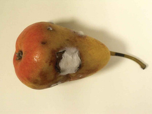
Infection of pears by Alternaria is most frequently found on fruit held in ordinary storage, but it seldom causes extensive loss of fruit in cold storage (Ogawa and English, Citation1991). Alternaria rot is characterized by round, brown to black, dry, firm, shallow lesions around skin breaks or at the calyx or stem depression. Advanced rots become spongy and the affected flesh is streaked with black firm shallow lesions. On pear, the stem decay appears as slow-moving, black lesions extending from the abscission zone toward the flesh. Dark-colored mycelium may be seen on the surface of lesions under moist conditions. At cold storage temperatures, the rot develops slowly, and lesions are usually less than 25 mm in diameter after 5 months. The lesions are often indistinguishable from those caused by Ulocladium spp. (Jones and Aldwinckle, Citation1990). For an example on alternaria rot, refer to .
Figure 3. Pear affected by Alternaria rot.
The causal organism is Alternaria alternata (Fr.) Keissler. Alternaria alternata is a weak pathogen and saprophyte, which can live on dead or debilitated tissue of many plants. It can infect fruit before or after harvest. However, fruit inoculated up to 7 weeks before harvest do not rot on the tree but develop symptoms within 2 months at 0 °C. The fungus infects fruit through weak or injured tissue associated with mechanical or chemical injury, scald, soft scald, physiological senescence, sunburn, and chilling injury (Jones and Aldwinckle, Citation1990).
A. alternata (Fr.) Keissler has been recorded on pears in Israel, Chile, Canada, and the U.S. A. kikuchiana S. Tanaka is reported to cause serious damage to pears in Japan and Korea (Snowdon, Citation1990). It has also been reported (Li et al., Citation2007) that Alternaria alternata can cause rot on “Pingguoli pear” (Pyrus bretschneri Rehd. Cv. Pingguoli), one of the most important cultivated crops in China. Alternaria rot on Pingguoli pear occurs after latent infection: fruit surface can be asymptomatic within 60 days of storage under cold conditions (0 °C, RH 85–90%), but black-grey hyphae could be seen in the lenticels or calyx tube of pears after 90 days of storage (Li et al., Citation2007).
Mucor rot
Mucor rot is a fungal disease of pears in the U.S., Canada, Australia, South Africa, and Europe, where it occurs less constantly than blue mold and grey mold (Jones and Aldwinckle, Citation1990). Mucor rot is generally not a major problem, particularly when good harvest management and water-sanitation practices at packing are implemented (WSU, 2015).
Mucor rot develops at the stem or calyx end of the fruit or at puncture wounds in the skin. Stem-end mucor rot is seen on ‘d’Anjou’ pears (WSU, 2015). The decayed area is light brown to brown with a sharp margin. The decayed tissue is very soft and juicy and can be readily separated from the healthy one. Grey mycelium with dark sporangia may appear on the decayed area. Mucor rot has also a sweet odor. Sporangiophores with sporangia may protrude through cracks in the skin. After about 2 months in cold storage at 0 °C, infected fruit completely decay, releasing large quantities of juice containing sporangiospores, which cause additional infections (Jones and Aldwinckle, Citation1990).
Mucor rot is caused primarily by Mucor piriformis E. Fischer. Other species of Mucor that have been reported to cause decay include M. mucedo P. Mich. Ex Saint-Amans, M. racemosus Fres., and M. strictus Hagem (Jones and Aldwinckle, Citation1990). Mucor piriformis is a soil-borne pathogen and survives in the orchard soil. The pathogen enters packing facilities through infested soils or organic debris adhering to field bins. Drench solutions and dump-tank water are the primary source of inoculum for fruit infection at drenching and packing (WSU, 2015).
Cladosporium rot
Cladosporium rot of apple and pear is associated with wounds or surface injuries to fruit that are accompanied by necrosis. Senescence of fruit in long-term storage may increase the incidence of the disease. Lesions are typically round to oval, less than 2 cm in diameter, dark brown to black, and slightly sunken. Internal diseased tissue is spongy and varies from light brown to black and from wet to dry. The disease is provocated by Cladosporium herbarum (Pers.) Link, a ubiquitous saprophyte found in air, on plant surfaces, and in decaying plant debris (Jones and Aldwinckle, Citation1990).
Coprinus rot
Coprinus rot has been found throughout the Pacific Northwest. It is caused by Coprinus psychromorbidus Redhead & Traquair, a low-temperature basidiomycete. It is often mistaken for bull’s eye rot but Coprinus rot lesions have light centers, which are lacking in early bull’s eye lesions. Fruit infection occurs during the last month before harvest. Lesions are circular, 0.5–25 mm in diameter, and sunken, with dark brown borders and light centers. The decayed tissues are firm and dry (WSU, Citation2015c; Jones and Aldwinckle, Citation1990).
Black rot and white rot
Botryosphaeria obtusa (Schw.) Shoemaker (syn. Physalospora obtusa (Schw.) Cooke) is responsible for black rot of pears, which has been reported in the U.K. and South Africa, but is of particular importance in the Eastern U.S. (Snowdon, Citation1990).
Infections on young fruit begin as reddish flecks that develop into purple pimples 0.10–1.0 mm in diameter. These lesions do not enlarge rapidly until the fruit begin to mature. Incipient infections on more mature fruit are often black, irregularly shaped, and surrounded by a red halo. As the lesions enlarge, they form a series of concentric rings alternating from black to brown, giving to the lesions a “frog-eye” appearance. The lesions remain firm and are not sunken (Biggs and Miller, Citation2004; Jones and Aldwinckle, Citation1990). Colonized mummified fruit are often located in close proximity to the black-rot infected fruit. Black rot often appears late in the season just before harvest as black spots on the fruit associated with mummified fruit left from chemical thinners (Biggs and Miller, Citation2004).
White rot fruit lesions usually become noticeable 4–6 weeks before harvest, although infection may have occurred earlier in the season. Lesions begin as small, often circular, slightly sunken, and brown to tan spots, which may be surrounded by a red halo. As lesions expand in diameter, the rotted area extends in a cylindrical manner toward the core (Jones and Aldwinckle, Citation1990).
In both cases, fruits are invaded via lenticels, mechanical injuries, and insect punctures, and in some cultivars through the calyx resulting in a blossom-end rot or core rot. Both fungi are checked by a storage temperature around 0 °C, but rotting is resumed at temperatures above 10 °C. The decay does not usually spread from fruit to fruit (Snowdon, Citation1990).
Cylindrocarpon rot
The fungus (Nectria galligena Bresad.) causes European canker of apple and pear trees, which occurs in all producer countries, but is of particular importance in the humid climates of northern Europe, the northwestern U.S., and New Zealand.
Lesions may occur at the calyx-end in the form of an “eye-rot,” or anywhere on the fruit surface. The rot is brown, sunken, and with a definite margin between healthy and infected tissue. Internally, the rotted tissue is soft and often has a striated appearance.
Infection can occur through the calyx-end, the stem-end, through insect wounds, or scab lesions, and the fruit may rot on the tree. Alternatively, the fungus may enter a lenticel and remain quiescent until after harvest, when the fruit becomes increasingly susceptible to rotting. The fungus grows best at approximately 20 °C and produces spores only in the presence light (Snowdon, Citation1990).
Phacidiopycnis rot
Phacidiopycnis rot is caused by Phacidiopycnis piri (Fuckel) Weindlymayr, anamorph of the discomycete Potebniamyces pyri (Berkeley & Broome) Dennis (Xiao and Boal, Citation2004). This disease has been recorded in Belgium, France, Italy, Switzerland, Germany, Norway, Denmark, and Russia (Snowdon, Citation1990), and more recently in the U.S., where it is more common on ‘d’Anjou’ pears (Xiao and Boal, Citation2004).
Phacidiopycnis rot causes three types of symptoms on pears: stem-end rot, clayx-end rot, and wound-associated rot originating from infection at the stem, calyx, and wound on the skin of the fruit, respectively (Liu and Xiao, Citation2005). In the early stage of symptom development, the decayed area appears water-soaked and its color varies with age. As the disease progresses, the aging decayed area turns brown and then black, but the margin of decayed area continues to have a water-soaked appearance. Under high relative humidity, the fungus forms white mycelium and pycnidia of the fungus are often formed on the decayed area at advanced stages, starting from the infection sites (WSU, 2015).
The firm leathery texture is somewhat similar to that of brown rot, but phacidiopycnis rot is distinguished by the formation of numerous characteristic greenish-grey bodies up to 1 mm across and clearly visible to the naked eye. These structures darken and exude abundant spores (conidia) in the form of creamy droplets or waxy coils (Snowdon, Citation1990).
In the early stage of the development, Phacidiopycnis rot can also be misdiagnosed as grey mold because of the similarity in symptoms, but the fruit flesh at the margin of Phacidiopycnis rot appears translucent and water soaked, whereas internal decayed flesh of grey mold usually appears brown (WSU, 2015).
Under high relative humidity, the fungus produces fluff mycelium on decayed fruit, which is able to infect surrounding healthy fruit, causing secondary infection through fruit-to-fruit contact (Xiao and Boal, Citation2004).
Aspergillus rots
Aspergillus rots of pears vary greatly in appearance, depending on the species of Aspergillus. Some cause firm and rather moist rots, others dry and leathery rots, while A. niger produces a soft watery rot. Aspergillus rot is associated only with fruits stored under warm conditions (Jones and Aldwinckle, Citation1990). In a recent study Aspergillus niger Van Tiegh was found to cause black mold rot of pear in Kashmir Valley, India (Parveen et al., Citation2014). The infected fruits were characterized by soft watery rot and their surface was covered with black conidial heads of the pathogen.
Optimal conditions for fungal growth in pear fruit
The colonization of any fungi infecting fruit products, as well as the production of mycotoxins, is dependent on an array of factors that are of intrinsic or extrinsic nature. Intrinsic factors include the initial contamination of fruit post-harvest and before storage and the type of contaminant. They also include fruit pH, water activity, as well as the texture and nutritional status of the fruit. As a matter of fact, pears have a low pH, a high water activity, and are a very good source of nutrients, making them an ideal medium for fungal growth (Wheeler et al., Citation1991). Features that are more extrinsic in nature include storage temperature, relative humidity, and the atmosphere composition present while packaging and storing the fruits.
Fruit is expected to be nutritious, forming a rich habitat for microorganisms. It is comprised of living tissues, thus invasion requires that fungal pathogens overcome barriers that the plant has evolved to prevent its death by infection. Consequently, there exists specificity between the fruit and different mold species, through which defense mechanisms present in the fruit are overcome, resulting in spoilage. Being acidic, fruits restrict spoilage predominantly to fungi; therefore, much importance is given to the study of fruit diseases caused by fungal infections (Pitt and Hocking, Citation1997).
Pears are now often sold in areas of the world far away from their production sites. Consequently, the need for retaining their quality, as well as the desire for extended shelf life, has increased (Labuza and Breene, Citation1989). The selection of post-harvest storage conditions for pears reflects the level of fruit spoilage by fungi, which in turn has an effect on fruit losses. There are seven principal physical and chemical factors that govern their spoilage. A summary of the optimal growth conditions for the fungi discussed in the previous sections is summarized in and their importance is described hereunder.
Table 2. Optimal growth conditions for the fungi.
Water activity and relative humidity
By definition, the water activity (aw) of a food is the ratio between the vapor pressure of the food itself, when in a completely undisturbed balance with the surrounding air, and the vapor pressure of distilled water under identical conditions. The water activity increases with temperature and it is a dominant environmental factor, which governs the food stability and spoilage, given that fungi need water to thrive (Department of Health, Education, and Welfare Public Health Service Food and Drug Administration, Citation2015). In an ideal state, microorganisms may contain 80% of water, which is obtained from food in which they grow and multiply.
Water activity does not directly translate to water content; in true fact aw is a measure of the unbound, free water in a system that is available to support the biological and chemical reactions. Hence, water activity is highly affected and is dependent on the micro-environmental levels in food materials. Two different foods may have the same absolute water content but may have very different aw values. This depends on the degree of free water available, which is otherwise bound to food constituents. Each microorganism has a critical aw, below which growth cannot occur. Yeasts and molds usually thrive at aw >0.62 (FAO, Citation2003). With water activities as high as 0.93 in most perishable fruits, such as pears, fungi together with other microorganisms have plenty of water available to thrive (Sandulachi, Citation2012). An overview of the water activity levels for a number of pear-spoilage fungi is given in . A. alternata is seen to have the widest aw range for positive growth, with a minimum of aw 0.84 and an optimum of 0.98–1.0. B. cinerea has a minimum growth requirement of aw 0.856 and an optimum of aw 0.981. Being more hydrophilic, P. expansum has a low of aw 0.895 and an optimum of aw 0.988. On the extreme is R. stolonifera, which is a hyrophile and has a minimum aw >0.9. M. laxa has an optimum of aw 0.99.
Furthermore, minimal changes in relative humidity (RH) of the environment where the food is stored can result in great differences in the rate of microorganism multiplication. This is because water activity is closely related to the relative humidity. Under equilibrium (where the product neither gains nor loses its natural moisture), aw = RH/100 (FAO, Citation2003).
A high RH is required for the protection of fruit from dehydration and weight loss. Consequently, this may encourage the development of fungal pathogens during storage through an enhanced condensation over the fruit surface. In fact, the increase in rate of decay may be attributed to the moisture held within wounds, lenticles, and stomata of the fruit, where fungal spores use this water for germination (Eckert, Citation1975). On the other hand, preventing the dehydration and maintaining the freshness of the fruit may decay less due to a reduced accumulation of dead tissue, which may serve as a good nutrient source for saprophytic fungi. Perishable crops, such as apples and pears, may tolerate relatively low levels of RH due to their well developed cuticle and epidermal layer. However, in 1978, Eckert saw that wounding ‘d’Anjou’ pears before inoculation with Penicillium expansum and Botrytis cinerea and storing them at 97% RH at –1.1 °C did result in successful germination and decay (Atanda et al., Citation2011). Overall, water is a basic main requirement for growth, metabolism, and plays a role in supporting a myriad of chemical reactions occurring in food products. Apart from supporting chemical reactions occurring inside the fruit, water supports the growth of fungi.
Hydrogen ion concentration
A pH that is lower than 5 poses a hostile environment for bacterial growth and thus allows space for fungi to flourish. Low pH conditions are typical environments in most fruits. Fungi are commonly tolerant to a pH range of 3–8 (Wheeler et al., Citation1991). Some conidial fungi are even capable of growing down to pH 2, and yeasts down to pH 1.5. However, as pH moves away from the optimum (pH 5), other growth limiting factors may have a synergistic effect together with pH in supporting fungal growth and promoting fruit spoilage. The ripening of fruit makes it very susceptible to fungal infections. This is partly because, as the fruit ripens, the pH starts to increase making the environment more supportive to fungal growth. However, other factors, such as the softening of the tissue leading to easy bruising and the build of sugars, also make the fruit more easily decayed by fungi as it ripens (Yates et al., Citation1967).
Temperature
After harvest, pears are usually washed and stored at chilled temperatures of 0 to 4 °C. Such a low temperature is assumed to play a preservative role as such conditions are less optimal for fungal growth and germination (refer to for a range of temperatures supporting growth of fungi). Nevertheless, species including Fusarium, Cladosporium, and Penicillium are reported to grow at temperatures as low as –7 °C (Pitt, Citation1979). Cooling fruit after harvest extends the storage life by reducing the rate of physiological change of the tissue and limiting the growth rate of spoilage fungi. Fruit can be kept out from the direct sun. This is a very low-cost method to cool the product. Furthermore, fruit can be harvested in the early morning so it is much cooler when transporting. For long-term storage, fruit is stored in refrigerators or freezers. Nevertheless, Botrytis cinerea is able to infect pears in cold and humid conditions, which is the typical scenario present in storage freezers. The fruit is infected when it is still in the field, before harvest, through very small wounds. An infected area on a pear can easily spread to a healthy pear during the storage period (Monroe, Citation2009). Freezing temperature should not be below 0 °C as fruit may then suffer from freezing injuries and consequently spoil quickly. When perishable fruits such as pears are refrigerated, only healthy looking products should be selected, as a low temperature does not destroy the pathogen present already on the fruit, but will only slow down its multiplication and activity. Moreover, it does not improve the quality of the product, but it only maintains it; hence, a damaged product will spoil much more quickly before a healthy one, even in refrigerated storage. Cooling should be started as quickly as possible after harvest, slowing down the development of disease-causing microbes. This might be a challenge in countries with a hot climate, especially in less-developed countries, where vehicles transporting raw fruit are not equipped by a good refrigeration system. A high temperature during transportation results in tissue softening and bruising, leading to rapid microbial attack and decay (Scott, Citation1957). Penicillium species, such as P. expansum, P. solitum, P.commune, P.verrucosum, P. chrysogenum, and P.regulosum, cause blue mold, which is usually initiated through the entry of Penicillium spp. spores in the fruit, leading to a watery lesion that can easily spread to healthy tissue at ambient temperature (Gock et al., Citation2003). Another problematic infection that may result from the damage of the pear tissue is the Rhizopus soft rot (Pierson et al., Citation1971). In fact, it usually occurs during the transportation, storage, and sale phases (Pitt and Hocking, Citation2009). Cladosporium is also associated with wounds or surface injuries of the pear fruit that leads to tissue necrosis (Jones and Aldwinckle, Citation1990).
Gas tension
The presence of atmospheric oxygen is both important for maintaining fruit freshness during storage and for the growth of spoilage fungi. Fruits are more susceptible to fungal disease during the post-harvest phase due to the increase in the respiration rate. Oxygen availability in air (21%) promotes the production of ethylene, a natural plant hormone that plays a central role in ripening. Oxygen also promotes browning reactions and pigment oxidation. Moreover, the most common spoilage fungi require oxygen for their growth. Food spoilage by filamentous fungi usually occurs under aerobic conditions, so decreasing the oxygen presence may limit fungal growth. However, in controlled atmosphere environments where oxygen levels may be as low as 2%, some fungal species can even be efficient oxygen scavengers and use up oxygen dissolved in the substrate, rather than atmospheric oxygen (Miller and Golding, Citation1949). A clear example is Penicillium expansum, which shows normal growth in 2.1% oxygen over its entire temperature range (Gock et al., Citation2003). Saccharomyces species together with other fermentative yeasts are capable of growth in the complete absence of oxygen (Yates et al., Citation1967). Some species of Mucor, Rhizopus, and Fusarium are able to cause yeast-like fermentative spoilage (Stolk and Dakin, Citation1966). Many other common food spoilage fungi are slightly inhibited when grown in nitrogen atmospheres containing approximately 1.0% oxygen (Hocking, Citation1989).
Fruits are more susceptible to fungal disease during the post-harvest phase due to the increase in the respiration rate. Oxygen availability in air (21%) promotes the production of ethylene, a natural plant hormone that plays a central role in ripening. Oxygen also promotes browning reactions and pigment oxidation. Moreover, most common spoilage fungi require oxygen for their growth.
Carbon dioxide, occurring at a percentage of 0.01% in air, is slightly corrosive in the presence of moisture when dissolving in water and producing carbonic acid. This increases the acidity of the solution and reduces the pH (Scott, Citation1957). Consequently, most food spoilage molds seem to be sensitive to high (0.5% to 2%) levels of carbon dioxide (Taniwaki et al., Citation2001).
Techniques used for the control of post-harvest decay manipulate the concentration of atmospheric gases in air by producing modified atmospheric packaging (MAP). This technique is used to prolong the shelf-life period of foods. Usually, the level of oxygen in the modified atmosphere is lowered, while the level of carbon dioxide is heightened. Such conditions decrease the respiration rate and therefore the product shelf-life is increased (Prasanna et al., Citation2007). The production of ethylene is reduced with higher carbon dioxide levels and lower oxygen. Moreover, ethylene-blocking molecules (1-methylcyclopropene) are added to the MAP to maintain fruit freshness (Escalona et al., Citation2006). Nitrogen is also used in the post-harvest control of packaged fruits. It does not support the growth of aerobic microorganisms and so does not support the growth of most fruit spoilage fungi (Sandhya, Citation2010).
Consistency of fruit
Fruit texture and consistency is dependent on the maturity of the fruit at harvest. The time of harvest will determine the quality of fruit during the post-harvest phase. Immature fruit may lack flavor and the texture is very hard. This is because at this stage fruit contains pectic materials known as protopectins, which have a very high molecular weight. As the fruit matures, the length of this polymer decreases and pectins become water soluble, leading to a softer and mushier texture of fruit.
On the other hand, overripe fruits are easily exposed to mechanical damage, making them highly susceptible to fungal invasion. This is why many fruits are harvested at the mature but unripe stage of development in order to ensure the best eating quality (Spotts and Peters, Citation1982).
Overripe pears may be susceptible to infections caused by blue mold (Penicillium spp.) and Rhizopus rot. The latter is a saprophyte and lives off dead tissue (Monroe, Citation2009; Pierson et al., Citation1971). On the other hand, immature, unripe, and uninjured pears are susceptible to Colletotrichum gloesporioides infection, which is the cause of Bitter rot (Jones and Aldwinckle, Citation1990; Pierson et al., Citation1971).
Other factors
These include the nutrient status and the specific solutes. Carbohydrate-rich substrates are very suitable for fungal growth, unlike proteinaceous foods, which serve as substrates for bacteria. Most molds are equally indifferent to nitrogen source, using nitrate, ammonium ions, or organic nitrogen sources with equal ease (Department of Health, Education, and Welfare Public Health Service Food and Drug Administration, Citation2015). The presence of specific solutes may pose additional effects on the growth of fungal species. Hocking and Pitt (Citation1979) showed that germination and growth of several Penicillum species was poorly affected when the aw medium was controlled with glucose–fructose, glycerol, or sodium chloride.
Conclusion
Physical and chemical factors determine the growth and germination of fruit fungi. Their definition may be useful to determine the fruit’s stability and ways to prevent its decay by proper storage conditions. As discussed, such factors act synergistically together and not in isolation. Further studies will require the in vivo assessments of the fungal growth under different environmental conditions.
Related Research Data
Literature cited
- Ames, G.K., and H. Born. 2012. Pears: Organic production. 13 August 2015. http://ucanr.edu/sites/placernevadasmallfarms/files/112366.pdf.
- Amiri, A., W. Chai, and G. Schnabel. 2011. Effect of nutrient status, pH, temperature and water potential on germination and growth of Rhizopus stolonifer and Gilbertella persicaria. J. Plant Pathol. 93(3):603–612.
- Atanda, S.A., P.O. Pessu, S. Agoda, I.U. Isong, and I. Ikotun. 2011. The concepts and problems of post-harvest food losses in perishable crops. African Journal of Food Science. 5(11):603–613.
- Biggs, A.R., and S.S. Miller. 2004. Relative susceptibility of selected apple cultivars to fruit rot caused by Botryosphaeria obtusa. HortScience 39(2):303–306.
- Brzonkalik, K., D. Hümmer, S. Syldatk, and A. Neumann. 2012. Influence of pH and carbon to nitrogen ratio on mycotoxin production by Alternaria alternate in submerged cultivation. AMB Exp. 2:28. DOI: 0.1186/2191-0855-2-28.
- Casals, C., I. Viñas, R. Torres, C. Griera, and J. Usall. 2010. Effect of temperature and water activity on in vitro germination of Monilinia spp. J. Appl. Microbiol. 108(1):47–54.
- Cotoras, M., C. García, and L. Mendoza. 2009. Botrytis cinerea isolates collected from grapes present different requirements for conidia germination. Mycologia 101( 3):287–295.
- Department of Health, Education, and Welfare Public Health Service Food and Drug Administration. 2015. Water activity (aw) in foods. http://www.fao.org/docrep/V5030e/V5030E0b.htm.
- Douglas, S.M. 2003. Disease control for home pear orchards. Connect Agr. Expt. Stat.
- Eckert, J.W. 1975. Postharvest pathology - general principles. In: E.B. Pantastico (ed.). Postharvest physiology, handling and utilization of tropical and subtropical fruits and vegetables. Avi Publ. Co., Westport, CT.
- El Arbi, B., E.L. Salem, I.S. Brahim, M.K. Issam, H. Lahoucine, and H. Abderraouf. 2014. Screening of actinomycete bacteria producing antifungal metabolites which could be used in biological control against a phytopathogenic fungus (Rhizopus Stolonifer). Amer. J. Biol. Life Sci. 2(4):84.
- Ellis, M.A. 2008. Phytophtora root and crown rot of fruit trees. Ohio State Univ. Ext.
- Escalona, V.H., B.E. Verlinden, S. Geysen, and B.N. Nicolai. 2006. Changes in respiration of fresh-cut butter head lettuce under controlled atmospheres using low and super atmospheric oxygen conditions with different carbon dioxide levels. Postharvest Biol. Technol. 39(1):48–55.
- Food and Agriculture Organization (FAO). 2003. Handling and preservation of fruits and vegetables by combined methods for rural areas. http://www.fao.org/docrep/005/y4358e/y4358e00.htm
- Gock, M.A., A.D. Hocking, J.L. Pitt, and P.G. Poulos. 2003. Influence of temperature, water activity and pH on growth of some xerophilic fungi. Int. J. Food Microbiol. 81(1):11–19.
- Gougouli, M., and K.P. Koutsoumanis. 2010. Modelling growth of Penicillium expansum and Aspergillus niger at constant and fluctuating temperature conditions. Int. J. Food Microbiol. 140(2):254–262.
- Gubler, D.W., B.G. Zoller, and R.A. Duncan. 2007. Diseases, p. 145. In: Pear production and handling material. University of California Agriculture and Natural Resources, Oakland, CA.
- Harris, J.E., and C. Dennis. 1980. Distribution of Mucor piriformis, Rhizopus sexualis, and R. stolonifer in relation to their spoilage of strawberries. Trans. Brit. Mycol. Soc. 75(3):445–450.
- Hartman, J., and B. Eshenaur. 2002. Powdery mildew. In: Plant pathology fact sheet. West Virginia University, Morgantown, WV.
- Henriquez, J.L., D. Sugar, and R.A. Spotts. 2004. Etiology of bull’s eye rot of pear caused by Neofabraea spp. in Oregon, Washington, and California. Plant Dis. 88(10):1134–1138.
- Henriquez, J.L., D. Sugar, and R.A. Spotts. 2008. Effects of environmental factors and cultural practices on bull’s eye rot of pear. Plant Dis. 92(3):421–424.
- Hocking, A.D. 1989. Responses of fungi to modified atmospheres. Fumigation and controlled atmosphere storage of grain. ACIAR Proceedings n. 25, Singapore, 14–18 Feb. 1989, p. 14–18.
- Hosen, M.I., A.U. Ahmed, and M.R. Islam. 2010. Physiological variability and in vitro antifungal activity against Botrytis cinerea causing Botrytis gray mold of chickpea (Cicer arietinum L.). Span. J. Agr. Res. 8( 3):750–756.
- Hubballi, M., S. Nakkeeran, T. Raguchander, T. Anand, and R. Samiyappan. 2010. Effect of environmental conditions on growth of Alternaria alternata causing leaf blight of noni. World J. Agr. Sci. 6(2):171–177.
- Jones, A.L., and H.S. Aldwinckle. 1990. Compendium of apple and pear diseases, 224 p. APS Press, Saint Paul, MN.
- Judet-Correia, D., S. Bollaert, A. Duquenne, C. Charpentier, M. Bensoussan, and P. Dantigny. 2010. Validation of a predictive model for the growth of Botrytis cinerea and Penicillium expansum on grape berries. Int. J. Food Microbiol. 142(1):106–113.
- Labuza, T., and W. Breene. 1989. Applications of “active packaging” for improvement of shelf‐life and nutritional quality of fresh and extended shelf‐life foods. J. Food Proc. Preserv. 13(1):1–69.
- Larsen, H., and L. Pokharel. 2011. Powdery mildews of stone fruit crops. Colorado State University. http://webdoc.agsci.colostate.edu/aes/wcrc/techbulletins/powdery%20mildews%20of%20fruit%20crops-western%20Colorado-2011-05.pdf
- Li, B., T. Lai, G. Qin, and S. Tian. 2009. Ambient pH stress inhibits spore germination of Penicillium expansum by impairing protein synthesis and folding: A proteomic-based study. J. Proteome Res. 9(1):298–307.
- Li, Y., Y. Bi, and L. An. 2007. An occurrence and latent infection of Alternaria rot of Pingguoli pear (Pyrus bretschneideri Rehd. cv. Pingguoli) fruits in Gansu, China. J. Phytopathol. 155(1):56–60.
- Liu, Q., and C. Xiao. 2005. Influence of nutrient and environmental factors on conidial germination of Potebniamyces pyri. Phytopathology 95(5):572–580.
- Mari, M., P. Bertolini, and G.C. Pratella. 2003. Non-conventional methods for the control of post-harvest pear diseases. J. Appl. Microbiol. 94:761–766.
- McManus, P. 1997. Apple and pear disorder: Sooty blotch and flyspeck. 13 August 2015. http://polk.uwex.edu/files/2014/02/Sooty-blotch-and-Flyspeck-A3173.pdf.
- Miller, D.D., and N. Golding. 1949. The gas requirements of molds. V. The minimum oxygen requirements for normal growth and for germination of six mold cultures 1, 2. J. Dairy Sci. 32(2):101–110.
- Monroe, A. 2009. Integrated pest management for Australian apples and pears: NSW Industry and Investment Management. 13 August 2015. http://www.dpi.nsw.gov.au/__data/assets/pdf_file/0009/321201/ipm-for-australian-apples-and-pears-complete.pdf.
- Ogawa, J.M., and H. English. 1991. Diseases of temperate zone tree fruit and nut crops, 464 p. UCANR Publications, Oakland, CA.
- Oviedo, M.S., M.L. Ramirez, G.G. Barros, and S.N. Chulze. 2011. Influence of water activity and temperature on growth and mycotoxin production by Alternaria alternata on irradiated soya beans. Int. J. Food Microbiol. 149(2):127–132.
- Palmer, L.T., and J. Riesselman. 1972. EC72-1855 Apple Diseases (1972). Historical Materials from University of Nebraska-Lincoln Extension. Paper 4191. http://digitalcommons.unl.edu/cgi/viewcontent.cgi?article=5192&context=extensionhist
- Parveen, S., A.H. Wani, M.Y. Bhat, S.A. Pala, and A.A. Ganie. 2014. Biology and management of Aspergillus niger Van Tiegh. causing black mold rot of pear (Pyrus communis L.) in Kashmir Valley, India. International Journal of Advanced Research 2(6):24–34.
- Pierson, C.F., M.J. Ceponis, and L.P. McColloch. 1971. Market diseases of apples, pears, and quinces, p. 1–96. In: Agriculture Handbook, vol. 376. Agricultural Research Service, U.S. Department of Agriculture, Washington, D.C.
- Pitt, J.I. 1979. The genus penicillium and its teleomorphic states eupenicillium and talaromyces. Mycologia 73(3):582–584.
- Pitt, J.I., and A.D. Hocking. 1997. Influence of solute and hydrogen ion concentration on the water relations of some xerophilic fungi. J. Gen. Microbiol. 101(1):35–40.
- Pitt, J.I., and A.D. Hocking. 2009. Spoilage of stored, processed and preserved foods, p. 401–421. In: Fungi and food spoilage. Springer, New York, NY.
- Prasanna, V., T.N. Prabha, and R.N. Tharanathan. 2007. Fruit ripening phenomena—An overview. Crit. Rev. Food Sci. Nutr. 47( 1):1–19.
- Pscheidt, J.W., and C.M. Ocamb. 2014. Pacific northwest plant disease management handbook. Oregon State Univ., Corvallis, OR.
- Rivard, C. 2007. Phytophtora cactorum. PP728 Pathogen Profile, North Carolina State University. https://www.cals.ncsu.edu/course/pp728/cactorum/Pcactorum.html
- Sandhya. 2010. Modified atmosphere packaging of fresh produce: Current status and future needs. LWT–Food Sci. Technol. 43(3):381–392.
- Sandulachi, E. 2012. Water activity concept and its role in food preservation. Technical University of Moldova. http://www.utm.md/meridian/2012/MI_4_2012/8_Art_Sandulachi_E_Water.pdf.
- Schipper, M.A., and J.A. Stalpers. 1984. A revision of the genus Rhizopus. Centraalbureau Voor Schimmelcultures, the Netherlands.
- Schubert, T.S. 1983. Bitter rot of apple. Plant Pathology Circular 248.
- Scott, W. 1957. Water relations of food spoilage microorganisms. Adv. Food Res. 7(1):83–127.
- Shim, J.O., K.D. Choi, K.D. Hahn, J.H. Lee, I.H. Hyun, T.S. Lee, I.K. Kyoung, P.L. Hai, and W.L. Min. 2002. Blue mold of pear caused by Penicillium aurantiogriseum in Korea. Mycobiology 30(2):105–106.
- Snowdon, A.L. 1990. Pome fruits, p. 170–216. In: A colour atlas of postharvest diseases and disorders of fruits and vegetables, volume 1. Wolfe Scientific, London, UK.
- Spotts, R.A., and S. Castagnoli. 2010. Pear scab in Oregon: Symptoms, disease cycle and management. Oregon State Univ. Ext. Serv. 13 August 2015. https://catalog.extension.oregonstate.edu/sites/catalog.extension.oregonstate.edu/files/project/pdf/em9003.pdf.
- Spotts, R.A., and L. Cervantes. 1991. Effect of temperature and wetness on infection of pear by Venturia pirina and the relationship between pre-harvest inoculation and storage scab. Plant Dis. 75(12):1204–1207.
- Spotts, R.A., and B.B. Peters. 1982. Use of surfactants with chlorine to pear decay control. Plant Dis. 66:725–777.
- Stolk, A.C., and J. Dakin. 1966. Moniliella, a new genus of moniliales. Antonie Van Leeuwenhoek 32(1):399–409.
- Sugar, D., and R. Spotts. 1992. Sources of inoculum of Phialophora malorum, causal agent of side rot of pear. Phytopathology 82(7):735–738.
- Sutton, T.B., H.S. Aldwinckle, A.M. Agnello, and J.F. Walgenbach. 2014. Compendium of apple and pear diseases and pests, 2nd ed. APS Press, Saint Paul, MN.
- Taba, S., K. Teruya, and Z. Moromizato. 2006. Rhizopus rot of balsam pear (Momordica charantia) by Rhizopus stolonifer var. stolonifer. Jap. J. Phytopathol 72:22–24.
- Taniwaki, M., A. Hocking, J. Pitt, and G. Fleet. 2001. Growth of fungi and mycotoxin production on cheese under modified atmospheres. Int. J. Food Microbiol. 68(1):125–133.
- The Royal Horticultural Society. 2015. Brown rot. https://www.rhs.org.uk/advice/profile?pid=114
- University of Illinois. 2000. RPD No. 942 - Gray-Mold Rot or Botrytis Blight of Vegetables. http://ipm.illinois.edu/diseases/series900/rpd942/
- University of Illinois. 2005a. Bitter Rot of Apple. https://ipm.illinois.edu/diseases/rpds/818.pdf
- University of Illinois. 2005b. Sooty Blotch and Flyspeck of Apple. https://ipm.illinois.edu/diseases/rpds/815.pdf
- Van der Zwet, T., and A.R. Briggs. 2004. Fabraea leaf spot. West Virginia Univ. Ext.
- Wheeler, K.A., B.F. Hurdman, and J. Pitt. 1991. Influence of pH on the growth of some toxigenic species of Aspergillus, Penicillium and Fusarium. Int. J. Food Microbiol. 12(2):141–149.
- Wilcox, W. 1994. Sooty blotch Gloeodes pomigena (Schwein.) Colby and flyspeck Shizothyrium pomi. 13 August 2015. http://nysipm.cornell.edu/factsheets/treefruit/diseases/sbfs/sbfs.pdf.
- Won, K., H. Bastiaanse, Y.K. Kim, J.H. Song, S.S Kang, H.C. Lee, K.H. Cho, L. Brewer, G. Singla, S.E. Gardiner, D. Chagné, and V.G.M. Bus. 2014. Genetic mapping of polygenic scab (Venturia pirina) resistance in an interspecific pear family. Mol. Breeding 34(4):2179–2189.
- Washington State University (WSU). 2015a. Postharvest Diseases of Apples and Pears. http://decay.tfrec.wsu.edu/displayPage.php?id=pathlab&pn=20
- Washington State University (WSU). 2015b. Tree Fruit Market Diseases. http://postharvest.tfrec.wsu.edu/market/pinkmold
- Washington State University (WSU). 2015c. Postharvest Diseases and Disorders of Apples and Pears. http://postharvest.tfrec.wsu.edu/pages/N7I3A
- Xiao, C. 2006. Postharvest fruit rots in d’Anjou pears caused by Botrytis cinerea, Potebniamyces pyri, and Sphaeropsis pyriputrescens. Plant Health Progress. 13 August 2015. http://www.plantmanagementnetwork.org/pub/php/diagnosticguide/2006/pears/.
- Xiao, C., and R. Boal. 2004. Prevalence and incidence of Phacidiopycnis rot in d’Anjou pears in Washington state. Plant Dis. 88(4):413–418.
- Yamak, F., T. Peever, G. Grove, and R. Boal. 2002. Occurrence and identification of Phytophthora spp. pathogenic to pear fruit in irrigation water in the Wenatchee River Valley of Washington State. Phytopathology 92(11):1210–1217.
- Yates, A., A. Seaman, and M. Woodbine. 1967. Growth of byssochlamys nivea in various carbon dioxide atmospheres. Can. J. Microbiol. 13(8):1120–1123.
- Yu, T., C. Yu, F. Chen, K. Sheng, T. Zhou, M. Zunun, O. Abudu, S. Yang, and X. Zheng. 2012. Integrated control of blue mold in pear fruit by combined application of chitosan, a biocontrol yeast and calcium chloride. Postharvest Biol. Technol. 69:49–53.
- Zong, Y., B. Li, and S. Tian. 2015. Effects of carbon, nitrogen and ambient pH on patulin production and related gene expression in Penicillium expansum. Int. J. Food Microbiol. 3(206):102–108. DOI: 10.1016/j.ijfoodmicro.2015.05.007.