
ABSTRACT
A micropropagation method has been developed for the first time for difficult to propagate small fruit plant medlar (Mespilus germanica L.). Embryonic axes excised from surface-disinfected and microbe-free seed(s), were inoculated on Murashige and Skoog (MS) medium for activation/germination. Axillary bud(s) derived from embryonic axes were used as explants, cultured on the MS medium supplemented with different concentrations of 6-benzylaminopurine (BA) and α-naphthaleneacetic acid (NAA). The 86.33% of the explant exhibited shoot induction within 59.00 days on media with 2.00 mg l–1 BA together with 0.50 mg l–1 NAA. The highest number (8.50/plantlet) of leaf, and node (7.53/plantlet) and the longest (7.26 cm/plantlet) root length were obtained on MS with 2.00 mg l–1 BA together with 1.00 mg l–1 NAA. Maximum number (6.30/plantlet) of shoot and root (5.66/plantlet) were regenerated on MS medium supplemented with 0.50 mg l–1 BA together with 1.00 mg l–1 NAA. Plantlets were transferred to pots filled with perlite and peat moss in equal proportions for acclimatization. Cent percent of the cloned plantlets survived under ex vitro conditions. The method described will be useful for rapid multiplication of medlar for commercial purposes.
Introduction
Medlar (Mespilus germanica L.) belonging to the Rosaceae family is a large shrub or small tree, native to the eastern Mediterranean area. This plant is cultivated for its medicinal and nutritional properties (Ercisli et al., Citation2012; Rop et al., Citation2011). This plant produces a low number of seeds containing live embryos that are the problems of seed and seed germinations. Plants are propagated by vegetative methods like cutting, layering, budding, and grafting. These methods are too slow to meet rapidly growing demands and the needs of large-scale plantations (Pati et al., Citation2013). Hard rooting of stem cuttings, time-consuming sucker propagation, and production of very few suckers over a long period of time are some of the most important problems for traditional propagation of medlar.
Micropropagation through tissue culture is a useful technique for the regeneration of large numbers of plants from explants of mother plants in a relatively short period and, crucially, without seasonal restrictions. Shoot culture through existing meristems may produce plants with little somaclonal variation (Ďurkovič, Citation2008). In vitro propagation of trees has been recognized as an important and efficient method for large-scale propagation and overcoming problems caused by heterogeneous seed production (Campbell et al., Citation2003; Nunes et al., Citation2018).
Commercial use of propagating woody plants through tissue culture is limited to some species due to high variability and plantlets’ survival during acclimatization (Nunes et al., Citation2018). Plant reproductive biology is the basis of improvement by conventional or biotechnological methods. Some woody plants are propagated through seed (Al-Safadi and Elias, Citation2011; Sharma, Citation2016; Shekhawat and Manokari, Citation2016) and zygotic embryo (Buendia-Gonzalez et al., Citation2012; Rai et al., Citation2007; Rout, Citation2005) under in vitro condition. Some researchers applied seedling-derived explants for in vitro proliferation (Shekhawat and Manokari, Citation2016; Wang et al., Citation2008; Yildirim, Citation2012). Shoot culture through existing meristems is the most popular method of in vitro propagation. This method is most popular in woody plants for shoot multiplication (Sharma, Citation2017).
Micropropagation of species belonging to the Rosaceae family has been reported by various researchers using axillary bud, node, callus, leaf, root, seed, runner, embryo, apical meristem, and other explants (Işıkalan et al., Citation2008; Pati et al., Citation2013). Axillary bud proliferation is important, and is induced to grow, and when desired, excised, rooted, and acclimatized (Read and Preece, Citation2014). A number of protocols have been reported for micropropagation of species belonging to the Rosaceae family (Işıkalan et al., Citation2008; Pati et al., Citation2013).
There is no published data on micropropagation of M. germanica L. Therefore, the purpose of the current research was to evaluate the effect of different concentrations of 6-benzylaminopurine (BA) and α-naphthaleneacetic acid (NAA), individually and in combination, on in vitro propagation of M. germanica L. using embryogenic axes and axillary buds as primary and secondary explants, respectively, by the direct organogenesis method, which was performed for the first time.
Materials and Methods
Experiment Conditions
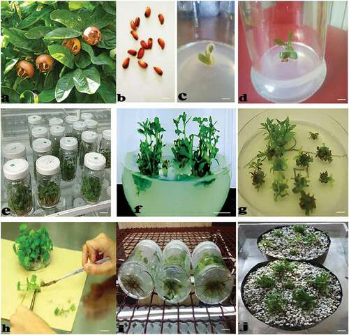
In this experiment, medlar (Mespilus germanica L.), a fruit tree species, was used as mother plants (). Medlar is a bisexual and self-pollinating species. Fruits of this species were collected from the forest trees of Hyrcanian forests in the northern part of Iran. Fruits were selected from healthy mature trees without any symptoms. These fruits were selected to use the embryonic axes of their seeds () as a primary explant. The experiments were performed in greenhouse and laboratory of the Hyrcan Agriculture and Biotechnology Research Institute, Amol city, Mazandaran province, the northern part of Iran, on 2017.
Figure 1. Micropropagation process of Mespilus germanica L. (a) mother plant containing fruits, (b) seeds, (c) germinated embryonic axis in culture medium without PGRs, (d) grown embryonic axis, (e) shoot multiplication through cultivation of axillary buds as secondary explants obtained from the germinated embryonic axes in media supplemented with different concentrations of BA and NAA, (f) produced shoots in medium enriched with 1.00 mg l–1 in combination with 0.50 mg l–1 BA after 45 days, (g) produced shoots in mentioned medium ready for sub-culture, (h) isolation of shoots for proliferation, (i) rooting of shoots, and (j) transfer of plantlets to the pots containing peat moss and perlite (1:1) (scale bar = 10 mm)
Explant Source and Sterilization
Morphologically-uniform and healthy seeds were washed thoroughly under running tap water for 10 min. with a few drops of washing liquid followed by fully rinsed with running tap water. Seeds were transferred to aseptic condition in a laminar air flow cabinet and were surface sterilized by dipping into 70% (v/v) ethanol for 2 min. followed by agitation for 10 min. in a sodium hypochlorite (NaOCl) solution containing 5% available chlorine and 0.05% (v/v) Tween-20, after which the seeds were then rinsed five times with sterile-distilled water. Sterilized seeds coats were broken, their embryonic axes were removed and used as a primary explant.
Culture Medium, Treatments and Measured Parameters
The surface-sterilized embryonic axes were inoculated on MS (Murashige and Skoog, Citation1962) medium () without plant growth regulators (PGRs). Axillary buds produced from germinated embryonic axes () were cultured in MS media supplemented with 0.00, 0.50, 1.00 and 2.00 mg l–1 of BA and NAA, individually and in combination (). Sucrose (3%) was used as carbon source and media were solidified with agar–agar at a concentration of 0.7% (w/v). The pH of the media was adjusted to 5.7 ± 0.10 using 1 N NaOH or 0.1 N HCl prior to autoclaving for 20 min. at 15 psi, 121°C. The cultures were maintained in a plant growth chamber at 20 ± 2°C under a 12-h photoperiod, with photosynthetic flux density (PFD) of approximately 50 μmol m−2 s−1 provided by cool white fluorescent lamps and 75–80% relative humidity (RH). Axillary buds were sub-cultured twice every 45 days in fresh media (). Roots were removed for sub-culturing the buds. After 90 days of last sub-culture, some growth parameters including plantlet height, shoot number, node number, leaf number, root length, root number, explants with shoot induction and time for shoot induction were determined.
Plantlets Acclimatization
Plantlets with well-developed roots were taken out from culture vessels and washed with sterilized distilled water to remove adherent nutrients. They were then planted in a mixture of perlite and peat moss in 1:1 (v/v) ratio. Plantlets were covered by thin plastic bags and transferred to a greenhouse at a temperature of 25 ± 1°C with 70–80% RH. These plantlets were watered regularly. Plantlets were exposed gradually to external environment by removing the plastic bags of the pots.
Experimental Design
The setup of the experiment was in a completely randomized design (CRD), and a minimum of 9 replicates per treatment were repeated three times. Data on the measured parameters were recorded from 144 explants after 90 days of last sub-culture. The results were expressed as mean ± SD of the experiments conducted thrice.
Data Analysis
The statistical analysis of data was performed using Statistical Package for Social Sciences (SPSS) v 16.0. Least significant difference (LSD) test at P < .05 was used to find out the significance of differences among the mean values. Correlation procedure of SPSS statistical software was used to calculate the correlation coefficients between features.
Results
Shoot Induction
Statistically significant differences were recognized between the means for explants with shoot induction and PGRs, individually and/or in combination. Means comparison of the effect of different concentrations of BA (), and NAA () on shoot induction and other measured parameters have been presented. shows that the highest shoot induction (77.50%) was induced using 2.00 mg l–1 BA. Also, through the use of different concentrations of NAA, the highest shoot induction (78.90%) was induced with 0.50 mg l–1 (). Explants with shoot induction in the media was changed significantly with the use of BA in combination with NAA (). Explants with shoot induction in MS basal media supplemented with 0.50 mg l–1 NAA together with 2.00 mg l–1 BA (86.33%), 0.50 mg l–1 NAA together with 0.50 mg l–1 BA (85.00%), and 1.00 mg l–1 NAA together with 0.50 mg l–1 BA (85.00%) was the maximum. Explants with shoot induction in medium supplemented with 0.50 mg l–1 BA without NAA (58.66%) was the minimum ().
Table 1. Mean comparison of the effect of different concentrations of BA on measured parameters of Mespilus germanica L. grown in vitro condition
Table 2. Mean comparison of the effect of different concentrations of NAA on measured parameters of Mespilus germanica L. grown in vitro condition
Table 3. Mean comparison of the effect of different concentrations of BA and NAA on measured parameters of Mespilus germanica L. grown in vitro condition
Time for Shoot Induction
Time for shoot induction in medium fortified with 1.00 mg l–1 NAA together with 0.50 mg l–1 BA was the minimum (59.00 day). Time for shoot induction in medium fortified with 0.50 mg l–1 NAA together with 0.50 mg l–1 BA was the maximum (96.66 day) (). There was a statistically significant difference among different concentrations of NAA (p ≤ 0.01), also NAA in combination with BA (p ≤ 0.05) and time for shoot induction. There was no significant difference between different concentrations of BA and time for shoot induction.
Plantlet Height
Differences in plantlet height in axillary buds grown under different concentrations of BA and NAA, individually or in combination were significant (p ≤ 0.01). Among all concentrations of BA used individually, highest and lowest plantlet height (6.14 and 3.40 cm, respectively) were induced in explants treated with 1.00 mg l–1 and control plantlets (). On the other hand, among all concentrations of NAA used individually, highest and lowest plantlet height (5.90 and 3.56 cm, respectively) were induced in plantlets treated with 1.00 mg l–1 and control plantlets (). Overall, the highest plantlet height (9.06 cm) was obtained in explants treated with 1.00 mg l–1 NAA in combination with 1.00 mg l–1 BA. Height (7.63 cm) was high in plantlets treated with 1.00 mg l–1 NAA in combination with 2.00 mg l–1 BA. The lowest plantlet height (2.23 cm) was obtained in untreated (control) axillary buds. All BA-free treatments stimulated plantlets growth less than 4.00 cm ().
Shoot Number
The high number of shoots was obtained in treatments containing BA in combination with NAA. The highest number of shoot (6.30 per explant) was observed when the explants were grown on medium supplemented with 1.00 mg l–1 NAA in combination with 0.50 mg l–1 BA. The treatment containing 0.50 mg l–1 NAA together with 2.00 mg l–1 BA stimulated more shoots than other treatments by inducing 5.50 shoots per explant after the mentioned above treatment (). There was no positive correlation between enhancing NAA and BA concentrations and increasing the number of shoot (). The data clearly showed that there was a significant difference between different concentrations of NAA and BA for the number of shoot.
Node Number
The highest number of node (7.53 per explant) was obtained when the explants were grown on medium enriched with 1.00 mg l–1 NAA together with 2.00 mg l–1 BA (). Treatment of 1.00 mg l–1 NAA together with 1.00 mg l–1 BA was suitable for increasing node number (6.60). The least node number (3.03) was calculated in explants treated with 1.00 mg l–1 NAA and control. Among all concentrations of BA used individually, maximum and minimum node number (4.80 and 3.85, respectively) were obtained in explants treated with 1.00 mg l–1 and control plantlets (). On the other hand, among all concentrations of NAA used individually, maximum and minimum node number (5.51 and 3.17) were induced in explants treated with 1.00 mg l–1 and untreated explants, respectively (). There was significant difference between treatments of NAA also NAA in combination with BA and node number (p ≤ 0.01). No significant difference was observed between BA and node number.
Leaf Number
The data clearly showed that leaf number is strongly affected by treatments of BA (p ≤ 0.05), NAA (p ≤ 0.01) and BA together with NAA (p ≤ 0.01). The highest number of leaf (8.50 and 8.43 per explant) was obtained in plantlets grown on media enriched with 1.00 mg l–1 NAA together with 2.00 mg l–1 BA and 1.00 mg l–1 NAA together with 1.00 mg l–1 BA, respectively (). The lowest number of leaf (3.53) was obtained in control plantlets. Among all concentrations of BA used individually, the highest and lowest number of leaf (6.53 and 5.60, respectively) were induced in explants grown on media enriched with 2.00 and 0.50 mg l–1 (). Also, among all concentrations of NAA used individually, the highest and lowest number of leaf (7.99 and 4.30, respectively) were induced in plantlets grown on media enriched with 1.00 mg l–1 and control plantlets ().
Root Length
The highest average of the root length (7.26 cm per explant) was obtained in plantlets treated with 1.00 mg l–1 NAA in combination with 2.00 mg l–1 BA. Root length (7.06 cm) was high in medium containing 2.00 mg l–1 NAA without BA (). The lowest average of the root length (4.20 cm) was measured with treatment of 2.00 mg l–1 BA without NAA. Concerning the root length induced by various levels of NAA used individually, the highest and lowest length (6.28 and 5.19 cm) were obtained in media containing 1.00 mg l–1 and control, respectively (). There was statistically significant difference among different concentrations of NAA also NAA in combination with BA and root length (p ≤ 0.01). There was no significant difference between different concentrations of BA and root length.
Root Number
Rooting efficiency was satisfactory using NAA at a concentration of 1.00 mg l–1 (, ). Maximum number of roots (5.66 and 5.63 per shoot) were produced in media fortified with 1.00 mg l–1 NAA together with 0.50 mg l–1 BA and 1.00 mg l–1 NAA together with 2.00 mg l–1 BA, respectively (). Shoots cultured on medium without PGRs produced the least roots (3.10). Concerning the root number produced with various levels of NAA and BA used individually, the highest and lowest number (5.00 and 3.53) were obtained in media fortified with 1.00 mg l–1 NAA and control, respectively (). Statistically significant difference was observed between the mean for root number and PGRs.
Ex Vitro Establishment of Plantlets
Well-developed plantlets were transferred to small plastic pots containing peat moss and perlite in 1:1 (v/v) ratio for ex vitro establishment. Then, plantlets were transferred to big plastic pots filled with the same medium (). The acclimatized plantlets were covered by thin plastic bags and transferred to the greenhouse with 100% establishment rate.
Discussion
Medlar has medicinal and nutritional properties. There are several reports on in vitro propagation of the Rosaceae family members (Pati et al., Citation2013) and many fruit trees and shrubs (Hassan and Zayed, Citation2018; Sharma, Citation2017), but there are no reports on in vitro propagation of M. germanica L. Based on the results presented in , during the individual use of BA and NAA, the highest shoot multiplication and root induction was obtained using a concentration of 1.00 mg l–1 of both BA and NAA. However, the presence of both auxin and cytokinin in the culture medium is necessary for maximum shoot multiplication and root induction (). The synergistic effect of auxin and cytokinin for improvement of shoot multiplication has been reported in a number of woody plants (Dalal et al., Citation2006; Dinesh et al., Citation2019; Fan et al., Citation2017; Kaewpoo and Te-chato, Citation2009; Kaviani and Negahdar, Citation2017; Mathur et al., Citation2002; Sharma, Citation2017; Siwach and Gill, Citation2011; Venkatachalam et al., Citation2015). Damiano et al. (Citation2008) showed that BA together with IBA at low concentrations induced the highest frequency of shoot multiplication in Myrtus communis L., Punica granatum L. and Morus alba L., three fruit trees. Similar findings were reported by Kaewpoo and Te-chato (Citation2009) in Jatropha curcas, a fruit tree. Application of 1.00 mg l–1 BAP together with 1.00 mg l–1 IBA was appropriate for in vitro multiplication of apple (Dalal et al., Citation2006). This finding is similar to our finding. The combination of BAP (3.0 mg l–1) and IBA (0.5 mg l–1) was found to be the most suitable for the highest shoot induction from nodal explants derived from in vitro seedlings of Bambusa arundinacea (Venkatachalam et al., Citation2015). A combination of cytokinin and auxin was also used for shoot multiplication in some other woody species such as Salvadora persica (Mathur et al., Citation2002) and Ficus religiosa (Siwach and Gill, Citation2011). Similar to our findings, some studies have shown that in a medium containing cytokinins individually or in combination with auxins, apical and axillary bud explants produced an appropriate number of shoots (Dinesh et al., Citation2019; Kaviani and Negahdar, Citation2017; Sharma, Citation2017; Sharma et al., Citation2017). In many woody species, cytokinins especially BAP and Kin were effective for shoot induction and multiplication (Sharma, Citation2017). Higher concentrations of cytokinins are inhibitory in some woody plants (Kaviani and Negahdar, Citation2017; Nair and Seeni, Citation2001; Tornero et al., Citation2000). Positive effect of other cytokinins such as TDZ, BA and Zeatin on shoot induction and multiplication were reported in some woody plants (Fan et al., Citation2017; Gonzalez-Rodriguez et al., Citation2010; Kereša et al., Citation2012; Marriott and Sarasan, Citation2010; Sharma et al., Citation2017; Yildirim, Citation2012). In Pistacia lentiscus L., 1 mg l–1 BA was used for shoot initiation and multiplication (Yildirim, Citation2012).
In current study, simultaneous use of BA and NAA increased the number of leaf and node. Simultaneous use of BA and NAA has been suggested for the successful propagation of several woody plants in vitro (Ďurkovič, Citation2008; Savita et al., Citation2010; Sharma et al., Citation2017; Thakur and Karnosky, Citation2007). Sharma et al. (Citation2017) showed that MS medium with 3.00 mg l−1 BA was optimum for shoot bud induction in Bauhinia racemosa Lam., a leguminous tree. Shoot multiplication was further enhanced on MS medium containing 0.25 mg l−1 of BA together with 0.10 mg l−1 of NAA. In Couroupita guianensis, a medicinally tree, maximum number of shoots were regenerated on MS medium enriched with 1.00 mg l−1 each of BAP and Kin together with 0.50 mg l−1 NAA (Mahipal et al., Citation2016). Contrary to our findings, the combination of cytokinins such as BA and various auxins such as IAA, IBA or NAA did not improve the shoot proliferation in some trees and shrubs species (Kalinina et al., Citation2007; Lu, Citation2005; Pruski et al., Citation2005). Type and optimum concentration of PGRs for maximum shoot multiplication depends on the type of species, the type of explants, and the content of indigenous PGRs.
We used embryonic axis as primary and axillary bud derived from embryonic axis as secondary explant. The most important advantage of using seeds and embryonic axis as the explants is that it is easier to disinfect than other organs. Plant organs created by the growth of these explants can be used as explants for in vitro propagation with more simplicity and less contamination. Some researchers applied seed and zygotic embryo for in vitro propagation of some woody plants (Al-Safadi and Elias, Citation2011; Buendia-Gonzalez et al., Citation2012; Chabukswar and Deodhar, Citation2005; Rai et al., Citation2007; Rout, Citation2005; Sharma, Citation2016; Shekhawat and Manokari, Citation2016). Also, seedling-derived explants were used for in vitro proliferation of some woody trees (Mahipal et al., Citation2016; Shekhawat and Manokari, Citation2016; Wang et al., Citation2008; Yildirim, Citation2012). Venkatachalam et al. (Citation2015) used nodal explants derived from in vitro seedlings of Bambusa arundinacea. Two-node stem explants derived from axillary shoots were used for shoot multiplication of blueberry (Fan et al., Citation2017). Shoot cultures were established from aseptically germinated seedlings of Pistacia lentiscus L. (Yildirim, Citation2012). The use of embryonic axis in hard-coat seeds as an explant is more appropriate than the seed itself. Lack of adequate and healthy seeds and having a hard seed coat were the two main reasons for using the embryonic axis as an explant in the present study.
In current study, NAA at a concentration of 1.00 mg l–1 (individually or in combination with 0.50 mg l–1 BA) successfully induced rooting of M. germanica. These two PGRs had synergic effect for root growth. Similar to our findings, NAA was effective in inducing roots of some woody species under in vitro condition (Ďurkovič, Citation2008; Hadziabdic et al., Citation2004; Kaviani and Negahdar, Citation2017; Savita et al., Citation2011; Thakur and Karnosky, Citation2007). Ďurkovič (Citation2008) demonstrated that NAA at 0.50 mg l–1 promoted in vitro adventitious rooting frequency up to 73.30% in Cornus mas, whereas IBA was not effective. Similar finding was reported by Thakur and Karnosky (Citation2007) on Ulmus. Kaviani and Negahdar (Citation2017) showed that NAA was clearly the more suitable auxin than IBA for in vitro rooting of Buxus hyrcana Pojark., a woody shrub. Also, proper rooting was induced in Citrus limon by NAA (Rathore et al., Citation2004). However, in many investigations, maximum roots were formed when medium was supplemented with IBA (Dinesh et al., Citation2019; Fan et al., Citation2017; Kereša et al., Citation2012; Mahipal et al., Citation2016; Nand et al., Citation2004; Prakash et al., Citation2006; Sharma and Vashistha, Citation2015c; Sulusoglu and Cavusoglu, Citation2013; Yildirim, Citation2012). Castillón and Cornish (Citation2000) revealed that IBA was most effective auxin for induction of roots than IAA and NAA in Parthenium argentatum, a woody desert shrub. Venkatachalam et al. (Citation2015) showed that among the three auxins (IAA, NAA and IBA) used for in vitro rooting of the cultured shoots of Bambusa arundinacea, IBA was the most suitable followed by NAA. In Couroupita guianensis, the multiplied shoots were rooted on medium supplemented with 2.50 mg l−1 IBA (Mahipal et al., Citation2016). In apple cv. ‘Topaz’, high rooting efficiency (68.70%), a high number of roots per shoot (6.60) and the best quality of shoots were obtained in rooting medium containing 2.00 mg l−1 of IBA (Kereša et al., Citation2012). The nature of auxin required, the needed concentration of exogenous auxins and the content of endogenous auxins for in vitro root regeneration are species-specific (Rathore et al., Citation2004).
In some woody plants, the presence of both cytokinin and auxin stimulated better rooting than when only one auxin was used (Kaviani and Negahdar, Citation2017; Savita et al., Citation2010; Sharma, Citation2017). In our study, 1.00 mg l–1 NAA together with 0.50 and 2.00 mg l–1 BA induced the maximum root number and length, respectively. Similar to our findings, Savita et al. (Citation2010) showed the maximum rooting percentage in citrus on medium containing 0.50 mg l–1 NAA in combination with 3.00 mg l–1 BA. In Buxus hyrcana, explants cultured on medium supplemented with 1.00 mg l–1 NAA in combination with 0.50 mg l–1 BAP produced the highest number of root per plantlet (7.63) (Kaviani and Negahdar, Citation2017).
Conclusion
An efficient in vitro propagation protocol discussed here for the first time can be applied for large-scale production of medlar (Mespilus germanica L.), using embryonic axes-derived axillary bud explants. In nature, sucker production for vegetative propagation is low and time-consuming. Furthermore, viability of seeds is very low in many of them due to the lack of healthy embryos. Also, the seed coat is thick and hard, and easier to disinfect. Embryonic axes-derived organs do not need to be disinfected, are younger, and are easier to differentiate. Therefore, in this study, after selecting healthy seeds, their embryonic axes were used as a primary explant. These embryonic axes grew in culture medium and produced an acceptable number of axillary buds. The axillary buds grew well in culture media containing both BA and NAA and had proper induction and growth of shoot and root. NAA at a concentration of 1.00 mg l–1 in combination with 0.50 mg l–1 BA induced the highest number of shoots and roots. Simultaneous production of shoots and roots has made this protocol more economical and shorter in time. The protocol can be used to increase the natural population of this fruit tree with nutritional and medicinal value.
Declaration of Interest Statement
The authors declare no conflict of interest.
Acknowledgments
Financial support by Rasht Branch, Islamic Azad University, grant no. 17/16/4/16034 is gratefully acknowledged.
Literature cited
- Al-Safadi, B., and R. Elias. 2011. Improvement of caper (Capparis spinosa L.) propagation using in vitro culture and gamma irradiation. Sci. Hort. 127:290–297. doi: https://doi.org/10.1016/j.scienta.2010.10.014.
- Buendia-Gonzalez, L., M.E. Estrada-Zuniga, J. Orozco-Villafuerte, F. Cruz-Sosa, and E.J. Vernon-Carter. 2012. Somatic embryogenesis of the heavy metal accumulator Prosopis laevigata. Plant Cell Tiss. Org. Cult. 108:287–296. doi: https://doi.org/10.1007/s11240-011-0042-4.
- Campbell, M.M., A.M. Brunner, H.M. Jones, and S.H. Strauss. 2003. Forestry’s fertile crescent: The application of biotechnology to forest trees. Plant Biotech. J. 1(3):141–154. doi: https://doi.org/10.1046/j.1467-7652.2003.00020.x.
- Castillón, J., and K.A. Cornish. 2000. Simplified protocol for micropropagation of guayule (Parthenium argentatum Gray). In Vitro Cell Dev. Biol. Plant. 36:215–219. doi: https://doi.org/10.1007/s11627-000-0040-4.
- Chabukswar, M.M., and M. Deodhar. 2005. Rooting and hardening of in vitro plantlets of Garcinia indica Chois. Ind. J. Biotech. 4:409–413.
- Dalal, M.A., B. Das, A.K. Sharma, M.A. Mir, and A.S. Sounduri. 2006. In vitro cloning of apple (Malus domestica Borkh) employing forced shoot tip cultures of M9 rootstock. Ind. J. Biotechnol. 5:543–550.
- Damiano, C., M.D.A. Padro, and A. Frattarelli. 2008. Propagation and establishment in vitro of myrtle (Myrtus communis L.), pomegranate (Punica granatum L.) and mulberry (Morus alba L.). Prop. Ornament. Plants 8:3–8.
- Dhar, U., J. Upreti, and I.D. Bhatt. 2000. Micropropagation of Pittosporum napaulensis (DC.) Rehder & Wilson – A rare, endemic Himalayan medicinal tree. Plant Cell Tiss. Org. Cult. 63:231–235. doi: https://doi.org/10.1023/A:1010610603893.
- Dinesh, R.M., A.K. Patel, J.B. Vibha, S. Shekhawat, and S.N. Shekhawat. 2019. Cloning of mature pomegranate (Punica granatum) cv. Jalore seedless via in vitro shoot production and ex vitro rooting. Vegetos. 32: 181–189. In Press. doi:https://doi.org/10.1007/s42535-019-00021-8.
- Ďurkovič, J. 2008. Micropropagation of mature Cornus mas ‘Macrocarpa’. Trees. 22:597–602. doi: https://doi.org/10.1007/s00468-008-0228-5.
- Ercisli, S., M. Sengul, H. Yildiz, D. Sener, B. Duralija, S. Voca, and D. Dujmovic Purgar. 2012. Phytochemical and antioxidant characteristics of medlar fruits (Mespilus germanica L.). J. Appl. Bot. Food Qual. 85:86–90.
- Fan, S., D. Jian, X. Wei, J. Chen, R.C. Beeson, Z. Zhou, and X. Wang. 2017. Micropropagation of blueberry ‘Bluejay’ and ‘Pink Lemonade’ through in vitro shoot culture. Sci. Hort 226(19):277–284. doi: https://doi.org/10.1016/j.scienta.2017.08.052.
- Gonzalez-Rodriguez, J.A., F. Ramirez-Garduza, M.L. Robert, A. O’Connor-Sanchez, and Y.J. Pena-Ramirez. 2010. Adventitious shoot induction from adult tissues of the tropical timber tree yellow Ipe primavera (Tabebuia donnell-smithii rose [Bignoniaceae]). In Vitro Cell. Dev. Biol. Plant. 46:411–421. doi: https://doi.org/10.1007/s11627-010-9304-9.
- Hadziabdic, D., R.N. Trigiano, S. Garton, and M.T. Windham. 2004. In vitro regeneration of Cornus kousa. Proc. Southern Nursery Assoc. Res. Confer. 49:356–358.
- Hassan, S.A.M., and N.S. Zayed. 2018. Factor controlling micropropagation of fruit trees: A review. Sci. Intl. 6(1):1–10. doi: https://doi.org/10.17311/sciintl.2018.1.10.
- Işıkalan, Ç., F. Adıyaman Akbaş, S. Naml, E. Tilkat, and D. Başaran. 2008. In vitro micropropagation of almond (Amygdalus communis L. cv. Nonpareil). Afr. J. Biotechnol 7( 1875–1880.):12.
- Kaewpoo, M., and S. Te-chato. 2009. Influence of explant types and plant growth regulators on multiple shoot formation from Jatropha curcas. Sci. Asia. 35:353–357. doi: https://doi.org/10.2306/scienceasia1513-1874.2009.35.353.
- Kalinina, A., C. Daniel, and W. Brown. 2007. Micropropagation of ornamental Prunus spp. and GF305 peach, a Prunus viral indicator. Plant Cell Rep. 26:927–935. doi: https://doi.org/10.1007/s00299-007-0315-x.
- Kaviani, B., and N. Negahdar. 2017. Propagation, micropropagation and cryopreservation of Buxus hyrcana Pojark., an endangered ornamental shrub. South Afr. J. Bot. 111:326–335. doi: https://doi.org/10.1016/j.sajb.2017.04.004.
- Kereša, S., A.M. Bošnjak, M. Barić, I.H. Jerčić, H. Šarčević, and A. Biško. 2012. Efficient axillary shoot proliferation and in vitro rooting of apple cv. ‘Topaz’. Notulae Botanicae Horti AgrobotaniciCluj-Napoca 40(1):113–118. doi: https://doi.org/10.15835/nbha4017211.
- Lu, M.C. 2005. Micropropagation of Vitis thunbergii Sieb. et Zucc., a medicinal herb, through high-frequency shoot tip culture. Sci. Hort. 107:64–69. doi: https://doi.org/10.1016/j.scienta.2005.05.014.
- Mahipal, S., N.S. Shekhawat, and M. Manokari. 2016. In vitro propagation, micromorphological studies and ex vitro rooting of cannon ball tree (Couroupita guianensis aubl.): A multipurpose threatened species. Physiol. Mol. Biol. Plants 22(1):131–142. doi: https://doi.org/10.1007/s12298-015-0335-x.
- Marriott, P., and V. Sarasan. 2010. Noval micropropagation and weaning methods for the integrated conservation of a critically endangered tree species, Medusagyne oppositifolia. In Vitro Cell. Dev. Biol.-Plant. 46:516–523. doi: https://doi.org/10.1007/s11627-010-9321-8.
- Mathur, S., G.S. Shekhawat, and A. Batra. 2002. Micropropagation of Salvadora persica Linn. via cotyledonary nodes. Indian J. Biotechnol. 1:197–200.
- Murashige, T., and F. Skoog. 1962. A revised medium for rapid growth and bioassays with tobacco tissue cultures. Physiol. Plant. 15:473–479. doi: https://doi.org/10.1111/j.1399-3054.1962.tb08052.x.
- Nair, L.G., and S. Seeni. 2001. Rapid in vitro multiplication and restoration of Celastrus paniculatus Willd. sub sp. paniculatus (Celastraceae), a medicinal woody climber. Indian J. Exp. Biol. 39:697–704.
- Nand, N., A. Roderick, and S. Drew Ashmore. 2004. Micropropagation of two Australian native fruit species, Davidsonia pruriens and Davidsonia jerseyana G. Harden & J.B. Williams. Plant Cell Tiss. Org. Cult. 77:193–201. doi: https://doi.org/10.1023/B:TICU.0000016826.22501.43.
- Nunes, S., D. Sousa, V.T. Pereira, S. Correia, L. Marum, C. Santos, and M.C. Dias. 2018. Efficient protocol for in vitro mass micropropagation of slash pine. In Vitro Cell. Dev. Biol. – Plant. 54(2):175–183. doi: https://doi.org/10.1007/s11627-018-9891-4.
- Pati, R., M. Mishra, and R. Chandra. 2013. Micropropagation in Fruit Crops. Central Institute for Subtropical Horticulture, Lucknow, India, p. 47.
- Prakash, E., S.V. Khan, P.S. Vivek Sreenivasa, T.J. Rao, and E.S. Meru. 2006. Micropropagation of red sanders (Pterocarpus santalinus L.) using mature nodal explants. J. For. Res. 11:329–335. doi: https://doi.org/10.1007/s10310-006-0230-y.
- Pruski, K.W., T. Astatkie, and J. Nowak. 2005. Tissue culture propagation of Mongolian cherry (Prunus fruticosa) and Nanking cherry (Prunus tomentosa). Plant Cell Tiss. Org. Cult. 82:207–211. doi: https://doi.org/10.1007/s11240-004-7836-6.
- Rai, M.K., N. Akhtar, and V.S. Jaiswal. 2007. Somatic embryogenesis and plant regeneration in Psidium guajava L. cv. Banarasi local. Sci. Hort. 113:129–133. doi: https://doi.org/10.1016/j.scienta.2007.02.010.
- Rathore, J.S., V. Rathore, N.S. Shekhawat, R.P. Singh, G. Liler, M. Phulwaria, and H.R. Dagla. 2004. Micropropagation of woody plants, p. 195–217. In: P.S. Srivastava, A. Narula, and S. Srivastava (eds.). Plant biotechnology and molecular markers. Anamaya Publishers, New Delhi, India.
- Read, P.E., and J.E. Preece. 2014. Cloning: Plants – micropropagation/tissue culture. Encyclopedia of agriculture and food systems. Food Science, Elsevier, 317–336.
- Rop, O., J. Sochor, T. Jurikova, O. Zitka, H. Skutkova, J. Mlcek, P. Salas, B. Krska, P. Babula, V. Adam, et al. 2011. Effect of five different stages of ripening on chemical compounds in medlar (Mespilus germanica L.). Molecules. 16:74–91. doi: https://doi.org/10.3390/molecules16010074.
- Rout, G.R. 2005. In vitro somatic embryogenesis in callus cultures of Azadirachta indica A. Juss.- a multipurpose tree. J. For. Res. 10:263–267. doi: https://doi.org/10.1007/s10310-004-0130-y.
- Savita, V., B. Singh, G.S. Virk, and A.K. Nagpal. 2011. An efficient plant regeneration protocol from callus cultures of Citrus jambhiri Lush. Physiol. Mol. Biol. Plants. 17:161–169. doi: https://doi.org/10.1007/s12298-011-0055-9.
- Savita, V., G. Virk, and A. Nagpal. 2010. Effect of explant type and different plant growth regulators on callus induction and plantlet regeneration in Citrus jambhiri Lush. Environ. We Intl. J. Sci. Technol. 5:97–106.
- Sharma, H. 2016. Cloning of economically important trees through micropropagation technique- A review. J. Global Res. Analysis. 4(2):146–151.
- Sharma, H. 2017. Role of growth regulators in micropropagation of woody plants - A review. Int. J. Adv. Res. 5:2378–2385. doi: https://doi.org/10.21474/IJAR01/3421.
- Sharma, H., and B.D. Vashistha. 2015c. In vitro plant regeneration through callus in Giloy (Tinospora cordifolia (Willd.) Miers ex Hook. f & Thoms.). Ind. J. Sci. 12(34):59–68.
- Sharma, U., V. Kataria, and N.S. Shekhawar. 2017. In vitro propagation, ex vitro rooting and leaf micromorphology of Bauhinia racemosa Lam.: A leguminous tree with medicinal values. Physiol. Mol. Biol. Plants 23(4):969–977. doi: https://doi.org/10.1007/s12298-017-0459-2.
- Shekhawat, M.S., and M. Manokari. 2016. In vitro propagation, micromorphological studies and ex vitro rooting of cannon ball tree (Couroupita guianensis aubl.): A multipurpose threatened species. Physiol. Mol. Biol. Plants 22(1):131–142. doi: https://doi.org/10.1007/s12298-015-0335-x.
- Siwach, P., and A.R. Gill. 2011. Enhanced shoot multiplication in Ficus religiosa L. in the presence of adenine sulphate, glutamine and phloroglucinol. Physiol. Mol. Biol. Plants. 17:271–280. doi: https://doi.org/10.1007/s12298-011-0074-6.
- Sulusoglu, M., and A. Cavusoglu. 2013. Micropropagation of cherry laurel Prunus laurocerasus L. J. Food Agric. Environ. 11:576–579.
- Thakur, R.C., and D.F. Karnosky. 2007. Micropropagation and germplasm conservation of Central Park Splendor Chinese elm (Ulmus parvifolia Jacq. ‘A/Ross Central Park’) trees. Plant Cell Rep. 26:1171–1177. doi: https://doi.org/10.1007/s00299-007-0334-7.
- Tornero, O.P., J.M. Lopez, J. Egea, and L. Burgos. 2000. Effect of basal medium and growth regulators on the in vitro propagation of apricot (Prunus armenica L.) cv. Canino. J. Hort. Sci. Biotechnol. 75:283–286. doi: https://doi.org/10.1080/14620316.2000.11511238.
- Venkatachalam, P., K. Kalaiarasi, and S. Sreeramanan. 2015. Influence of plant growth regulators (PGRs) and various additives on in vitro plant propagation of Bambusa arundinacea (Retz.) Wild: A recalcitrant bamboo species. J. Gene. Engineer. Biotech. 13:193–200. doi: https://doi.org/10.1016/j.jgeb.2015.09.006.
- Wang, B., D.X. Peng, Z.X. Sun, N. Zhang, and S.M. Gao. 2008. In vitro plant regeneration from seedling-derived explants of ramie [Boehmeria nivea (L.) Gaud]. In Vitro Cell. Dev. Biol.-Plant. 44:105–111.
- Yildirim, H. 2012. Micropropagation of Pistacia lentiscus L. from axenic seedling-derived explants. Sci. Hort. 137(1):29–35. doi: https://doi.org/10.1016/j.scienta.2012.01.020.