
Abstract
Maintaining mitochondrial dynamics and proper execution of mitophagy is crucial for sustaining cellular health. Defects in these processes have been linked to cardiovascular diseases and neurodegeneration. In a recent publication, we reported that the mitochondrial division dynamin protein DNM1L/Drp1 and the E3 ubiquitin ligase PARK2/Parkin work in a synergistic manner to maintain mitochondrial function and structural integrity in the mouse heart and brain.
Mitochondria are highly dynamic structures that constantly divide and fuse to meet cellular energy demands. High-energy consuming cells found in the heart and brain are especially rich in mitochondria. Dysfunctional mitochondrial division and fusion processes in these cells may lead to heart abnormalities and neurodegenerative disorders. Recently, we investigated the role of mitochondrial dynamics in 2 cell types: cardiomyocytes in the heart that have low mitochondrial division rates and Purkinje neurons (PNs) in the cerebellum, which have high mitochondrial division rates.
To understand the role of DNM1L in the heart, mice lacking DNM1L specifically in the heart were generated using the Cre recombinase system. MyH6-Cre transgenic mice were bred with Dnm1l flox mice, leading to the loss of DNM1L during the early postnatal days. DNM1L deficiency results in decreased heart contraction and beat rate, ultimately causing lethality between P9 and P11. Thus, DNM1L is critically important for sustaining heart function and animal survival. In these animals heart failure is likely caused by decreased mitochondrial respiration in Dnm1l knockout (KO) cardiomyocytes. Histological activity assays, oxygen consumption measurements, staining and biochemical activity analysis revealed a decrease in the activity of the electron transport chain complexes in such cells.
To determine the effect of a lack of mitochondrial division on cardiomyocyte morphology, we characterized the ultrastructure of control and Dnm1l KO cardiomyocytes using conventional transmission electron microscopy, tomographic electron microscopy, and immunofluorescence microscopy. We observed that control cardiomyocytes have short tubular mitochondria whereas Dnm1l KO cardiomyocytes have interconnected and enlarged spherical mitochondria. Interestingly, oxidized proteins and lipids are enriched in the enlarged spherical mitochondria of Dnm1l KO cardiomyocytes as revealed by immunofluorescence microscopy using anti-HNE antibodies. In contrast, the elongated tubular mitochondria do not appear to accumulate oxidative damage. These observations together with our previous studies suggest that large spherical mitochondria likely result from a combination of defective mitochondrial division and an accumulation of oxidative damage. Although mitochondrial morphology is altered in Dnm1l KO cardiomyocytes, no activation of cell death pathways is detected.
To understand why mitochondria accumulate oxidative damage in Dnm1l KO cardiomyocytes, we examined mitophagy. We found striking accumulation of a mitophagy receptor protein, SQSTM1/p62, on the surface of enlarged spherical mitochondria along with ubiquitinated proteins. Interestingly, no difference is observed in LC3-II levels. It is likely that mitophagy is incompletely executed in Dnm1l KO cardiomyocytes because the ubiquitin-tagged mitochondria do not colocalize with lysosomes. We suggest that mitochondrial division deficiency leads to inefficient mitophagy, causing accumulation of mitochondria with oxidative damage and respiration defects. The loss of function likely promotes further attempts of mitophagy and recruitment of ubiquitin E3 ligases and SQSTM1 to damaged mitochondria.
PARK2 is an E3 ubiquitin ligase that is recruited to dysfunctional mitochondria and ubiquitinates mitochondrial outer membrane proteins. Defects in PARK2 can cause sporadic and familial Parkinson disease. Therefore, understanding the function of PARK2 has recently become a high priority in the field of mitochondrial quality control. To test the involvement of PARK2 in mitochondrial protein ubiquitination in Dnm1l KO cardiomyocytes, we crossed Park2 KO mice with Myh6-Dnm1l KO mice. Surprisingly, we found that mitochondrial ubiquitination and SQSTM1 accumulation are not decreased in Dnm1l Park2 double-KO cardiomyocytes. These results led to at least 2 hypotheses. First, PARK2 is not involved in mitophagy that is promoted by DNM1L-mediated mitochondrial division. Second, PARK2 does function in DNM1L-dependent mitophagy, but in the absence of PARK2 other E3 ligases functionally compensate for its loss. Supporting the first hypothesis, when we analyzed the effect of dual loss of PARK2 and DNM1L, both defects in mitochondrial respiration and heart function increase in the double-knockout mice. Park2 KO mice do not have cardiac defects or neurodegenerative phenotypes; however, upon the loss of mitochondrial division, PARK2 becomes critical for the maintenance of mitochondrial integrity and heart function.
To determine how general our findings are, we also generated double-knockout PARK2 and DNM1L cerebellar Purkinje neurons (PNs) in vivo. Understanding the functional relationship between DNM1L and PARK2 in neurons is very important because defects in PARK2 cause neurodegenerative Parkinson disease. We have previously shown that the loss of DNM1L causes mitochondrial respiratory defects and neurodegeneration in PNs. Our new study shows that these phenotypes are exacerbated by the additional loss of PARK2, that is, in Dnm1l Park2 KO PNs, although the single Park2 KO PNs are indistinguishable from control PNs. These results further support the notion that DNM1L and PARK2 are necessary for maintaining mitochondrial function and neuronal survival in a synergistic fashion. PARK2 becomes critical when mitochondrial division is lost, suggesting that the onset and progression of Parkinson disease may correlate with age-dependent decreases in mitochondrial division.
Accumulation of mitochondrial ubiquitination in the absence of DNM1L appear to be cell-type specific. We observe no accumulation of ubiquitinated proteins on mitochondria in Dnm1l KO embryonic fibroblasts. This could be explained by several mechanisms including decreased ubiquitination activity, rapid degradation of ubiquitinated proteins by proteasomes, removal of ubiquitin by deubiquitinating enzymes, or DNM1L-independent mitophagy. Supportive of the last model, we found that mitophagy is mildly decreased in Dnm1l KO mouse embryonic fibroblasts by using a biosensor called mito-Keima, which measures the delivery of mitochondria to lysosomes using live cell imaging. Consistent with this, Dnm1l KO mouse embryonic fibroblasts show normal mitochondrial respiration. However, similar to cardiomyocytes and PNs, when both DNM1L and PARK2 are absent, mitophagy dramatically decreases and mitochondrial respiration is strikingly lowered, resulting in defects in cell proliferation. Furthermore, mitochondrial DNA is aggregated in the portion of mitochondria with enlarged morphology. Therefore, synergistic roles of DNM1L and PARK2 are universal among different cell types.
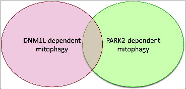
It has been proposed that PARK2 ubiquitinates mitochondrial proteins, whereas DNM1L initiates mitochondrial division, thereby stimulating engulfment of defective mitochondria within phagophores. Our new study suggests a model in which there are at least 2 types of mitophagy; a DNM1L-dependent pathway and a PARK2-dependent pathway (). These pathways synergistically function in the homeostasis of mitochondria and in cell proliferation, function, and survival. Our data hint at the existence of novel E3 ligases involved in the DNM1L-dependent mitophagy pathway. We also suggest that the PARK2-dependent pathway involves DNM1L-independent mitochondrial division. It would be of great interest to determine how these 2 pathways interact. Another important mechanistic question is which mitochondrial proteins are ubiquitinated in DNM1L-dependent mitophagy and what is the function of their ubiquitination. In our future studies, we hope to identify these key components to significantly advance our understanding of the DNM1L-dependent mitophagy pathway.
Figure 1. Model for 2 interacting mitophagy pathways: DNM1L and PARK2- dependent mitophagy. Two mitophagy pathway synergistically maintain mitochondrial homeostasis and cellular function and viability; each can operate in part independently, but they also overlap.
Funding
This work was supported by NIH grants GM084015 (MI), GM089853 (HS), and NS084154 (HS) and by the Mirowski Discovery Fund (HS).