
ABSTRACT
As a PhD student I explored macroautophagy/autophagy induced by starvation in Drosophila melanogaster using different microscopy techniques. The beauty and complexity of this process impressed me so deeply that I felt the need to paint it. Thus, I made 2 oil paintings based on my own scientific work, representing the autophagy mechanism at different scales with diverse artistic resources. The first painting, called Autophagy 1, is inspired by fluorescence confocal microscopy images. Therefore, saturated colors predominate in the composition. The second one is an oil on canvas titled Autophagy 2 which reflects autophagy at a smaller scale. This painting depicts this process as revealed by transmission electron microscopy, employing mainly a gray scale of colors. I performed these works with the intention to catch the essence of this biological process, conveying scientific ideas through art. My paintings are not intended only for the scientific community but also for the general public, as an instrument of enjoyment and popularization of science.
Introduction
Scientific artwork at the service of people
To understand a new idea or fact, normally we have the almost intuitive tendency to draw it. It is generally considered that our understanding can increase by the act of trying to put what we are learning on paper [Citation1]. Besides, drawing is a really good way to convey our understanding or interpretation to others in an efficient manner. To represent a phenomenon involves a high level of comprehension of the fact, and it can help others to understand it better or observe it from a different perspective.
From the beginning of modern biology, scientists have used drawing to represent nature, depicting the life forms observed through dissection and microscopy [Citation2]. Even in the present day, drawing and painting are considered fundamental tools to depict and synthesize scientific observations and discoveries, showing clearly and precisely the information to the scientific community. Considering that an efficient transmission is connected with the acute observation and deep understanding of the analyzed specimens, other means such as photography may not be appropriate to communicate this message. Instead, scientific illustration idealizes its subjects and orders the information in a way in which photography cannot [Citation3,Citation4].
Besides its communicative function, scientific artwork usually presents artistic qualities with an enormous esthetic value, albeit it is not clear if the merits belong to the artist or to nature itself. Very often the reality exceeds the capacity of our imagination as a consequence of the endless forms that biological diversity can take. Sadly, I am convinced that the exposition of the beauty of natural life does not reach the general public as an instrument of pleasure and science popularization. Instead, it is usually restricted to the scientific community, which employs the specific language of scientific illustration. A possible exception would be the naturalistic illustration, which aims to represent the specimens in their environments. This discipline could be considered more affordable to the general public because it can take more artistic license than scientific illustration [Citation5]. However, this kind of illustration is often used to depict mainly animals and plants, and it rarely focuses in conveying the complexity of the subcellular world and its biological processes. Thus, the magnificent subcellular universe remains veiled to the general public.
Traveling toward subcellular painting
Besides my scientific training, I had been drawing and painting for years. And I have done this not only to better represent my specimens; I did it because I wanted to be able to express my appreciation of life and nature in an artistic way.
During my PhD work I studied the autophagy process in the fruit fly Drosophila melanogaster. Autophagy can be defined as a degradative pathway by which cells carry out self-digestion of their cellular material under diverse stress conditions or during normal growth and development [Citation6,Citation7]. In particular, I focused on the study of autophagy induced by starvation in fat body cells from third instar larvae using different microscopy approaches. These cells constitute a triglyceride and glycogen storage tissue analogous to the liver of vertebrates [Citation8,Citation9]. The beauty and complexity that I found in the obtained images affected me so deeply that I felt the need to paint them.
A few years ago, during a scientific meeting, my former PhD co-advisor gave a lecture about our results. She had the kindness to show a fragment of one of my autophagic paintings during her talk, using the image as a cover. I do not remember now if it was at the beginning or at the ending of the lecture, but I remember she remarked that I was not only a co-author of the discussed work, but also the artist behind that painting.
After that, during the following recess, a colleague approached me and declared that he did not know anything about my artistic facet. “I didn’t know you were such a science fanatic! You must have spent so much time making that painting!” he added. The idea of being a science fanatic, similar to a religious fanatic, horrified me. I felt ridiculous and misunderstood and I even stopped painting for a while. For me this anecdote illustrates fairly well that we do not live in a fully scientific time yet. Undoubtedly our time counts as being partly scientific with all kinds of technological advances, but it is not a time in which scientific discoveries or facts are enmeshed with the view of life of ordinary people. Biological facts such as that our bodies are formed by an inconceivable amount of molecules which in turn form an incredible number of cells with different functions, for example, can only marvel people, and I deeply believe that it makes us appreciate life deeper.
Richard Feynman, in his fabulous book What do you care what other people think? discussed this idea in the following way: “I have a friend who’s an artist, and he sometimes takes a view which I don’t agree with. He’ll hold up a flower and say, ‘Look how beautiful it is,’ and I’ll agree. But then he’ll say, ‘I, as an artist, can see how beautiful a flower is. But you, as a scientist, take it all apart and it becomes dull.’ I think he’s kind of nutty … There are all kinds of interesting questions that come from a knowledge of science, which only adds to the excitement and mystery and awe of a flower. It only adds. I don’t understand how it subtracts.” [Citation10] English biologist Richard Dawkins had a similar reasoning, referring to this issue with these inspiring words: “There are those who fear reason as cold, bleak, cheerless, unpoetic. That’s not just untrue; it’s the very opposite of true. Science is the poetry of reality.” [Citation11]. I sincerely hope that my exploration of the subcellular world through paintings will contribute to build bridges between science and the general public.
Results and discussion
Autophagy 1
In order to expose the beauty and complexity of the autophagy mechanism, I made 2 paintings ( and ) based on my own lab work as a PhD student studying Drosophila melanogaster. The paintings are not in any way copies of photographs; they are inventions inspired by my observations and planned carefully. The first one, called Autophagy 1 (see ) is an oil on canvas in which I used visual resources motivated by fluorescence confocal microscopy images.
Figure 1. Autophagy 1. Oil on canvas (15.7 x 19.7 inches), which depicts a field of fat body cells from a starved third instar larva of Drosophila melanogaster as seen in fluorescence confocal microscopy photographs.
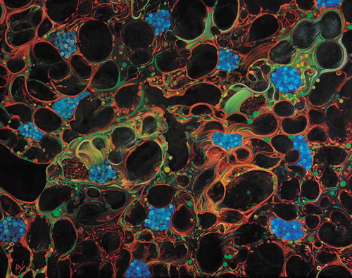
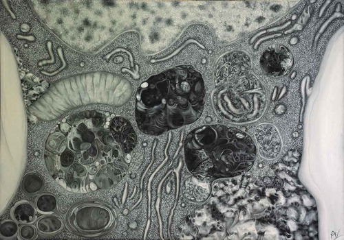
Figure 2. Autophagy 2. Oil on canvas (13.8 x 19.7 inches) that represents a transmission electron image of one fat body cell from a starved third instar larva of Drosophila melanogaster.
The scene is dominated by bright red, orange, green and yellow hues on a black background disrupted by the bounded presence of blue tones. These colors were used to depict a field of fat body cells from a starved third instar larva, a tissue in which the process is widely characterized [Citation8]. The cells of the fat body, on which the painting is inspired, had a polygonal shape and were 40–60 μm in length.
The tissue was made as if it was seen with a magnification close to 600x. The black areas of the cells represent lipid droplets, abundant compartments of fat body cells, and the nuclei appear in blue, as if they were DAPI-stained. Blue ovals in the nuclei depict polytene chromosomes typically present in dipterans [Citation12].
The number of cells in the painting could be inferred by counting the nuclei, which are approximately thirteen in number. Even though cell limits were not shown, they were subtly suggested, giving to the image an intricate effect that highlights the complexity of autophagy.
If the painting is analyzed carefully, 2 mostly green cells can be seen disposed in a diagonal line of the image (from left-below to right-above), among a background of red cells. This difference in the colors of the cells is based on the mosaicism usually observed when genes are expressed using the Gal4/UAS system in D. melanogaster organs, providing an additional sensation of directionality to the composition. Further, black regions (lipid droplets) of the painting confer to the scene a tridimensional sense, allowing the beholder to imagine a volume of tissue that fades into darkness.
The main intention of the painting is to show the behavior of the autophagic proteins Atg8 and zda (zonda) [Citation13], when the process is induced by starvation. During autophagy a membranous cistern called the phagophore buds from a cup-shaped structure associated with the endoplasmic reticulum (ER) known as an omegasome [Citation14,Citation15]. Afterward, the phagophore expands and finally seals, producing an autophagosome: a double-membrane organelle where cytoplasmic components are sequestered [Citation16]. After its formation, the autophagosome undergoes successive fusions with late endosomes and lysosomes, acquiring degradative enzymes and becoming a novel vesicle called an autolysosome, in which the engulfed material is degraded [Citation17].
It is well known that Atg8 shows a cytoplasmic distribution in fed conditions and it is incorporated into the phagophore membrane when larvae are subjected to starvation. Then, it remains associated with autophagic structures during the mechanism of progression [Citation8]. In contrast, zda displays a presumably ER distribution in well-fed larvae, and shortly after the onset of starvation, it nucleates in ER-associated foci. When the phagophore expands and seals, giving rise to the autophagosome, zda is incorporated into its membrane, overlapping with Atg8 [Citation13]. Considering this, in Autophagy 1 I show Atg8 distribution (represented in green) as seen in starved tissues: mostly in puncta (phagophore, autophagosome and autolysosome), but also in the cytoplasm. zda (shown in red) is mainly localized in foci, too (omegasome, autophagosome and autolysosome), but presents an additional reticular distribution (likely ER). Several orange and yellow tones are used to represent different degrees of colocalization between both proteins.
If the painting is observed forgetting for a moment what it represents, a feeling of fluency can be appreciated; as if the cytoplasm was liquid with suspended particles dragged by the current. This is essentially an esthetic resource used to make the composition more intriguing and attractive.
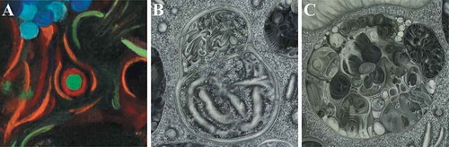
Undoubtedly, the main highlight of my PhD research was to be able to locate zda in the omegasome of D. melanogaster. Considering that the omegasome can be visualized as a ring or omega-shaped structure (with 1 μm in maximum diameter on average) when labeled with specific markers [Citation14], I decided to introduce in Autophagy 1 different examples of omegasomes decorated with zda, which can be found if the painting is observed carefully. These omegasomes generally are green on their concave side, representing Atg8-positive phagophores, which emerge from them (see details in ).
Figure 3. Magnified views of diverse autophagic structures painted in Autophagy 1 and Autophagy 2. (A) An example of the numerous omegasome representations shown in Autophagy 1. The omegasomes were painted as red circles (corresponding to the zda protein) with phagophore structures in their inside shown as green. (B,C) Magnifications of one of the many autophagosomes (B) and autolysosomes (C) depicted in Autophagy 2.
Autophagy 2
Different facets of autophagy must be studied at different scales. Therefore, most confocal microscopy studies are complemented with transmission electron microscopy analyses, which reveal the morphology of autophagic structures as well as their natural environment and their position among other cellular components [Citation18]. Analogously, I wanted to reflect the beauty of the autophagic mechanism at this smaller scale, too. Thus, the second painting, Autophagy 2 (), represents the autophagy process with a higher level of magnification than Autophagy 1. This painting was made using a gray scale of tones as if the tissue was seen through a transmission electron microscope with a magnification close to 30,000x. The image illustrates with great detail a small portion of a fat body cell, in which a fraction of the nucleus was located in the upper edge of the canvas and 2 portions of lipid droplets (white areas) were positioned at the sides, framing the composition. Glycogen deposits that are typically present in fat body cells [Citation19,Citation20] were depicted in the bottom right (juxtaposed to the right lipid droplet) and, to a lesser extent, in the upper margin (adjoined to the left lipid droplet). If the image is observed carefully, figures that represent different organelles can be recognized: a mitochondrion with its recognizable cristae at the top left, and abundant tube-like membranes of ER throughout the image. The cytoplasm was made with a dotted appearance, pointing out the crowding effect produced by the enormous amount of cytoplasmic ribosomes and other cellular components. Besides, in the bottom left of the canvas some curious concentric elements represent multi-lamellar membrane structures. I decided to include these figures in the composition because frequently during my PhD work I recognized the presence of this kind of structure and I have always been fascinated with its enigmatic nature.
The main elements of the canvas correspond to autophagic structures with different maturation states and diverse cytoplasmic constituents sequestered, which were distributed in strategic visual points following a diagonal line of the image. As in Autophagy 1, this line was positioned from left-below to right-above, giving to the image a feeling of flow in this direction. In this way, both paintings were created with a similar organization in order to emphasize that they belong to the same artistic series; one that illustrates the same biological process.
The autophagic structures were made with the intention of showing the enormous complexity and the strength of this catabolic process, trying to imagine an intricate and surreal small universe inside each vesicle. In sum, 6 autophagic structures were illustrated: 2 autophagosomes (with a length of 0.5–1.5 μm) were located on the right side and 4 autolysosomes (near 0.5- to 2-μm long) were placed in the center and sides of the canvas. The autophagosomes can be recognized first by the presence of 2 sets of lines delimiting their structures. These surrounding lines represent 2 parallel lipid bilayers separated by an electron-translucent space, which form the bordering double membrane characteristic of the autophagosomes. Typically, each lipid bilayer appears under electron microscopy as 2 dark bands: the 2 leaflets of the bilayer [Citation21]. Second, autophagosomes are denoted by their clearer interior, which depicts the electron-translucent content composed of cytosol and organelles. Particularly, one of these autophagosomes was made as if it contained ribosomes and ER membrane morphologically intact inside it (see ). In contrast, the autolysosomes were made using darker tones, which represent an electron-dense content composed by hydrolytic enzymes and cellular material at different stages of degradation. Surrounding them, only one set of lines has been drawn, instead of 2 as in the case of autophagosomes, to indicate that autolysosomes show only one limiting membrane.
The patterns and textures inside autolysosomes were partially inspired by the amazing work of H. R. Giger, who created surreal monochromatic landscapes. Inside these vesicles all kinds of imaginary figures can be appreciated, which bring to mind a mixture of organs and embryonic structures (see ).
If Autophagy 2 is analyzed closely, some vesicles (2 in particular) are composed of different domains clearly distinguishable, representing autophagic structures that were formed by the fusion of other preexisting vesicles. One of them represents the fusion between an autophogosome and an autolysosome as an example of a heterotypic fusion, whereas the other depicts a mixture of 2 autophagosomes (), representing homotypic fusion.
Final remarks
I hope that my paintings could help to express scientific concepts in an attractive way, awakening interest not only in a scientific environment but also in the general public. Both paintings were made with the intention of transmitting something that cannot be seen in photographs: the feeling of being an explorer in a wild, huge and unknown universe, which is at the same time so small that it is encompassed in every live being that ever existed. I have experienced this sensation when I saw this beautiful subcellular world for the first time, and my expectation is to be able to catch this essence and put it on a canvas.
Once again Feynman found the right words to express this feeling: “I wanted very much to learn to draw, for a reason that I kept to myself: I wanted to convey an emotion I have about the beauty of the world. (…) It’s an appreciation of the mathematical beauty of nature, of how she works inside (…) a feeling of how dramatic and wonderful it is.” [Citation22].
I have the same need.
Materials and methods
Composition
The challenge of making art based on scientific results lays in the need to represent the elements of study using artistic resources without losing the rigor of scientific observations. Being a scientist besides being an artist allowed me to know what artistic license I could take and what biological elements I had to show with high precision, in order to convey clearly the underlying essence of the represented biological process.
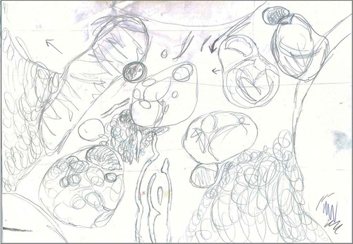
In this creative process the first step was sketching the original idea, being able to order the elements of the composition in a harmonious and attractive way. In line with this, shows the design on which Autophagy 2 is based. Usually, the final composition diverges from the sketch as a consequence of the natural development of the original idea. This is indeed the case with Autophagy 2, where some discrepancies can be identified if the painting () and the preliminary drawing () are compared carefully. Even so, essential characteristics of the painting, such as the presence of the main autophagic structures or the sense of directionality, were already present in the sketch.
Figure 4. Sketch made in pencil showing the original design behind Autophagy 2. The final painting partially differs from this preliminary drawing due to the creative process.
Materials and techniques
After choosing the canvas dimensions, 15.7 × 19.7 inches for Autophagy 1 and 13.8 × 19.7 inches for Autophagy 2, the sketches were drawn on these supports.
In both paintings I decided to employ oil colors because they take longer to dry than other media. Thus, color mixtures could be preserved longer and be reused without the need to make new mixtures every time. This makes it possible to create otherwise laborious textures relatively quickly. lists the colors used in each painting.
Table 1. Oil colors used in each painting
The thinner utilized in both artworks was a mix of turpentine (Eterna Company), linseed oil (Alba S.A.) and dammar varnish (Alba S.A.) in a 3:1:1 proportion.
Autophagy 1
At the moment of starting to paint, the areas of the canvas reserved for lipid droplets were initially delimited using diluted black. After the paint was dry, I added the base color for the cytoplasm of the cells. In this instance, then, the painting looked like a map, with different cells and elements glimpsed by a layer of plain base color. It was only after building this “color map” that complex textures could be added to the painting by the inclusion of increasingly elaborated layers of paint.
In the final product, hues of green and red predominate, which produce an important contrast effect due to their being complementary colors. The strong intensity of blue tones used to make the nuclei forces the viewer to focus initially on these elements, which disrupt the texture of the cytoplasm. Chromosomes are depicted as ovals with circular and delicate brushstrokes inside the nuclei.
Autophagy 2
Each element in Autophagy 2 is characterized by a particular design, for which several strategies were used, albeit they are difficult to deduce by seeing only the painting. In general the whole painting was performed using only the gray scale, where black was the darkest value and white the lightest one. However, in some cases it was helpful to add in the paint mix a pinch of a different color to prevent the painting from becoming too uniform. In line with this, the color mixture employed to make the cytoplasm was generated by adding some blue, while the nucleus texture was made using a bit of yellow. Besides, some areas inside the autolysosome shown in were generated using a bit of red in the mix.
The technique employed in the cytoplasm consisted of painting 3 sequential layers of dots over a background of gray mixed with a bit of blue. The dotted layers were made with gray, dark gray and light gray tones, after which the dots were randomly blurred conferring to the cytoplasm a less rigid and structured appearance.
The nucleus was painted with a similar strategy, but using a light yellow background and broader brushstrokes with a messy appearance. The glycogen deposits, conversely, were made by blurring the paint with the thinner mix mentioned above, which produces an effect of smoothness. Finally, the lipid bilayers with their constituent phospholipids were depicted using very thin brushes (with few bristles) and a hands-free magnifying glass with a 1.8x magnification.
Acknowledgments
I would like to thank my husband, Lautaro Gándara, who is always a source of inspiration and guidance both in my artistic and scientific life. It was his wonderful vision of the world that motivated me to paint, giving me the courage to express myself. I am extremely grateful to Mariana Melani and Cecilia D’Alessio, who always believed in my artistic ability and supported my development in the art world. I want to express my gratitude to the photographer Alejandro Valko, who had the kindness to photograph my paintings. Alejandro shared with me his knowledge of composition theory, helping me significantly to improve my work. Also I express my thanks to Nadia Ailén de los Santos and Maribel Gándara for making a careful reading of this manuscript, contributing with ideas that helped to improve it. Last but not least, I am very grateful to Daniel Klionsky for his gentle words on the eve of the Buenos Aires Conference on Autophagy 2017. His comments encouraged me to continue with my artistic work and to write this article.
Disclosure statement
No potential conflict of interest was reported by the author.
References
- Pyle CM. Art as science: scientific illustration, 1490–1670 in drawing, woodcut and copper plate. Endeavour. 2000;24(2):69–75.
- Sykes A. The art of scientific illustration: examples from poultry. Worlds Poult Sci J. 1995;51(3):327–335.
- Hodges ER. Scientific illustration: a working relationship between the scientist and artist. BioScience. 1989;39(2):104–111.
- Illustraciencia. What is scientific illustration? [cited 20 Jun 2019]. Available from: http://illustraciencia.info/en/que-es-la-ilustracion-cientifica/
- Illustraciencia. What is naturalist illustration? [cited 20 Jun 2019]. Available from: http://illustraciencia.info/en/que-es-la-ilustracion-naturalista/.
- Yang Z, Klionsky DJ. Mammalian autophagy: core molecular machinery and signaling regulation. Curr Opin Cell Biol. 2010;22(2):124–131.
- Boya P, Reggiori F, Codogno P. Emerging regulation and functions of autophagy. Nat Cell Biol. 2013;15(7):713.
- Scott RC, Schuldiner O, Neufeld TP. Role and regulation of starvation-induced autophagy in the Drosophila fat body. Dev Cell. 2004;7(2):167–178.
- Arrese EL, Soulages JL. Insect fat body: energy, metabolism, and regulation. Annu Rev Entomol. 2010;55:207–225.
- Feynman RP, Leighton R. What do you care what other people think?”: further adventures of a curious character. New York: WW Norton & Company; 2001.
- Dawkins R, Slaves to Superstition, in The Enemies of Reason. August 13, 2007.
- Orr-Weaver TL. When bigger is better: the role of polyploidy in organogenesis. Trends Genet. 2015;31(6):307–315.
- Melani M, Valko, A, Romero, N M, et al. Zonda is a novel early component of the autophagy pathway in Drosophila. Mol Biol Cell. 2017;28(22):3070–3081.
- Axe EL, Walker S A, Manifava M, et al. Autophagosome formation from membrane compartments enriched in phosphatidylinositol 3-phosphate and dynamically connected to the endoplasmic reticulum. J Cell Biol. 2008;182(4):685–701.
- Karanasios E, Ktistakis NT. Live-cell imaging for the assessment of the dynamics of autophagosome formation: focus on early steps. Methods. 2015;75:54–60.
- Mizushima N, Levine B, Cuervo A M, et al. Autophagy fights disease through cellular self-digestion. nature. 2008;451(7182):1069.
- Eskelinen E-L. Maturation of autophagic vacuoles in mammalian cells. Autophagy. 2005;1(1):1–10.
- Klionsky DJ, Abdalla F C, Abeliovich H, et al. Guidelines for the use and interpretation of assays for monitoring autophagy. Autophagy. 2016;12(1):1–222.
- Butterworth FM, Forrest EC. Ultrastructure of the preparative phase of cell death in the larval fat body of Drosophila melanogaster. Tissue Cell. 1984;16(2):237–250.
- Butterworth FM, Bownes M, Burde VS. Genetically modified yolk proteins precipitate in the adult Drosophila fat body. J Cell Biol. 1991;112(4):727–737.
- Robertson JD. The molecular structure and contact relationships of cell membranes. Prog Biophys Mol Biol. 1960;10:343–418.
- Feynman RP, Leighton R. “ Surely you’re joking, Mr. Feynman!”: adventures of a curious character. New York: Random House; 1992.