
Abstract
Introduction
Lead poisoning in childhood remains an important public health concern. We highlight the radiological findings in a patient with a high blood lead concentration.
Case summary
A 7-year-old girl presented to hospital with abdominal pain, nausea, and asthenia. Laboratory tests showed severe hypochromic microcytic anemia, punctate basophilic stippling of erythrocytes, and a blood lead concentration of 880 µg/L (4.3 µmol/L).
Images
Radiographs of the femur, tibia, and fibula demonstrated dense metaphyseal bands (“lead lines”). On cranial computed tomography, we observed multiple speck-like and curvilinear hyperdensities involving subcortical regions, putamen, and left cerebellar hemisphere.
Conclusion
In patients with lead poisoning, imaging of the brain and bones may show characteristic features. These imaging findings may point to the diagnosis of lead toxicity when these radiographic findings are discovered during the evaluation of vague complaints such as abdominal pain or mental status changes or when a blood lead concentration is not readily available.
Introduction
Lead poisoning in childhood remains an important public health concern [Citation1]. Once in the body, lead can accumulate in various organs, including the bones and central nervous system [Citation2]. We present the radiological findings in a patient with a high blood lead concentration.
Case summary
A 7-year-old girl presented to the hospital because of intermittent abdominal pain present for about 40 days, and nausea and asthenia for 15 days. Laboratory tests showed severe hypochromic microcytic anemia (hemoglobin concentration 75 g/L, mean corpuscular volume 78.90 fL, mean corpuscular hemoglobin 23.6 pg, red blood cell distribution width 19.7%), punctate basophilic stippling of erythrocytes, and a blood lead concentration of 880 µg/L (4.3 µmol/L). The child’s mother suspected that a foreign body had been ingested about forty days previously when complaints of abdominal pain began.
Images
Multiple speck-like and curvilinear hyperdensities were found on axial brain computed tomography involving the subcortical regions, the putamen (), and the left cerebellar hemisphere (). Radiographs of the knees () demonstrated bands of increased density at the metaphyses ("lead lines").
Figure 1. Axial brain computed tomography. Multiple hyperattenuating foci in the subcortical regions (white arrows) and putamen (dotted white arrows).
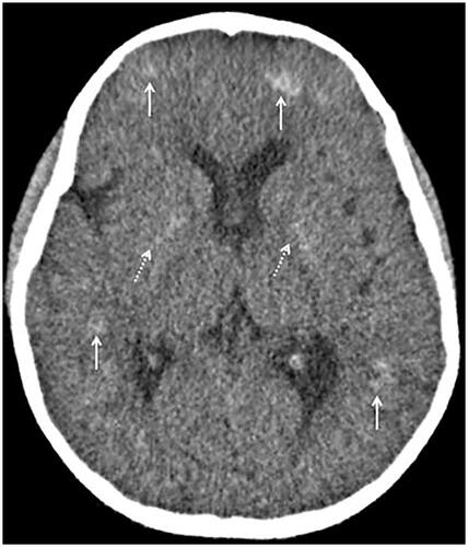
Figure 2. Axial brain computed tomography. Hyperattenuating foci in the left cerebellar hemisphere (white arrow).
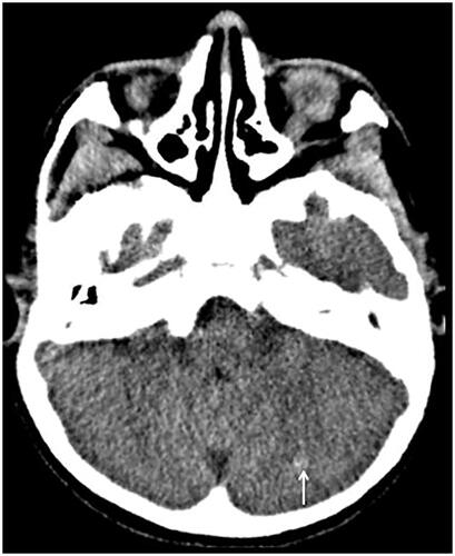
Figure 3. Antero-posterior radiograph of the knees demonstrates a thick, radiopaque metaphyseal band across the femur (black arrows), tibia (white arrows), and fibula (dotted white arrows).
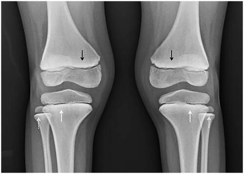
Lead absorption depends on concentration, exposure time, and individual criteria such as age and physiological status. In the blood, lead has a half-life of 1–2 months; in the bone, it is 20–27 years [Citation1]. Long bone radiographs can show increased density of the metaphysis (growth plate) or "lead lines". These findings are due to the deposition of lead ions within the hydroxyapatite crystals of calcifying bone, which inhibits osteoclastic remodeling, while osteoblast activity is preserved. This results in an increased number and thickness of trabeculae within the metaphysis and increased density on radiographs [Citation3].
The proximal part of the fibula is a critical key point for the differential diagnosis of lead poisoning [Citation4]. Sclerotic lines in the distal femoral and proximal tibial metaphyses can be seen in healthy children. The presence of dense transverse bands in the metaphyses of the proximal fibula and other non-weight-bearing bones can differentiate normal radiographic findings from evidence of lead toxicity [Citation4,Citation5].
Computed tomographic findings of cerebral and cerebellar hyperdensities have been described in patients with elevated blood lead concentrations [Citation6]. These hyperdensities can be punctate, curvilinear, dotted, or diffuse and are located in subcortical regions, basal ganglia, vermis, and cerebellum [Citation7,Citation8]. Subclinical neurological findings such as cognitive impairment are comparatively more common in pediatric patients, and the inverse association between lead exposure and intelligence quotient (IQ) score is well-established [Citation9].
Conclusion
In patients with lead poisoning, imaging of the brain and bones may show characteristic features. These imaging findings may point to the diagnosis of lead poisoning when these radiographic findings are discovered during the evaluation of vague complaints such as abdominal pain or mental status changes or when a blood lead concentration is not readily available.
Acknowledgment
The authors reported no funding associated with this study.
Disclosure statement
No potential conflict of interest was reported by the authors.
Additional information
Funding
References
- Hauptman M, Bruccoleri R, Woolf AD. An update on childhood lead poisoning. Clin Pediatr Emerg Med. 2017;18(3):181–192. doi: 10.1016/j.cpem.2017.07.010.
- Sahu JK, Sharma S, Kamate M, et al. Lead encephalopathy in an infant mimicking a neurometabolic disorder. J Child Neurol. 2010;25(3):390–392. doi: 10.1177/0883073809338625.
- Rabber SA. The dense metapyseal band sign. Radiology. 1999;211:773–774.
- Blickman JG, Wilkinson RH, Graef JW. The radiologic "lead band" revisited. AJR Am J Roentgenol. 1986;146(2):245–247. doi: 10.2214/ajr.146.2.245.
- Laor T, Jaramillo D. Metaphyseal abnormalities in children: pathophysiology and radiologic appearance. AJR Am J Roentgenol. 1993;161(5):1029–1036. doi: 10.2214/ajr.161.5.8273604.
- Tüzün M, Tüzün D, Salan A, et al. Lead encephalopaty: CT and MR findings. J Comput Assist Tomogr. 2002;26(3):479–481. doi: 10.1097/00004728-200205000-00029.
- Slassi Sennou A, Souirti Z, Messouak O, et al. Intoxication au plomb et hypersignaux des noyaux gris centraux [Lead poisoning and basal ganglia hyperintensities]. Rev Neurol. 2012;168(2):198–199. doi: 10.1016/j.neurol.2011.05.011.
- Mani J, Chaudhary N, Kanjalkar M, et al. Cerebellar ataxia due to lead encephalopathy in adult. J Neurol Neurosurg Psychiatry. 1998;65(5):797–798. doi: 10.1136/jnnp.65.5.797.
- Hou S, Yuan L, Jin P, et al. A clinical study of the effects of lead poisoning on the intelligence and neurobehavioral abilities of children. Theor Biol Med Model. 2013;10(1):13. doi: 10.1186/1742-4682-10-13.