1 Relevance of Animal Studies to Human Toxicology
Elcombe CR. CXR Biosciences, Dundee and Biomedical Research Centre, University of Dundee, Dundee, UK.
Introduction: This presentation will review the strengths and weaknesses of traditional animal studies in estimating the potential toxicity of chemicals in humans. Additionally, novel animal models for safety evaluation and risk assessment will be described. Toxicity studies: These involve experiments on laboratory animals and are designed to identify potential hazard, and thereby determine safety or estimate the risk following human exposure. The outcome is associated with some degree of uncertainty, no matter how much evidence is gathered. It must be remembered, however, that as with all toxic effects, the dose or amount of exposure is critical. For example, in testing for carcinogenicity, to decide on the risk that a particular carcinogen poses, it is important to determine how much of the chemical will cause how many cases of cancer in a specified population. Laboratory animal studies provide a way of detecting the carcinogenicity of a large number of chemicals, and can provide numerical values for cancer risks. Carcinogenicity studies: These are performed on laboratory animals and rely on the paradigm that exposure of small groups of animals to high dose levels of compound can be used as a surrogate for the exposure of large numbers of people to small quantities of compound. However, the relevance of the animal high-dose results to low doses or to humans is not always clear. Extrapolations from such animal studies to humans are, at times, confounded by species-specific pharmacokinetics and metabolism, high-dose dependent nonlinear pharmacokinetics, species differences in drug metabolism pathways, and species-specific mechanisms of response (e.g. male rat specific α-2u globulin nephropathy and kidney tumours; peroxisome proliferation and liver tumours in rodents). This means that future efforts at increasing the accuracy of safety assessment will need to focus on understanding mechanisms of toxicity, and will utilize new technologies such as toxicogenomics and novel animal models (transgenic, knockout and humanized). Such methodologies will be necessary to evaluate potential toxicity in susceptible human subpopulations (e.g. due to genetic polymorphisms and disease states). Drug metabolism and toxicity: The cytochrome P450 enzymes are a major determinant of drug metabolism and toxicity. They comprise a multigene family of proteins in animals and man and hence, the deletion of all the P450 genes individually is inconceivable. However, in order to function all the cytochrome P450 isozymes receive electrons from the electron donor cytochrome P450 reductase. A model has been developed where P450 reductase in the liver is conditionally deleted (HRNTM, Hepatic Reductase Null) and, as a consequence, cytochrome P450 activity is abrogated. This model can be applied as a mechanistic problem solving tool in safety or metabolic studies. For example, it can be used to establish whether toxic effects are due to parent compound or its metabolites. Compounds, such as paracetamol, that are metabolically activated by cytochrome P450 leading to hepatotoxicity do not exhibit hepatotoxicity in the HRNTM mouse. A number of different kinetic parameters can determine toxic response; for example, maximum circulating drug (or metabolite) concentration (Cmax) or the overall exposure (area under the curve; AUC). Understanding the toxicokinetic properties of a compound is a key factor in the design of clinical trials. Such studies are of particular importance in cancer chemotherapy since the therapeutic window is often narrow. Pass et al. (1) have demonstrated the power of the HRNTM models for studies of this nature by investigating the toxicokinetics of the antitumour agent, cyclophosphamide (CPA). P450s metabolise CPA to 4-hydroxycyclophosphamide (4-OH-CPA), which is responsible for both antitumour effects and toxicity (myelosuppression). In wild type animals, the Cmax and AUC for the production of the 4-OH-CPA at different doses were closely correlated with each other, so the elucidation of which parameter is most important in the myelotoxicity of CPA was not possible. However, the altered metabolism in the HRNTM mouse allowed these parameters to be clearly dissociated. In the HRNTM mice the Cmax was greatly reduced and the AUC remained very similar. Plotting AUC or Cmax of 4-OH-CPA against the level of myelosuppression demonstrated a close correlation with the Cmax but not with AUC. The antitumour effects of cyclophosphamide are reported to correlate with the overall exposure, i.e. AUC. These data suggest that the way to maximise the therapeutic benefit of this compound is to use it as an infusion rather than a bolus injection. Quite clearly such information prior to the instigation of Phase I trials could be extremely valuable and could make the difference between drug success and failure. Metabolism and toxicity testing relies on laboratory animals and human cell lines or tissues in culture. Each of these has drawbacks, however, in that animal metabolism can differ significantly from human metabolism while culture systems are unable to reflect whole body metabolism. Under development are new strains of “humanised” transgenic mice in which enzymes of the cytochrome P450 family (“P450s”) and a variety of transcription factors involved in the regulation of metabolism are replaced by their human counterparts in the liver. These animals are already showing utility in safety assessment. For example, the ligand-mediated activation of the (Peroxisome Proliferator Activated Receptor) in rats αtranscription factor PPAR and mice leads to hepatomegaly that is characterized by hypertrophy (due to the proliferation of peroxisomes and smooth endoplasmic reticulum) and hyperplasia (increased hepatocyte proliferation). Long-term administration of “peroxisome proliferators” leads to the development of liver tumours. When “peroxisome null mice, hepatomegaly is notαproliferator” ligands are administered to PPAR null mouseαobserved and liver tumours do not develop. Humanization of the PPAR gene leads to a liver that exhibits theαby insertion of the human PPAR ligands (2). These αhypertrophic, but not the hyperplastic effects of PPAR species differences data for ligand responsiveness contribute to the belief that peroxisome proliferators do not pose a carcinogenic hazard to humans. References: 1. Pass GJ, et al. Cancer Res 2005; 65:4211–4217. 2. Cheung C, et al. Cancer Res 2004; 64:3849–3854.
2 Research Methods in Occupational Toxicology
Wilks MF. Syngenta Crop Protection AG, Basel, Switzerland.
Background: Chemically-induced occupational illnesses are prevented by the control of exposure in the workplace. This requires an understanding of the underlying hazard (toxicology) as well as dose-response relationships, and to compare these with intensity, frequency and duration of exposure. Occupational toxicology is primarily concerned with the interaction of chemical and physical stressors in the work environment with biological systems (1). The aim is to provide information which can be used in risk assessment, risk management, medical surveillance and preventative programmes. It is important to remember that the work environment (and hence exposure) is very heterogeneous and occupational toxicology, therefore, has to cater for settings as diverse as mining, large chemical plants, cottage industries, trades and crafts, service industries or agriculture, to name but a few. Consequently, research methods and needs are equally diverse. Research methods: While toxicological research is usually laboratory-based, occupational toxicology is reliant on the generation of human data. Human studies fall broadly into two categories: those which are used to generate a hypothesis, and those which test a hypothesis (2). Hypothesis-generating studies are usually of a descriptive nature. They include single or multiple case reports, reports of adverse reactions and studies which demonstrate a correlation between the occurrence of illness and the existence of an occupational hazard. In contrast, hypothesis-testing studies are observational, looking at individuals or small groups of people. These studies are analysed using established statistical methods to determine if an association exists between presence of an exposure hazard and the occurrence of illness. Often, the hypothesis examined has been previously generated by a descriptive study. Examples include two widely used types of epidemiological studies: case-control and cohort studies. The nature of these studies can be either retrospective, i.e. taking an individual's or population group's health status or disease incidence and trying to correlate it with past exposure, or prospective, where the development of the disease is followed in exposed and non-exposed subjects over time, possibly over many years. Finally, studies can be categorised in terms of whether an intervention is carried out, such as withdrawing a subject from exposure, or whether observations are being made without interventions (3). Common problems: Occupational toxicology studies are at their most powerful if they include adequate information on both exposure and health effects. Such an ideal is rarely achieved; most studies concerned with exposure measurement look at relatively low levels of exposure over short periods which are unlikely to result in short-term health effects. Conversely, most epidemiological studies looking at health effects from chemical exposure suffer from a lack of verifiable exposure information, especially where there is a long latency period between exposure and onset of illness. Exposure can be determined by means of ambient or personal monitoring, a subset of the latter being biological monitoring. More recently, the discipline of ‘molecular epidemiology’ has added insights on metabolic pathways, enzymes and genes to biological and biochemical measurements of chemically-exposed populations and individuals. The choice of a method for exposure assessment depends principally on the purpose of the investigation, the chemical involved, the conditions at the workplace, and the resources available. Unfortunately, many epidemiological studies rely on indirect exposure estimates from questionnaire-based interviews or job exposure matrices. Even if exposure can be verified, let alone quantified, the question remains whether a reported association with an illness represents a true cause-effect relationship. While this may be obvious for specific exposures and rare disease processes, e.g. in the case of vinyl chloride and haemangiosarcoma of the liver (4), the vast majority of reported associations would benefit from a more rigorous examination along established scientific concepts such as Bradford Hill's ‘viewpoints’ (5) to make a judgement concerning causality. Conclusions: Occupational toxicology is at the interface between toxicology and epidemiology and its research methods can be applied to risk assessment and management of chemical exposure, and thus to the prevention of occupational diseases. Human data are at the centre of occupational toxicology research, which is at its most powerful when it leads to an accurate assessment of exposure-response relationships according to sound scientific criteria. References: 1. Olajos EJ, Salem H. Occupational Toxicology. In: Ballantyne B, Marrs T, Syversen T, eds. General and Applied Toxicology. 2nd ed. London, UK: Macmillan, 1999:1453–1471. 2. Cone JE, Reeve GR, Landrigan PJ. Clinical and Epidemiological Studies. In: Tardiff RG, Rodricks JV, eds. Toxic Substances and Human Risk – Principles of Data Interpretation. New York, USA: Plenum Press, 1987:95–120. 3. Wilks MF, Minton NA. Toxicity data obtained from human studies. In: Ballantyne B, Marrs T, Syversen T, eds. General and Applied Toxicology. 2nd ed. London, UK: Macmillan, 1999:453–470. 4. Creech JL, Johnson MN. Angiosarcoma of liver in the manufacture of polyvinyl chloride. J Occup Med 1974; 16:150–151. 5. Hill AB. The environment and disease. Association or causation? Proc R Soc Med 1965; 58:295–300.
3 Statistical Methods in Toxicology
Juurlink DN. University of Toronto, Toronto, Ontario, Canada.
An understanding of statistical principles is essential to conduct and interpret research in clinical toxicology. This address provides an overview of important concepts and common analytical techniques using examples from the toxicology literature. Elements of statistical inference: At the heart of all inferential statistics is the notion of a signal-to-noise ratio, where the signal is the important relationship and the noise is background variation. A higher signal-to-noise ratio imparts a higher degree of statistical significance and a lower P value. Ubiquitous in biomedical research, P values reflect Type I (alpha) error – the probability that the observed results could have occurred by chance. It is sometimes helpful to consider alpha error as a measure of one's willingness to accept a false positive conclusion. The opposite phenomenon – concluding that no difference exists when in fact one does – is termed Type II (beta) error, and is directly related to the concept of power. Common statistical procedures: The t-test is used to compare the means of two groups, and is based on the ratio of the difference between groups (the signal) to the standard error of the difference (noise). Despite its computational ease, the t-test is not appropriate for multiple groups or when individuals in the two groups are matched. One-way analysis of variance (ANOVA) deals with more than two groups, and again involves a signal (reflective of differences between the group means) and noise (reflecting variation within the groups). Studies involving repeated measures (eg. pre-post studies on the same individuals), often utilize a paired t-test or repeated measures ANOVA. Their main advantage is greater efficiency due to removal of within-subject variability. The chi-squared test is commonly used to describe categorical frequency data, and simply expresses how different the observed values in a table's cells are from those expected by chance. Several variations on the chi-squared test exist for special circumstances and will be discussed. The kappa statistic is used to characterize the extent of agreement beyond chance, and generally ranges from 0 to 1. Finally, regression techniques are commonly used in toxicology research. Simple regression allows assessment of the relationship between a single independent variable and a dependent variable, and involves fitting an ‘optimal’ regression line that minimizes the departure of data points from the line (residuals). Multiple regression denotes the assessment of multiple independent variables. The type of multiple regression used depends on the nature of the independent variable, including linear regression (continuous outcomes), logistic regression (dichotomous outcomes), and Poisson regression (rates or counts). Several underappreciated phenomena can thwart multiple regression, including the addition of too many terms to the model (overfitting) and unrecognized correlation between predictor variables (collinearity).
4 Studying the Epidemiology of Poisoning
Dawson AH. SACTRC University of Peradeniya, Kandy, Sri Lanka.
The epidemiology of poisoning is dependent upon the acquisition of clinical data. The design and quality assurance of the instrument that is used to acquire the data and the environment in which it is applied is critical to the use and applicability of the data. There are a number of pragmatic issues related to what data is possible to be collected and what perspective the data collector might have (e.g. institutional vs. community-based or public health). Finally, the use of individual datasets is often linked to other pre-existing datasets; for each of these linkages the perspective and limitations of those datasets needs to be appreciated. In general, databases may serve a mix of functions ranging from surveillance (in which known data items associated with poisoning are collected, e.g. known symptoms of a poisoning) to more extensive explorative databases (in which many data items are collected even if they are not known to relate to the poisoning; such data sets might help extend the knowledge about a syndrome). The collection of the data may be seen as obligatory, e.g. a clinical database that collects all the cases presenting to a hospital within a defined region, or all calls to a poison centre versus voluntary, e.g. voluntary adverse reaction reporting schemes, collation of literature. Comprehensive Clinical Databases are useful at an institutional level and, providing the source population is well defined and representative, they may be useful at a more regional or national level. These databases allow faster and more responsive iterative development, and it is possible to maintain a high degree of quality control for data collection and entry. They have the capacity to produce new clinical data. They are also potentially time-consuming and expensive, especially if they are not integrated into clinical care and existing hospital systems. In practice, it is often easier to commence with a limited clinical database and build from that position. At the other end of the spectrum is data generated from poisons information systems, such as TESS or adverse drug reporting systems. Such datasets produce good information about the potential importance of various poisonings and have the advantage of collecting some community-based information. They are easily integrated into existing systems, but their nature is essentially an opt in systems dependent upon someone generating an enquiry and providing clinical information. As such, there may be a bias toward new events or drugs. A potential disadvantage is that they may only ask questions about symptoms that are already known and may be insensitive to minor events or intermediate markers of toxicity. Finally, the move to internet-based poisons information queries gives an opportunity for passive collection of information based on hits and geographic mapping. Examples of some studies utilising datasets will explored to identify their limitations and strengths. References: 1. Buckley NA, Whyte IM, Dawson AH, et al. Pheniramine: A much abused drug. Med J Aust 1994; 160:188–192. 2. Buckley NA, Dawson AH, Whyte IM, et al. Greater toxicity in overdose of dothiepin than of other tricyclic antidepressants. Lancet 1994; 343:159–162. 3. Buckley NA, Whyte IM, Dawson AH, et al. Correlations between prescriptions and drugs taken in self-poisoning. Med J Aust 1995; 162:194–197.
5 Observational Research in Clinical Toxicology
Buckley NA. Clinical Pharmacology & Toxicology, Australian National University Medical School, ACT, Australia.
Most current evidence for management in clinical toxicology comes from observational data rather than randomised clinical trials. Thus, we commonly use relatively weak evidence to draw conclusions about the efficacy and safety of diagnostic and treatment interventions. Designing and comparing studies producing such evidence requires a strong appreciation of the weaknesses of these study designs and the best ways to analyse these data. Areas where observational data have been largely relied on are all diagnostic and prognostic criteria and most treatments for poisoning with pharmaceuticals (e.g. paracetamol, psychotropic and anticonvulsant drug poisoning). For example, commonly used assessments, such as ECG risk stratification for tricyclic antidepressant poisoning, clinical and laboratory prognostic criteria for paraquat poisoning, and the paracetamol nomogram, have all been the products of observational research. This is also true for many commonly used interventions, such as urinary alkalinisation for salicylate and chlorphenoxy herbicide poisoning, alkalinisation for antidepressant-induced arrhythmias or acetylcysteine in early paracetamol poisoning (although animal studies may support these human data). Even when efficacy data are available from RCTs, the numbers of patients required to provide adequate safety data on interventions has nearly always been generated by observational research (e.g. acetylcysteine, multiple dose activated charcoal, DigiFab). Similarly, observational data often provides the best available evidence for treatment in vulnerable populations (e.g. children, pregnancy), rare conditions and for cost-effectiveness. There are many pervasive and unavoidable sources of bias in observational research, particularly where data are collected retrospectively. A single study is rarely sufficient to give any degree of certainty. Adding to the uncertainty, many diagnostic and prognostic studies do not report confidence intervals or other quantitative estimates of error. Post-hoc selection of outcomes or criteria is very common and will routinely overestimate the extent of association and examples of bold claims that have been unable to be reproduced elsewhere, abound in the literature. However, while such studies may often need to be interpreted conservatively and replicated if possible, it should be remembered that they are still a few rungs up the level of evidence from expert opinion. The major limitations to the range of associations and interventions that may be studied are the need to achieve adequate power to study uncommon outcomes or poisonings, and the ability to replicate findings at other centres using similar methodology. The expansion of prospective data collection to many centres has the potential to overcome these obstacles. Observational research serves to fill the gaps in evidence by complementing, not replacing, RCTs – especially in situations where RCTs are ‘unnecessary, inappropriate, impossible or inadequate.’ References: 1. Buckley NA. Poisoning and epidemiology: ‘Toxicoepidemiology.’ Clin Exp Pharmacol Physiol 1998; 25:195–203. 2. Black N. Why we need observational studies to evaluate the effectiveness of health care. Br Med J 1996; 312:1213–1215. 3. McKee M, Britton A, Black N, et al. Methods in health services research. Interpreting the evidence: choosing between randomised and non-randomised studies. Br Med J 1999; 319:312–315. 4. Buckley NA, Whyte IM, O’Connell D, et al. Oral or intravenous N-acetylcysteine: which is the treatment of choice for acetaminophen (paracetamol) poisoning. J Toxicol Clin Toxicol 1999; 37:759–767. 5. Bailey B, Buckley N, Amre DK. A meta-analysis of prognostic indicators to predict seizures, arrhythmias or death after tricyclic antidepressant overdose J Toxicol Clin Toxicol 2004; 6:877–888. 6. Proudfoot AT, Krenzelok EP, Vale JA. Position Paper on urine alkalinization. J Toxicol Clin Toxicol 2004; 42:1–26. 7. Hickey AR, Wenger TL, Carpenter VP, et al. Digoxin Immune Fab therapy in the management of digitalis intoxication: safety and efficacy results of an observational surveillance study. J Am Coll Cardiol 1991; 17:590–598. 8. Dorrington CL, Johnson DW, Brant R. The frequency of complications associated with the use of multiple-dose activated charcoal. Ann Emerg Med 2003; 41:370–377.
6 Trial Problems in Clinical Toxicology
Thiermann H (1), Worek F (1), Eyer P (2), Zilker T (3), Szinicz L (1). 1. Bundeswehr Institute of Pharmacology and Toxicology, Munich, Germany; 2. Walther-Straub-Institute of Pharmacology and Toxicology, Munich, Germany; and 3. Toxicological Department of 2nd Medical Clinic, Technical University, Munich, Germany.
Poisoning represents about 1%–3% of all emergency department visits and is the cause of 11% of all injury deaths in the US (1). In Asia and the Western Pacific, it is estimated that about 300,000–400,000 people are dying of pesticide poisoning each year (2). Accordingly, the need and potential for research in clinical toxicology is enormous. To provide as much “evidence based medicine” as possible in clinical toxicology, performance of clinical studies is mandatory. Generally, clinical trials should be conducted according to the internationally accepted ICH (International Conference on Harmonization of Technical Requirements for Registration of Pharmaceuticals for Human Use) – GCP (Good Clinical Practice) Guidelines (3). Although GCP Guidelines are primarily directed to facilitate acceptance of clinical data for licensing by regulatory authorities, clinical studies on antidote effectiveness should also be performed according to this quality standard, even if the antidote is already licensed. However, the transfer of everyday clinical toxicological practice into ICP-GCP guidelines-conforming clinical studies may frequently touch limits. The safety of the trial subjects is one of the decisive aspects in a clinical trial. To be able to perform a reliable risk benefit assessment, all information on the risks by the trial has to be available, e.g. in the investigational plan and investigator's brochure. Based on this information, adverse events and serious adverse events have to be assessed in the background of a highly complex clinical situation when a poison causes dramatic effects, (e.g. respiratory arrest, cardiovascular failure). Serious adverse events have to be reported to the responsible institutional review board / independent ethics committee (IRB/IEC). According to the GCP Guidelines, this institution should safeguard the rights, safety, and well being of all trial subjects. This includes the obligation to make sure that patients are informed on all pertinent aspects of the clinical trial (e.g. the investigation product, possible risks), and have to be asked to sign informed consent after discussion. Such a procedure is frequently a problem, since the study subject may be unable to provide informed consent (e.g. artificial ventilation, unconsciousness). In such a situation, an alternative procedure (e.g. informed consent of a legally acceptable representative) has to be worked out in cooperation with the IRB/IEC. Generally the approval of the IRB/IEC is also requested for the assurance that has to be contracted for the trial subjects and the registration of the trial at the authorities in most countries. Another problem that has to be discussed with the IRB/IEC is the use of different treatment regimens, especially in randomised controlled trials. Including a placebo arm may arise ethical problems, and comparison with standard therapy is frequently not possible because of lacking a “gold standard.” A further issue may be the strongly recommended quality control procedures in a busy toxicological department. A well-organized infrastructure and a well-trained staff are essential for adequate performance and documentation according to the guidelines. Quality control measures, such as monitoring and auditing, will expand the physician's load, already burdened by treatment and assessment of clinical courses, but are essential to guarantee credibility of the data produced. These problems can be demonstrated in clinical research on oxime effectiveness in organophosphate (OP) poisoning. Here, although oximes have been introduced in clinical therapy decades ago (4,5), only a few randomised controlled trials have been performed (6), and the usefulness of oximes is, at present, still a matter of debate (7–13). There is no wonder about that uncertainty, since the patient usually presents unconscious and requires immediate life-supporting therapy. Time of poisoning, kind and amount of the poison, other diseases as well as primary care, may vary widely and are often unknown. Intensive care includes atropinization, artificial ventilation and sedation, fluid resuscitation, antagonization of cardiovascular failure, and stress ulcus prophylaxis. Complications, such as aspiration, may require antibiotics. All of these variables may confound the result when comparing oxime effectiveness. Next, oxime effects are difficult to detect, because the patient is atropinized, unconscious and artificially ventilated. Hence, improvement of cholinergic transmission at muscarinergic sites is obscured, calling for objective parameters to assess effective reactivation of acetylcholinesterase (AChE) by oximes. To this end, a laboratory diagnostic battery was introduced, including determination of red blood cell AChE, estimation of reactivatability and the presence of persistent anticholinesterases. The validity of these surrogate markers was fostered by an objective clinical parameter, the neuromuscular transmission evaluated by electrophysiological methods. To come to a deeper understanding of oxime effectiveness or failure, plasma concentrations of the oxime, the parent organophosphorus compound and the ultimate inhibiting oxon were determined. Such an approach allows a more refined judgement of the antidote effectiveness than the sole calculation of case fatality ratios, particularly in small groups. References: 1. Donovan JW, Burkhart KK, Brent J. General management of the critically poisoned patient. In:Brent J, Wallace KL, Burkhart KK, Phillips SD, Donovan JW, editors. Critical care toxicology: Diagnosis and management of the critically poisoned patient. Philadelphia: Elsevier Mosby, 2005:1–11. 2. Buckley NA, Karalliedde L, Dawson A, et al. Where is the evidence for treatments used in pesticide poisoning? Is clinical toxicology fiddling while the developing world burns? J Toxicol Clin Toxicol 2004; 42:113–116. 3. The European Agency for the Evaluation of Medicinal Products. ICH topic E 6 guideline for good clinical practice, Note for guidance on good clinical practice (CPMP/ICH/135/95). http://www.emea.eu.int. 2002. 4. Wilson IB, Ginsburg S. A powerful reactivator of alkylphosphate-inhibited acetylcholinesterase. Biochem Biophys Acta 1955; 18:168–170. 5. Luettringhaus A, Hagedorn I. Quartäre Hydroxyiminomethyl-pyridiniumsalze. Das Dichlorid des Bis-[4-hydroxyiminomethyl-pyridinium-(1)-methyl]äthers (“LüH6”), ein neuer Reaktivator der durch organische Phosphorsäureester gehemmten Acetylcholin-Esterase. Arzneimittelforsch 1964; 14:1–5. 6. Eddleston M, Szinicz L, Eyer P, et al. Oximes in acute organophosphorus pesticide poisoning: a systematic review of clinical trials. Q J Med 2002; 95:275–283. 7. de Silva HJ, Wijewickrema R, Senanayake N. Does pralidoxime affect outcome of management in acute organophosphorus poisoning? Lancet 1992; 339:1136–1138. 8. Vale JA. Oximes-useless and harmful? Przegl Lek 1995; 52:201. 9. Worek F, Backer M, Thiermann H et al. Reappraisal of indications and limitations of oxime therapy in organophosphate poisoning. Hum Exp Toxicol 1997; 16:466–472. 10. Thiermann H, Mast U, Klimmek R, et al. Cholinesterase status, pharmacokinetics and laboratory findings during obidoxime therapy in organophosphate poisoned patients. Hum Exp Toxicol 1997; 16:473–480. 11. Thiermann H, Szinicz L, Eyer F, et al. Modern strategies in therapy of organophosphate poisoning. Toxicol Lett 1999; 107:233–239. 12. Eyer P. The role of oximes in the management of organophosphorus pesticide poisoning. Toxicol Rev 2003; 22:165–190. 13. Cherian MA, Roshini C, Visalakshi J, et al. Biochemical and clinical profile after organophosphorus poisoning-a placebo-controlled trial using pralidoxime. J Assoc Physicians India 2005; 53:427–431.
7 Innovative Methods for Small Clinical Trials
Hoppu K. Poison Information Centre, Helsinki University Central Hospital, Helsinki, Finland.
The development of clinical trials methodology has been focused on large clinical trials, because they provide a better chance of showing significant treatment effect than small trials. The interest in development of medicines for treatment of small populations like patients with rare diseases, has highlighted the need for methods adapted to small clinical trials (1). Poisonings as a group are not rare but prevalence of poisoning by almost any substance is comparable to a rare disease. The clinical toxicology guidelines have demonstrated the need for good clinical trials in this field. The methods proposed for small clinical trials may be useful for trials in clinical toxicology. Basically, these methods are intended to increase the efficiency of design and analysis of the studies. These methods are also applicable for studies in large populations, but are rarely used because of increased complexity. The aim of a clinical trial, whether large or small, is either learning or confirming (2). A learning study is performed in a heterogeneous group of subjects and the analysis is focused on estimation. A confirming study builds on the results of learning study/studies, is performed in a homogenous group of subjects, and is designed to test the question of whether a treatment has an effect. Generally, a larger sample size and/or a smaller variance will result in higher levels of statistical significance and narrower confidence intervals. Variability (in patient characteristics, toxicokinetics, toxicodynamics, dose and timings) is a threat for success in clinical trials. Efficient study design and analysis requires a clear understanding of all these potential sources of variability. The aim is to obtain an unbiased estimate of the effect of the treatment being investigated compared to placebo or another active compound/treatment, making randomized allocation necessary. The goal of obtaining an unbiased estimate of the size of effect is valid for studies in small populations and in large trials. A stratified randomization procedure combined with suitably stratified/modeled analysis can greatly increase the efficacy of a trial, if any strong prognostic factors for the outcome exist. Covariate-adaptive methods, also called dynamic methods, may be used instead of stratification. The principle of such methods is that each new allocation may lead to imbalances between the groups with respect to measured covariates. These methods aim at correcting that imbalance by changing the probability of the next patient of being allocated to one particular treatment group, based on the characteristics of patients already assigned and on the characteristics of the patient to be assigned. These types of approaches are not strictly ‘random,’ and conventional statistical methods cannot be used for data-analysis. Covariate-adaptive methods are likely to be suitable when randomization should be stratified, but there are too many factors to make stratification feasible. Another approach are the response-adaptive methods, which instead of changing the allocation of patients to treatments to achieve a balance of baseline covariates, change the allocation ratio based on which treatment appears to be ‘best.’ As soon as one treatment appears better, allocation of new patients is biased in favor of that treatment. Such methods require that outcome data is available quickly. The analysis of the results of these types of studies may be very complex. Although there are no statistical methods particularly intended for small samples, some methodologies exist which may be helpful. Sequential designs aim at demonstrating ‘statistical significance’ if a treatment is genuinely superior to control and generally reduces the required sample size. Designs can be ‘open-ended’ continuing to recruit patients until a reliable positive or negative conclusion about the treatments can be made, or ‘closed’ and have a predefined fixed upper limit to recruitment, but may stop before this is reached. Sequential designs, similar to response-adaptive designs, require treatment alternatives to be available quickly relative to patient recruitment rate. The results as they accumulate will begin to approach a stopping boundary, favoring one treatment or the other, or else will indicate no meaningful difference between the treatments. Any trial should be designed and conducted to minimize bio-noise (sum of avoidable and unavoidable non-systematic errors in the design and conduct of a trial). It usually leads to a bias towards failing to show a difference between treatments (a common problem in clinical toxicology). In a large trial, increasing sample size can usually reduce the impact of noise-to-effect ratio. In small trials, bio-noise may become a severe problem, so that minimsation of avoidable errors is important. Unreliability of one particular outcome can be avoided by choosing another clinically meaningful outcome, as long as it is done prior to the start of the study. Studies with small sample size are often perceived to be rather simple to analyze. The reality, however, is that for ‘simple’ situations complex approached using most efficient and informative analytical methods should be applied. Many of these methods involve statistical ‘modeling.’ Adjustment for baseline variables may greatly improve the efficiency of analysis. The factors used to stratify the randomization should be used to stratify the analysis. Including prognostic variables in a model can greatly enhance the precision of a treatment effect. Use of Bayesian methods is a further source of ‘adding assumptions’ to a data, a way of formally combining knowledge from previous data or prior ‘beliefs’ with data from a study. However, proper measures should be taken to ensure that conclusions from small studies are reasonably data-dependent and not entirely belief-dependent. Conclusions: The clinical toxicologist, both as a researcher and a reader of research reports, may prefer simple study designs and analysis of data. In human poisonings, many confounding variables cause variability. Simple designs may not be able to show treatment effects in small trials. The use of innovative study designs and most efficient and informative analytical methods, may increase the likelihood of success. References: 1. EMEA (CHMP/EWP83561/2005) Guideline on Clinical trials in Small Populations (http://www.emea.eu.int/pdfs/human/ewp/8356105en.pdf) 2. Sheiner LB. Learning versus confirming in clinical drug development. Clin Pharmacol Ther 1997; 61:275–291.
8 Is This Patient Poisoned? A Systematic Approach to the Diagnosis of Poisoning in Patients with Nonspecific Acute Presentations
Hoffman RS. New York City Poison Center, New York, NY, USA.
Objectives: One of the more difficult clinical tasks is distinguishing poisoning from other clinical etiologies in patients who are unable or unwilling to provide a history. Many patients with severe poisoning demonstrate clinical signs and symptoms that resemble common medical disorders. In the absence of an empty pill bottle, a suicide note, or other patients presenting with similar illness, these more common medical diagnosis are usually considered first. Often, when this occurs, the unrecognized poisoning continues to evolve and the potential for an optimal patient outcome is lost (1). The use of a systematic approach to the undifferentiated patient that provides a rapid and safe assessment will allow for the correct diagnosis and management. Methods: This abstract will examine three major area of overlap: A) cardiovascular compromise; B) metabolic acidosis; C) reduced level of consciousness. Results: Both electrocardiographic and hemodynamic alterations provide clues regarding cardiovascular poisoning. In general, dysrhythmias are more common in people with underlying cardiovascular disease. In addition, it is predictable which dysrhythmias should be associated with hypoperfusion. Poisoning should be suspected when a young patient presents with any dysrhythmia. Also, a mental status that is inappropriate for the blood pressure (deep sedation or coma with an acceptable or borderline blood pressure), or any abnormal physical sign (pupils, skin, bowel or bladder) should suggest poisoning. Although not well studied, the only ECG abnormality that is highly suggestive of poisoning is a terminal 40 msec rightward access deviation of the QRS complex (2). In the proper setting, this finding has a diagnostic accuracy of over 90%. However, it can not be applied to children, or patients with lung disease. When a wide complex rhythm is present, and poisoning is suspected, the use of type IA and IC antiarrhythmic agents becomes contraindicated, and hypertonic sodium bicarbonate becomes the drug of choice. An additional consideration is torsade des pointes, where a majority of cases are drug-related. Vital signs can also be helpful when there is a dissociation between expected changes in pulse and blood pressure. The normal physiologic response to hypotension is tachycardia; when bradycardia is paired with hypotension a cardiotoxin should be suspected. Likewise, hypertension with bradycardia (in the absence of herniation) suggests a potent vasoconstrictor. Metabolic acidosis typically accompanies most severe medical illness and is usually associated with either a brisk ketosis or an elevated lactate. Once diabetic ketoacidosis has been excluded, and the lactate is insufficient to account for the anion gap, a toxic etiology should be considered. Additionally, extremely high lactates and extremely low pH's in the setting of reasonable perfusion almost always suggest a toxin. Early investigations for toxic alcohols may be warranted when the clinical presentation does not suggest another cause and empiric use of alcohol dehydrogenase inhibitors are warranted. The osmol gap has many false positive and false negative results and should be used with caution, if ever (3). A depressed level of consciousness may be among the most clinically challenging differential diagnoses, in that it encompasses infectious, vascular, traumatic, toxic and metabolic etiologies, among others. The major findings that distinguish poisoning relate to a dissociation of the neuroaxis. That is, with structural events, depression of consciousness it typically associated with changes in cranial nerves (especially pupils), respiration, and long track (motor and/or sensory) abnormalities. The hallmarks of poisoning include stupor or coma with preserved cranial nerve function and a lack of focality. Likewise, when pupils fail to respond to light, the level of consciousness and other manifestations of the examination are often inconsistent with herniation syndromes. Reflexive use of a “coma cocktail” is generally not warranted, as predictive criteria exist to identify most patients who will respond (4). Similarly, there is little utility for routine drug screening for drugs of abuse as clinical examination and surrogate markers (ECG) are usually sufficient to identify the cause. Conclusion: In the absence of a clear history, the most useful method to identify patients who are poisoned involves a thorough physical examination, a detailed evaluation of the ECG, and an evaluation of acid-base status. The judicious use of selected reversal agents (e.g. naloxone, flumazenil, and physostigmine) and therapeutic agents (e.g. hypertonic sodium bicarbonate or alcohol dehydrogenase inhibitors) will help assure the correct diagnosis and management. References: 1. Anderson RJ, Potts DE, Gabow PA, et al. Unrecognized adult salicylate intoxication. Ann Intern Med 1976; 85:745–748. 2. Niemann JT, Bessen HA, Rothstein RJ, et al. Electrocardiographic criteria for tricyclic antidepressant cardiotoxicity. Am J Cardiol 1986; 57:1154–1159. 3. Hoffman RS, Smilkstein MJ, Howland MA, et al. Osmol gaps revisited: normal values and limitations. J Toxicol Clin Toxicol 1993; 31:81–93. 4. Hoffman RS, Goldfrank LR: The poisoned patient with altered consciousness. Controversies in the use of a ‘coma cocktail.’ JAMA 1995; 274:562–569.
9 Clinical Versus Laboratory Findings in the Diagnosis of Acute Poisoning
Heyerdahl F, Hovda KE, Bjornaas MA, Ekeberg O, Jacobsen D. Department of Acute Medicine, Ullevaal University Hospital, Oslo, Norway.
Objective: Poisoned patients often present with impaired consciousness. Diagnosing the correct toxic agent by anamnesis may therefore be difficult. Methods: One year prospective multicenter study including all patients =16 years admitted to the four emergency departments in Oslo with a main diagnosis of acute poisoning. Physicians completed a standardized form, and blood samples were drawn for toxicological screening (laboratory analysis) of eight different substances. These results were retrospectively regarded as the correct diagnosis and they were unavailable to the physicians when diagnosing. The quality of the clinical assessment was then compared to the laboratory findings. There were 951 admissions, of which 652 blood samples were drawn (69%). The patients without blood samples were excluded from the calculations. Results: See .
TABLE 1
For the substances most commonly suspected, the sensitivity was good for paracetamol and ethanol, and lower for benzodiazepines and opiates. NPV was relatively low for benzodiazepines, and PPV was low for cocaine and ecstasy. Conclusion: Correct diagnosis of the acute poisoned patient is difficult to obtain. The more frequently found toxic agents, such as ethanol and paracetamol, had a higher agreement between the clinical assessment and the analyses, compared to the less frequently found cocaine and ecstasy. The use of benzodiazepines is frequently underestimated, as found here. Despite possible inaccuracy of the laboratory analyses, and variations in half-life of the measured substances, these results visualize the problems of detecting the correct toxic agents in these patients.
10 Clinical Application of Poisons Severity Scoring Systems
Hantson P. Cliniques St-Luc, Université catholique de Louvain, Brussels, Belgium.
Background: In comparison with other diseases, acute poisoning is currently characterised by a low mortality and morbidity rate. This means that any database can only be built from a limited number of patients and it could be a bias for the statistical analysis. It appears also that the mortality can be directly due to the toxin (lung failure after paraquat poisoning), or can be considered as a late consequence of poisoning (aspiration pneumonia, rhabdomyolysis). The use of scores to predict toxic-related mortality is therefore questionable. A Medline search from 1998 to 2005 using key words “poisoning” and “severity scores”, shows that the only scoring system that was used occasionally in poisoned patients admitted in the ICU is the Acute Physiology and Chronic Health Evaluation II score (APACHE II). Also during the annual EAPCCT meetings, a few communications are discussing the role and usefulness of poisoning severity scores in the daily clinical practice. The Poisoning Severity Score (PSS) was elaborated and tested during a project running 1991–1994. It has been developed jointly by the EAPCCT, the International Programme on Chemical Safety and the European Commission. At this time, 14 poison control centres from various countries (2 from Asia, 1 from New Zealand, 1 from Northern USA, 2 from Southern USA and 8 from Europe). Nine different toxic agents were considered originally: amatoxins, corrosive substances, ethylene glycol, organophosphates, paracetamol, petroleum distillates, snake venom, and tricyclic antidepressants. The grades of severity were (0) none, (1) minor, (2) moderate, (3) severe, and (4) fatal poisoning. The concordance among centres was satisfactory. This score is mainly regarded as an outcome score and is often used retrospectively. It was not intended to predict mortality or morbidity in the ICU. Since the introduction of this score, the profile of poisoning has significantly changed in the different countries. The general experience is that the proportion of patients admitted to the ICU has significantly decreased. The length of ICU stay is usually less than 24 hours. The toxins involved are also somewhat different; severe tricyclic antidepressants poisoning is now less frequently encountered. Simultaneously, over the last few years, many scoring models have been developed to describe the severity of illness of intensive care patients or to predict the outcome of intensive care. The Sequential Organ Failure Assessment score (SOFA) was planned to quantify the severity of the patient's illness based on the degree of organ dysfunction serially over time. Six organ systems are investigated with a grading from 0 to 4 points according to the degree of dysfunction. With a total of 12 variables, the SOFA score contains fewer variables than most other ICU severity of illness scoring systems. Accuracy and reliability has been demonstrated in different settings. Some of the older physiologic scoring systems like the APACHE II and the Simplified Acute Physiology II scores (SAPS II) have been recently updated. The objective was to increase the performance of risk adjustment. An extremely large number of patients were included in the databases, but the number of poisoned patients in the database is probably extremely low. The limitations of the general scores have been outlined in several papers. The assessment of neurological dysfunction is usually based on the Glasgow Coma Scale (GCS). This score is frequently misinterpreted by ICU physicians with an underestimation due to the use of sedative drugs during mechanical ventilation. Particularly in the case of acute poisoning, the GCS is not appropriate for grading the severity of the disease. This is especially the case for alcohol-intoxicated patients. Other simple scores (Alert Verbal Pain Unconscious [AVPU]) have been proposed, but with the same limitations. The major problem is that consciousness level can fluctuate rapidly in some patients, and serial assessments are then required. Neither the GCS nor the AVPU score were included in the PSS. Comparisons between general scores and specific scores have been made in some particular settings: sepsis, nosocomial infections, pneumonia, etc. This was never really done for poisoned patients. A limited number of publications are dealing with the value of APACHE II or SAPS II scores in patients requiring ICU admission after acute poisoning by specific substances (mainly organophosphates and paraquat). In one paper on organophosphate poisoning, a comparison was established between APACHE II and SAPS II. The severity of paracetamol was also assessed in some publications by the APACHE II score. One of the limitations of the general or specific scores is that they are mainly based on signs or symptoms, while the treatment measures, as such, are not considered. From the ICU physician's perspective, severity criteria for specific interventions in selected cases of poisoning seem more helpful than general severity scores. For example, criteria for liver transplantation following severe paracetamol poisoning are routinely used in the ICU. Criteria have also been developed to guide the use of extracorporeal hemodynamic support in case of poisoning by some cardiotropic agents. Conclusion: In conclusion, poisoning severity scores are usually based on a small database of poisoned patients. They can not be used to predict mortality in the ICU, but are probably helpful to grade retrospectively the severity of poisoning. Some of the toxins included in the initial analysis are less frequently represented at the present time. No comparison exists between the PSS and the other physiologic scoring systems. For most of the ICU physicians, it appears more important to define severity criteria, not only to predict mortality, but also to improve specific interventions.
11 The Development of the Japanese Simplified Poisoning Severity Score (PSS)
Okumura T, Ohhashi N, Kuroki Y, Iizuka F, Endo Y, Yoshioka T. Japan Poison Information Center, Amakubo, Tsukuba, Japan.
Background: Japan Poison Information Center (JPIC) collects clinical poisoning information by questionnaire from hospitals throughout Japan. The Poisoning Severity Score: PSS is a well-established international index for scoring poisoning and is recommended by the International Program on Chemical Safty (IPCS), the European Association of Poisons Centres and Clinical Toxicologists (EAPCCT), and the European Commission (EC). However, given that the PSS is relatively complicated for doctors who have not specialized in poisoning, return rates of the questionnaires has been low. We therefore developed the Japanese Simplified PSS (JSPSS) based on the PSS. Objective: To confirm the effectiveness of the JSPSS compared to the PSS. Method: We developed the JSPSS by simplifying the PSS and focusing on target organs. We then identified the most prevalent poisoning agents in Japan as acetaminophen, salicylate, paraquat, cyclic antidepressant, and organophosphate. In instances of acetaminophen poisoning, the minor criteria include nausea and vomiting, moderate criteria include AST, ALT < 1500 IU/l, and severe criteria include AST, ALT> 1500 IU/l, elevated ammonia, coagulopathy and bilirubin>5 mg/dl. In cases of salicylate poisoning, minor criteria include pH> 7.25 and a Japan Coma Scale score of JCS-I (eyes are open), moderate criteria include pH 7.15 < pH < 7.24, convulsions and JCS-II (conscious with stimulation), and a severe criteria where pH < 7.15, patients present with status epilepticus and JCS-III (remains unconscious with stimulation). In instances of paraquat poisoning, minor criteria include minor oral erosion, moderate criteria include respiratory failure without tracheal intubation, 2 mg/dl <serum creatinine <5 mg/dl, oral erosion with difficulty swallowing, and severe criteria include respiratory failure with tracheal intubation and serum creatinine>5 mg/dl. For cyclic antidepressant poisoning, minor criteria include JCS-I, moderate criteria include JCS-II, convulsion, 1st degree AV(atrioventicular) block, 2nd degree type 1 AV block, and severe criteria include JCS-III, status epilepticus, 2nd degree type 2 block, 3rd degree AV block. For organophosphate poisoning, minor criteria include muscarinic effects and JCS-I, moderate criteria include respiratory failure without the necessity for tracheal intubation, seizure and JCS-II. Severe criteria include respiratory failure with tracheal intubation, status epileptics, muscle weakness and JCS-III. We compared the JSPSS with the PSS using the JPIC database. Results: The findings obtained with the JSPSS were found to closely corroborate those obtained using the PSS. The processing time is decreased and, relative to the PSS, an increase in the response rate for questionnaires is anticipated. However, the limitation exists that JSPSS is not suitable for multiple agents’ poisoning and poisoning agents whose pathophysiology is unclear. Conclusion: The JSPSS is a simplified and robust derivative of the PSS.
12 Oral Methotrexate Overdose
Balit CR, Daly FFS, Little M, Murray L. NSW Poisons Information Centre, The Children's Hospital, Westmead, Australia.
Background: Methotrexate (MTX) is a folate antagonist used in the management of rheumatoid arthritis and as an antimetabolite in cancer chemotherapy. MTX toxicity following parenteral administration is well described, predictable on the basis of timed serum concentrations, and preventable by the administration of folinic acid. Although MTX toxicity has never been reported following single oral overdose, many authorities recommend treatment with folinic acid. The objective of this study was to describe the clinical course following acute MTX ingestion and the utilization of MTX levels in refining the risk assessment. Methods: Following institution of a standard management protocol for acute MTX ingestion at the NSW Poisons information Centre, data was prospectively collected on all acute intentional or unintentional ingestions of methotrexate reported to the centre. The protocol withheld immediate administration of folinic acid for ingestion of < 500 mg in adults or < 5 mg/kg in children, provided results of timed plasma MTX concentrations could be obtained within 24 hours. Follow-up full blood count was recommended for all patients at 7 days. Results: Between March 2004 and November 2005, 13 patients were reported to the centre: three paediatric unintentional and 9 adult intentional ingestions. One patient was lost to follow-up. Accidental ingestions: doses ranged from 2.5–12.5 mg. No child developed symptoms or required any treatment. Intentional ingestions: median ingested dose was 300 mg (Range 40–1000 mg). below demonstrates the MTX levels for the intentional ingestions. No patient demonstrated any symptoms and no patient had a serum MTX level greater than that predicting toxicity (5 micromoles/L at 6 hours post-ingestion). Many patients were given folinic acid by the treating physician despite PIC recommendations. Three patients had follow-up full blood counts at 7 days post ingestion; all were normal. Further cases are being recruited. Conclusions: Single acute overdose of MTX does not result in serum MTX concentrations associated with toxicity. It is likely that folinic acid therapy can be safely withheld for ingestions of less than 500 mg in adults.
TABLE 1
13 Reconsidering the Treatment of Sodium Channel Blocker Toxicity
Seger DL. Vanderbilt University Medical Center, Nashville, TN, USA.
The voltage-gated cardiac sodium channel (NaCh) is opened and closed by changes in membrane potential caused by ion movement in and out of the cell (reflected by the action potential). Membrane potential determines the functional state (open, inactivated, resting) of the channel. NaCh blocking drugs bind to the open and inactivated channels (with little affinity for resting channels) to slow recovery and prolong conduction. Tachycardia increases NaCh block as more channels are open or inactivated in each unit of time, allowing more drug to bind to the channel. Overdose of NaCh blocking drugs are said to cause intraventricular conduction delay (IVCD), ventricular dysrhythmia, hypotension, or bradycardia. Clinically, ivcd and hypotension are most frequently seen and dysrhytmias are rare. In normal myocardium, the upslope of phase 0 of the action potential reflects the rapid conduction rate through the ventricles and QRS is narrow. NaCh blockers slow the conduction rate and depress the upslope of phase 0 which prolongs the QRS. Prolonged QRS has been proposed as a prognostic marker of impending cardiotoxicity, and treatment has been recommended based on QRS prolongation and aimed at narrowing the QRS. Current data does not support the prognostic accuracy of QRS prolongation. Heart rate is seldom considered in assessment of toxicity, but animal evidence indicates that concurrent widened QRS and tachycardia increase sodium channel block. Other parameters must be considered in determining indications for treatment. Administration of sodium bicarbonate (NaHCO3) until the serum pH is 7.5 has been recommended for QRS widening (in an attempt to prevent hypotension) and for hypotension or arrhythmia. Changes in sodium concentration and pH may ameliorate hemodynamic instability, depending on the properties of the ingested NaCH blocker. In vitro, increasing sodium concentration and pH increase the upslope of phase 0 of the action potential, and may change the affinity of the NaCh and drug for each other. However, the majority of the time when NaHCO3 is administered, serum alkalinization is not documented and probably not achieved. NaHCO3 may have deleterious effects in certain settings. One must wonder if the current method of alleged alkalinization impacts outcome. We need to reassess specific treatment approaches for each NaCh blocking drug. We may need to be more discriminating with treatments determined by the differing properties of the NaCh blocker. Catecholamine pressors may not be efficacious in the hypotensive young patient with a NaCh blocker overdose. Healthy adrenals respond with outpouring of endogenous catecholamines and receptors are sensitive. Administering exogenous catecholamines is frequently of no benefit. Glucagon has been administered to treat hypotension in this setting. As it is usually administered following other drugs, it is difficult to determine efficacy, although case reports indicate that it improves hypotension. Glucagon increases Vmax of phase 0 of the action potential, and shifts the membrane response curve to the left, increasing intraventricular conduction. Perhaps glucagon (rather than NaHCO3 or in addition to NaHCO3) should be considered for the initial treatment of concomitant widened QRS and tachycardia. References: 1. Critical Care 2003; 7:R101–107. 2. J Toxicol 2003; 41:331–338. 3. J Pharmacol Exp Ther 1993; 264:1190. 4. Clin Pharm Ther 1975; 18:22–30.
14 Poisoning with Potassium Channel Blockers
Thomas SHL. Newcastle Hospitals NHS Trust and University of Newcastle, Newcastle, UK.
Introduction: Repolarisation of the myocardium occurs when slow outward movement of potassium through specialist channels located in the cell membrane outstrips entry of sodium and calcium. The most important potassium channel is the rapidly activating component of the delayed rectified channel (IKr). However, others make important contributions (e.g. IKs, IK1, IK(ATP), IK(Ach) and IKUr). Potassium channels generally consist of an alpha subunit containing 4 proteins, which form a transmembrane pore. This may allow conductance of potassium according to the conformation of these proteins which, in turn, depends on membrane potential. The function of the alpha subunit is modified by a beta subunit which is in close proximity (e.g. MiRP1 for IKr, minK for IKs). Drugs modify potassium channel function by binding to high affinity sites within the pore, preventing opening and thus delaying repolarisation. These effects are stereoselective and dose-related. Patients who are poisoned with drugs that prolong repolarisation are more likely to suffer arrhythmia than those receiving therapeutic doses (1,2). Electrophysiological effects: Prolongation of the cardiac repolarisation prolongs the duration of the cardiac action potential and refractory period. Although a useful (Class III) antidysrhythmic action, excessive prolongation may promote arrhythmias via two mechanisms: (1) areas of the myocardium may vary in their sensitivities to drug effects, resulting in an increase in dispersion of repolarisation. Consequently, some parts of the ventricle will be refractory when others are not, and impulses can wander across the myocardium as they find myocytes that are nonrefractory; this can precipitate re-entrant ventricular tachyarrhythmias; (2) delay to repolarisation may be sufficient that calcium channels may no longer be refractory while the cell is still depolarized. This may allow secondary entry of calcium, which results in early after depolarizations (EAP), especially in Purkinje tissue and M cells, which can, if repetitive, trigger tachyarrhythmias. ECG Effects: Prolongation of repolarisation results in lengthening of the QT interval on the ECG, i.e. the interval between the start of the QRS complex and the end of the T wave. The QT interval is inversely related to heart rate and a correction is needed to quantify underlying drug RR interval is most effects. The Bazett formula corrected QT interval is commonly used and is adequate for general clinical use, although other corrections may be more precise. All corrections may be inaccurate in the presence of significant tachycardia. Pro-arrhythmia: When drug-induced prolongation of cardiac repolarisation is sufficiently severe, there is a risk of the arrhythmia termed torsade de pointes. This polymorphic ventricular arrhythmia is characterised by a continuously changing QRS axis and occurs in short bursts, often precipitated by a ventricular extrasystole synchronous to a prolonged T wave. During the episode of ventricular tachycardia the resulting QT interval shortening usually results in spontaneous termination of the arrhythmia and the patient will suffer transient syncope or dizziness, sometimes associated with hypoxic convulsions. However, the arrhythmia may degenerate to ventricular fibrillation and cause sudden death. Torsade is most likely to occur when the underlying heart rate is slow, because under these circumstances repolarisation is most prolonged. Most episodes occur when the QTc is> 500 msec (3). Drug-induced QT interval prolongation: Large numbers of drugs and other toxins can prolong cardiac repolarisation (). This has been a challenging issue for drug regulatory bodies because these actions have resulted in serious adverse effects in patients receiving drugs and therapeutic doses. A number of drugs have been withdrawn from the market or had their prescribing significantly altered because of this drug safety concern. Susceptible patients: Patients may have pre-existing abnormal repolarisation because of congenital long QT syndromes or other polymorphisms affecting cardiac ion channels. Torsade is also more likely to be precipitated in patients with frequent ventricular extrasystoles or bradycardia and in those with hypoxia, hypokalaemia, hypomagnesaemia or hypocalcaemia. Structural abnormalities of the heart that increase risk include left ventricular dysfunction, hypertrophy or ischaemia. Older patients and those taking digoxin or diuretics also appear at increased risk. Importantly, torsade is more common in women that in men because of thier longer QTc during the reproductive years. Furthermore, the extent that drugs prolong QT interval may be greater in women. Because of electrolytre abnormalities and the predominance of females, repolarisation abnormalies are often present in anorexia nervosa. Torsade in poisoning: Since drug-induced repolarisation abnormalities are dose related, torsade will be a complication of poisoning with agents that have this effect. However, it is not a frequent arrhythmia in this context because overdose with implicated drugs is not common, and because poisoned patients often have an underlying sinus tachycardia. Poisoned patients with QT prolongation should undergo ECG monitoring to detect bursts of torsade. Since assessment of QT interval is difficult from monitor leads, a 12 lead ECG should be performed at intervals until the QTc is less than 500 ms. Treatment: Torsade is best treated with intravenous magnesium sulphate 2 g (20 ml of 10% solution) given slowly iv and repeated one or twice if needed (1). Hypoxia, pH and electrolyte abnormalities should be corrected. If these measures fail, the arrhythmia may be aborted by increasing heart rate using isopretenolol or electrical pacing. Conventional antiarrhythmic drugs, particularly those that further prolong the duration of the action potential (e.g. quinidine, amiodarone) should usually be avoided. References: 1. Viskin S. Long QT syndromes and torsade de pointes. Lancet 1999; 354:1625–1633. 2, Tristani-Firouzi M, Chen J, Mitcheson JS, Sanguinetti MC. Molecular biology of K+ channels and their role in cardiac arrhythmias. Am J Med 2001; 110:50–59. 3. Stratmann HG, Kennedy HL. Torsades de pointes associated with drugs and toxins; recognition and management. Am Heart J 1987; 113:1470–1482.
TABLE 1 Examples of drugs causing repolarisation delay
15 Acute Intentional Self – Poisoning with Glyphosate – Containing Herbicides
Roberts DM (1,2), Buckley NA (2). 1. South Asian Clinical Toxicology Research Collaboration, N. Central Province Clinical Units, Sri Lanka; 2. Medical School, Australian National University, Canberra, Australia.
Glyphosate is one of the most widely used herbicides. Previous case series of acute poisoning with glyphosate-containing herbicides described severe toxicity and death in 8–16% of patients. Based on these studies, systems have been developed to classify the severity of toxicity (1). Objective: To describe the clinical outcomes of acute intentional self-poisoning with glyphosate-containing herbicides in a large, multicentre prospective cohort study in general hospitals in Sri Lanka. Case series: 286 patients with a history of acute poisoning with glyphosate-containing herbicide were admitted to study hospitals from 2002–2005. The majority of patients had ingested the concentrate formulation. A simplified system () was used to classify patients on the basis of clinical outcomes. All patients were managed with supportive care. There were 10 deaths giving a case fatality ratio of 3.5%; another 3 patients died following ingestion of both paraquat and glyphosate, where paraquat appeared to induce death. Predictors of death were increased age and male gender, while admission blood pressure, level of consciousness and time to presentation did not appear to influence outcomes (). Conclusion: The incidence of severe toxicity and death in this case series is low compared to that described in other studies. Patients with limited clinical toxicity on admission may still die or develop severe toxicity. More research is required to determine useful measures for risk-assessment in patients with intentional self-poisoning with glyphosate-containing herbicides. Reference: Bradberry SM, Proudfoot AT, Vale JA. Glyphosate poisoning. Toxicol Rev 2004; 23:159–167.
TABLE 1
TABLE 2
16 Human Toxicity of Pesticides in Self–Poisoning
Eddleston M (1), Dawson AH (2), Buckley NA (3). 1. South Asian Clinical Toxicology Research Collaboration, Centre for Tropical Med, Oxford University; 2. Department of Clinical Medicine, University of Peradeniya, Sri Lanka; 3. Department of Clinical Pharmacology & Toxicology, Canberra Clinical School, Australia.
Objective: Pesticide self-poisoning kills around 300,000 people every year in rural parts of the Asia Pacific region. The relative toxicity of pesticides is currently classified according to their toxicity in rats. We set up a large prospective cohort in rural Sri Lanka to determine whether animal toxicity was a good reflection of human toxicity. Case series: We recruited over 10,000 acute self-poisoned patients to the cohort, of whom around 60% had ingested pesticides. Treated following a standardised protocol, the case fatality varied from more than 60% for the herbicide paraquat to zero for many of the newer pesticides introduced in the last 20 years. Relative animal toxicity did not reflect human toxicity in many cases – for example, amongst the herbicides, the case fatality of propanil poisoning (rat acute oral LD50 around 1400 mg/kg) was over 11%, while the case fatality for the apparently more toxic 4-chloro-2-methylphenoxyacetic acid (MCPA; rat acute oral LD50 around 700 mg/kg) was just 5%. Similarly, the case fatality for organophosphorus or carbamate pesticide poisoning varied from around 25% in dimethoate poisoned patients to 2.4% in diazinon poisoned patients without close relation to rat toxicity. Conclusion: We found much variation in case fatality ratio for pesticide self-poisoning that was not always predicted from animal studies. Pesticides introduced more recently into agricultural practice caused few deaths. It should be possible to use this data to perform a cost analysis of the effect of changing agricultural practice from using the more toxic older pesticides to the safer newer pesticides on self-harm deaths. Such information will be useful for public health campaigns to reduce deaths from self-harm.
17 Louis Roche Lecture: Human Studies in Clinical Toxicology
Meulenbelt J. National Poisons Control Centre, National Institute for Public Health and the Environment, Bilthoven, The Netherlands.
The full text of the 2006 Louis Roche lecture will be published in full in due course.
18 What is the Role of Bicarbonate in the Management of Acidosis in the Poisoned Patient?
Mégarbane B, Baud F. Réanimation Médicale et Toxicologique, and INSERM U705, Hôpital Lariboisière, Paris, France.
Objectives: Metabolic acidosis is a frequent complication in acute poisonings, related to a toxic or nontoxic origin (1). Moreover, the degree of acidosis most consistently correlates with poisoning severity and outcome. Many toxicants may induce metabolic acidosis through varying mechanisms. Ingested mineral acids (toilet bowl cleaners or battery acids) or salicylate may serve as sources of exogenous acid. Toxic alcohols, including ethylene glycol and methanol, are usual causes of exogenous organic acid production. Lactic acidosis may result from interference with mitochondrial functions, like in cyanide or metformine poisonings or with antiretroviral (reverse transcriptase nucleoside inhibitors) medications. Poisonings associated with profound cardiovascular collapse, mainly following cardiotropic drug ingestion in relation to suicide attempts, also induce metabolic acidosis related to organ hypoperfusion and anaerobic cellular metabolism. Finally, toxic acute renal failure usually results in acid-base disorders. For many years, administration of sodium bicarbonate solution, even in large quantities, was considered as the universal antidote of all metabolic acidosis. In fact, correction of metabolic disorders-induced decreased arterial pH using bicarbonate appeared as a logical therapy. However, in 1985, Graf and colleagues demonstrated that administration of bicarbonate worsen intracellular acidosis (2). Thus, the role of bicarbonate may be questionable in the management of poisoning-related acidosis. Our objective here was to analyze the remaining and adequate place of bicarbonate therapy in acute poisonings. Methods: Review of the international literature, including experimental and clinical studies. Results: The corner treatment for all acid-base disorders is the correction of the underlying cause. Thus, medical history review, physical examination, and laboratory evaluation are warranted to determine the etiology of the acid-base disturbance. Reliance on simple calculation of the anion or osmolal gap may lead to misdiagnosis and non-indicated administration of bicarbonate solutions. Only a careful determination of the metabolic determinants of acidosis, including the PaCO2, the strong ion difference, and the total weak acid concentration will determine the need for an adequate bicarbonate therapy (3). In fact, sodium bicarbonate improves acid-base balance only by increasing sodium relative to chloride in metabolic acidosis. Thus, if the observed disorder is characterized by decreased or normal sodium, then administration of sodium bicarbonate may be warranted. In the field of toxicology, other indications of sodium bicarbonate are proposed. Urine alkalinization increases the renal elimination (by producing urine with a pH ≥ 7.5) of chlorpropamide, 2,4-dichlorophenoxyacetic acid, diflunisal, fluoride, mecoprop, methotrexate, phenobarbital and salicylates (4). This therapy appears especially appropriate in the case of associated metabolic acidosis. Based on volunteer and clinical studies, urine alkalinization should be considered as first line treatment for patients with moderately severe salicylate poisoning who do not meet the criteria for hemodialysis. Similarly, molar sodium bicarbonate is proposed to improve ventricular conduction in sodium-channel blocking agents, like cyclic antidepressants, cocaine, or Vaughan-Williams class I antidysrhythmics (including flecainides) (5–9). The exact mechanism of bicarbonate action in cardiovascular status improvement is not clear, whether it is due fluid resuscitation, metabolic acidosis correction, or sodium loading resulting in an increase in the cellular transmembrane sodium gradient. However, both experimental and case reports suggest the benefit of sodium bicarbonate infusion in these poisonings, although one animal randomized study suggested that hypertonic sodium chloride is more effective than sodium bicarbonate in tricyclic antidepressant poisoning, enhancing the role of sodium loading rather than alkalinisation or acidosis compensation (10). In organophosphosphorous pesticide poisonings, the benefit of sodium bicarbonate is still debated. There is insufficient evidence to support the use of plasma alkalinisation and further research is required to determine the method of alkalinisation that will optimise outcomes and the regimen that will produce the target arterial pH of 7.50 (11). More recently, higher doses of sodium bicarbonate, proposed to sufficiently alkalinize blood, appeared to be beneficial (12). In ethylene glycol and methanol poisonings, the AACT/EAPCCT practice guidelines recommend the use of sodium bicarbonate if arterial pH is below 7.3 (13,14). High doses may be required to achieve correction, particularly if alcohol dehydrogenase has not yet been inhibited. Adding bicarbonate to the dialysate during hemodialysis may also be helpful to restore the serum bicarbonate concentration, but efforts to correct acidosis should not wait for dialysis. In methanol poisoning, correction of the acidosis reduces the ratio of formic acid to formate. Undissociated formic acid is 3 more potent as an inhibitor of mitochondrial cytochrome oxidase, the final step in mitochondrial electron transport (15). The resultant anaeorobic glycolysis produces a lactic acidosis that contributes significantly to the late acidosis associated with severe methanol poisoning. Thus, improvement in acidosis not only corrects general acid-base balance, but may specifically improve outcome. Dramatic improvement may be obtained by correcting acidosis, particularly regarding ocular injury (16). Patients experiencing complete recovery of vision have more rapid correction of acidosis than those who did not (17). Moreover, amelioration of acidosis may enhance formate elimination. In a case report, correction of arterial pH before antidotal therapy increased formate elimination (18). Otherwise, in toxic alcohol poisonings, hemodialysis should be considered in case of severe metabolic acidosis with an elevated anion gap, which reflects the presence of an elevated concentration of alcohol-derived toxic metabolites (19). Various criteria of alcohol-induced acidosis severity have been proposed in the literature, including an initial arterial pH <7.25, a drop in pH>0.05 resulting in a pH outside the normal range, an instability to maintain arterial pH>7.3, or a decrease in bicarbonate concentration>5 mmol/l despite bicarbonate therapy. Alternative treatments to bicarbonate have been investigated; however, their availability is still limited in many countries. THAM (tris-hydroxymethyl-aminomethane) has significant advantages over bicarbonate inasmuch as the carbon dioxide produced by the combination of bicarbonate and acids is buffered by the carbonate. Recent studies have shown promising results in treating acidosis during acute lung injury (20). However, no data is available in poisoinings. Dichloroacetate has been also proposed as an alternative treatment of lactic acidosis in biguanide-induced lactic acidosis (21). The ultimate technique to correct acidosis remains hemodialysis, which is able not only to correct plasma bicarbonate but also ion abnormalities, including hyperkalemia, with a little risk of worsening intracellular acidosis. Hemodialysis is also able to remove all kinds of dialyzable endogenous (lactate or pyruvate) or exogenous (ethylene glycol, methanol, and their metabolites) molecules from the plasma compartment. Conclusions: Instead of being the universal treatment of acidosis, sodium bicarbonate infusion should be limited to restricted cases, based on the precise quantitative analysis of the acid-base disturbances. In all poisoned cases, bicarbonates should be completed with the usual supportive, antidotal and etiological treatments. References: 1. Borron SW. In: Brent J, et al. (Eds). Critical Care Toxicology, Elsevier Mosby, 2005. 2. Graf H, et al. Science 1985; 227:754–756. 3. Kellum JA. Kidney Int 1998; 66:S81–S86. 4. Proudfoot AT, et al. J Toxicol Clin Toxicol 2004; 42:1–26. 5. Chouty F, et al. Presse Med 1987; 16:808–810. 6. Kolecki PF, et al. Crit Care Clin 1997; 13:829–848. 7. Wilson LD, et al. J Toxicol Clin Toxicol; 41:777–788. 8. Seger DL, et al. J Toxicol Clin Toxicol 2003; 41:331–338. 9. Blackman K, et al. Emerg Med 2001; 13:204–210. 10. McCabe JL, et al. Ann Emerg Med 1998; 32:329–333. 11. Roberts D, et al. Cochrane Database Syst Rev 2005, CD004897. 12. Balali-Mood M, et al. Clin Toxicol 2005; 43:571–574. 13. Barceloux DG, et al. J Toxicol Clin Toxicol 2002; 40:415–445. 14. Barceloux DG, et al. J Toxicol Clin Toxicol 1999; 37:537–560. 15. Liesivuori J, et al. Pharmacol Toxicol 1991; 69:157–163. 16. Bennett JL, et al. Medicine (Baltim) 1953; 32:431–463. 17. Liu JJ, et al. J Toxicol Clin Toxicol 1998; 36:175–181. 18. Jacobsen D, et al. Med Toxicol 1988; 3:418–423. 19. Mégarbane B, et al. Intens Care Med 2005; 31:189–195. 20. Kallet RH, et al. Am J Respir Crit Care Med 2000; 161:1149–1153. 21. Preiser J, et al. Anesthesiology 1998; 89:267–268.
19 Six Months Experience of a National Consultant Toxicologist Rota in the UK
Good AM (1), Thomas SHL (2), Thompson JP (3), Vale JA (4), Bateman DN (1). 1. NPIS Edinburgh, Royal Infirmary, Edinburgh, UK; 2. NPIS Newcastle, Wolfson Unit, Newcastle, UK; 3. NPIS Cardiff, Llandough Hospital, Penarth, UK; 4. NPIS Birmingham, City Hospital, Birmingham, UK.
Background: The UK National Poisons Information Service (NPIS) provides poisons information through TOXBASE (Internet database) and a 24-hour telephone advice line staffed by specialists in poisons information with consultant referral for more complicated cases. Following implementation of the European Working Time Directive the UK NPIS moved to a national on-call rota from 1 May 2005 with 14 consultants and specialist registrars from four centres (Birmingham, Cardiff, Edinburgh and Newcastle) providing out of hours cover (18:00 to 09:00 Mon-Fri, weekends and public holidays) for UK and Ireland. A nationally agreed protocol is used to determine when information officers should refer enquiries. Method: Centres provided details of enquiries that were referred to a consultant out of hours, and these were analysed for frequency and time of referrals, enquirer type, agents involved and specific problems with this service. Results: 599 referrals were available for analysis. The number of referrals averaged 2.4/day (range 0–9) for weekdays/public holidays and 5.2/day(0.13) at weekends. 213 (35.6%) referrals were made between 23:00 and 07:00. 96.5% of referrals came from hospitals (Emergency Department 62.6%, medical 12.8%, HDU/ITU 8.1%, paediatrics 5.4%, others 11.1%). Where the caller was known, 85.1% of calls referred were from doctors and 13.5% from nurses. 77.0% of enquiries involved drugs (paracetamol 135, 22.5%). Common groups were drugs of abuse (86), cardiac drugs (69), benzodiazepines (37), SSRIs (33), anticonvulsants (31). Other common referrals were chemicals (10.3%; household, agrochemical, industrial), flora/fauna (3.5%) and unknown (7.5%). The principal reasons for referral were specific management advice (310, 55.0%), general management advice (178, 31.6%), diagnosis (31, 5.5%), toxicological analysis support (28, 5.0%), reassurance/therapeutic advice (17, 3.0%). Specific management advice included advice on dose and appropriateness of antidote, on haemodialysis or haemofiltration, and on management of hypotension and cardiac arrhythmias. Problems identified included: Difficulty in reaching the doctor making the enquiry; length of time required for some enquiries; and multiple calls about one patient where advice given was not followed. The referral protocol was revised to address some of these issues. A common enquiry related to patients who had received the full course of N-acetylcysteine for paracetamol overdose but whose LFTs were mildly deranged and the TOXBASE protocol was changed to cover this. Conclusions: A national rota has advantages in ensuring compliance with the European Working Time Directive for centres with few toxicologists. Most referrals were for specific management issues. Analysis of referrals can produce data to improve protocols, management advice and reduce the need for referrals.
20 International Prospective Study of Practice into the Ventilatory Management of the Poisoned Patients
Bédry R (1), Nouts C (1), GUISSET O (2). 1. Polyvalent and Toxicological Intensive Care Unit, Clinique Mutualiste, Pessac, France; 2. Medical Intensive Care Unit, Saint-André Hospital, Bordeaux, France.
Very few studies were carried out on the ventilatory support of poisoned patients. US statistics (TESS) show prevalence from 3% of admissions in Intensive Care Units (ICU) and annual mortality is 700 patients. The number not specified of patients having profited from a ventilatory support. In France, suicide attempts are the principal cause of nontraumatic coma among patients < 35 years old. In 1993, out of 727 patients poisoned and hospitalized in the Emergency Department of the Bichat hospital in Paris, 61 (8%) were admitted in the ICU and 12 (1.6%) were placed under mechanical ventilation (1). In front of this absence of data on the management of poisoned patients requiring a ventilatory support, a French-speaking European multicentric study was carried out in June 2005 in France, Belgium and Switzerland. Objectives: 1) to describe the ventilatory support of poisoned patients in the pre-hospital and hospital phase, and 2) to specify the management of the inhalation pneumoniae in ICU. Inclusion criteria: Adult poisoned patients, hospitalized in ICU in France, French-speaking Belgium or Switzerland, and requiring a ventilatory support. Method: Prospective and multicentric epidemiologic study on practice. The period of inclusion is one month (01–30 June, 2005). Studied variables were: age, sex, medical history, poisons involved, symptomatology indicating a ventilatory support, presence of a bronchial inhalation, and characteristic of the ventilatory support. Two types of questionnaire were sent to each ward having agreed to take part: one to fill for each patient included, and another specifying the proportion of intoxications needing or not a pulmonary support in the department for the period of inclusion. Results: Among the 257 departments contacted (104 pre-hospital department called SAMU, and 153 ICU) in France, Belgium and Switzerland French-speaking people, 84 (32.68%) agreed to take part (36 SAMU and 48 ICU). Pre-hospital management: 1851 patients were managed (11.17% of the 16567 medicalized patients), of which 101 was intubated (5.45% of the intoxications and 2.52% of the intubated patients). 149 files were collected in this study, including 78 women, age: 46.46 + 15. 4 years, and 70 men, age: 40.55 + 14.9 years (and a patient of sex not specified). The delay between the intoxication and the arrival of the medical team on site was of 4.77 + 7.89 hours (median: 2 hours). The poisons were mainly medications (109: 73%) with benzodiazepines (74.3%), neuroleptics (39.4%) and serotonin reuptake inhibitors (24.7%). The central neurological signs prevail (115 patients: 77.18%), with primarily a hypotonic coma (80%). Mean Glasgow Coma Scale (GCS) was 7 (median: 6). 42.3% of the patients show a respiratory failure, with mainly a inhalation (46%). Cardiovascular disorders were present in 30.8%, and were dominated by a collapse (58.7%). Five patients deceased (3.35%). Invasive ventilation was carried out in 94 cases (63% of the 149 patients), by the means of an oro-tracheal intubation (99.9%) after a “crash” induction (62.7%). Sedation was maintained during transport in 86% of the cases after induction. Patients admitted in ICU: 275 intoxicated patients were admitted (11.5% of the 2,394 hospitalized in these departments) of which 141 required a ventilatory support (51.3% of the poisoned patients and 11.2% of the 1,260 patients under ventilatory support). Among the 164 received files: the patients were 86 women, age: 46.25 + 14.53 years, and 78 men, age: 41.16 + 11.89 years. Mean SAPS II score (known about 119 patients) was of 42 + 16.2. The median delay of admission in the services was 4.5 h after the intoxication. The drugs most often involved were benzodiazepines (66.46%), neuroleptics (38.4%) and carbamates (27.4%). Alcohol is associated in 31% of cases. Neurological symptoms were prominent (93.4%), generally related to a hypotonic coma (84.2% of the neurological disorders), with a mean GCS of 6 + 3. Respiratory disorders (43.3%) were mainly related to inhalation (42.2%). The hemodynamic disturbancies (65 cases, 39.6% of the patients) were dominated by a cardiovascular collapse (81.5%). An invasive mechanical ventilation was necessary in 93.3% of the admitted patients, by the means of an oro-tracheal intubation (94.1%) following an anaesthetic induction (77.7%). Sedation was continued during 38 hours (median: 12 h). Artificial ventilation, mainly by assisted-controled ventilation was sustained for 2.7 days (median: 2 days). Finally the mean hospitalization stay in ICU was 4.54 days, and mortality accounted for 3.6% of the 164 patients. 25% of the 164 patients presented a bronchial inhalation on admission. A pneumopathy was treated by antibiotics (40.8%), generally in the first 24 h with an aminopenicillin, and for 7 days. Conclusion: As far as we know these data represent the first statistics on severe intoxications and their management in European French-speaking countries, excepted Switzerland. Reference: 1. Staikowsky F. et al. Voluntary drug intoxications in an emergency ward. Presse Med 1995, 24:1296–1300.
21 Management of Temperature Disturbances Due to Intoxications
Zilker T. Toxikologische Abteilung, Klinikum Rechts der Isar der Technischen Universität München, Germany.
Introduction: Normal body temperature is around 37.0°C, although this varies with the time of the day. The definition of fever is arbitrary. The Society of Critical Care Medicine has defined fever as a body temperature>38.3°C. Etiology: There are many causes of fever both infectious and non-infectious. Those induced by intoxication belong to the noninfectious causes of fever but they can easily be caused by both origins as infection, for example, after aspiration due to reduction in consciousness can easily happen in those intoxications that create fever by itself. Most noninfectious cases of fever induce temperatures <38.9°C. Exceptions of this include drug fever, transfusion reactions, adrenal insufficiency, thyroid storm, neuroleptic malignant syndrome (NMS), heat stroke and malignant hyperthermia (MH). Fever>41°C is usually noninfectious in origin. Toxins can induce fever by a series of mechanisms: 1. Adrenergic fever: psychomotor agitation with muscular heat production plus vasoconstriction with the inability to dissipate heat. 2. Antidopaminergic fever: dopamin modulates the heat-regulatory centres in the hypothalamus. Too little dopamine following the cessation of antiparkinson treatment or antidopaminergic treatment can induce central fever. 3. Anticholinergic fever: anticholinergic agents inhibit sweating reducing the ability to dissipate excess body heat. Excess motor activity or muscle tone in anticholinergic syndrome increases body heat even more. 4. Serotonergic fever: serotonin exhibits many different effects on autonomic functions. Depending on the stimulation of specific 5-HT (Serotonin) receptors it can increase or decrease temperature. 5 HT2a receptor stimulation leads to fever whereas 5 HT1a leads to hypothermia. 5. Uncoupling of oxidative phosphorylation: toxins that lead to uncoupling of oxidative phosphorylation migrate across the inner mitochondrial membrane, bypassing the normal flow of protons in the electron transport chain that lead to the production of ATP. Instead of ATP, heat is produced from the exothermic reaction of H2 +1/2 O2®H2O. 6. Malignant hyperthermia: malignant hyperthermia (MH) is a rare genetic disorder that manifests following treatment with anesthetic agents due to a mutation in the gene for the skeleton muscle ryanodine receptor. 7. Drug-induced fever, which can be due to hypersensitivity or idiosyncratic reactions. Differential treatment: Treatment of the temperature disturbances is guided by the diagnosis and the mechanism behind. Ad 1: Adrenergic fever: the drugs that can induce this sort of temperature elevation are cocaine and amphetamines + designer drugs as well as monoamine oxidase inhibitors (MAOIs). Hyperthermia in cocaine intoxication is a sign of severe poisoning and correlates with a fatal outcome. This is why hyperthermia in these circumstances needs rigorous treatment. Temperature over 41°C has to be treated by external cooling up to immersion in an ice water bath. Sedation with benzodiazepines can reduce the temperature by reducing agitation and muscle activity. A neuromuscular blocker is Very effective of the non-depolarising type, plus ventilatory support. As a rule, fluid resuscitation with cooled infusion fluids is of importance. Antipyretics and dantrolene are useless in cocaine-induced hyperthermia. As intoxication with amphetamines and their derivatives are similar to cocaine intoxications, the treatment is the same with one exception: that dantrolene may be useful in hyperpyrexia. The same is true for intoxications with MAOIs. Ad 2: Antidopaminergic fever: the typical disorder following this mechanism is the NMS. Its incidence is estimated at approximately in 0.2% of patients treated with neuroleptics. The blockade of dopamine receptors in the hypothalamus is thought to lead to impaired heat dissipation and the blockade of dopamine receptors in the corpus striatum is thought to cause muscular rigidity, generating heat. This combination leads in association with the decrease in heat dissipation to hyperthermia in NMS. Temperature varies from mild elevations to hyperthermia with 41°C and higher. The management of NMS focuses primarily on discontinuation of neuroleptic medication and symptomatic treatment, including fluid and electrolyte replacement and physical cooling. Pharmacological treatment comprises mainly of dantrolene and bromocriptine. Amantadine, cardidopa and levodopa were also used with some success. For all these treatment forms we have no controlled studies. Benzodiazepines have shown to be efficient in NMS treatment. Benzodiazepines not only decrease neuromuscular agitation, but also inhibit glutamergic neurotransmission. Administration of benzodiazepines early in the course of the illness can halt progression to the fulminant hyperthermic syndrome. Bromocriptine and dantrolene have to be tapered to avoid recrudescence. Obviously it is not always necessary to use dantrolene which is expensive and very tedious to administer. Pancuronium can do it as well. So it is the relaxation and not the specific dantrolene effect on intracellular calcium flux that reduces hyperthermia. Ad 3: Anticholinergic fever: anticholinergic agents that can induce hyperthermia especially in environments with high ambient temperatures belong to manifold different groups of substances. The most prominent ones are: antispasmodics, antihistamines, antiulcer, antiparkinson, neuroleptic and cyclic antidepressant drugs, as well as ingredients of plants (belladonna alkaloides) and mushrooms. There is no clear pharmaceutical treatment for fever due to an anticholinergic syndrome. Treating the patient with sedation for his agitation using benzodiazepines may be enough to reduce temperature. In rare cases hyperthermia (>40°C) may result in direct tissue injury and must be treated aggressively. Methods of rapid cooling have to be applied. Dantrolene or other selected muscle relaxants may be used. Physostigmine which is nearly banned in Anglo-Saxon areas is used with some success in Germany. It allows the patient to perspire and may be helpful in lowering temperature. Ad 4: Serotonergic fever: there are different pathways by which excess 5-HT receptor stimulation can occur: a) increased serotonin synthesis induced by the precursor L-Tryptophan; b) increased serotonin release by stimulation through amphetamines and cocaine, decreased metabolism through the action of monoamine oxidase inhibitors or amphetamine metaboites; c) inhibiton of serotonin reuptake by TCAs, SSRIs some opioids or other antidepressants; d) direct serotonin receptor agonists like buspirone, LSD and mescaline; e) dopamine agonists like amantadine, bromocriptine, bupropion and L-dopa; and f) non-specific serotonergic agents like lithium. Generally, not just one of these substances but a combination of two or more agents that increase serotonergic stimulation by different mechanisms provoke serotonin syndrome (SS). The most common drug combination causing SS are MAOs and SSRIs, MAOs and TCAs, SSRIs and tryptophan, pethidine and dextrometorphan in combination with SSRIs, and last, but not least, TCAs in combination with lithium. SS is characterised by a rapid onset of altered mental status, autonomic dysfunction, neuromuscular abnormalities and elevated temperature. As hyperthermia is the most significant complication of SS and is seen mainly together with muscle rigidity it seems obvious that it should be treated rapidly and aggressively. Benzodiazepines improved muscle rigidity and should be used first, non-depolarising relaxants like pancuronium come next. To treat SS in general, true antagonists at the 5-HT receptors have been used which are chlorpromazine, methylsergide, propranolol and cyproheptadine. Cyproheptadine seems to be the most effective antiserotonergic drug in humans, but it is only available in oral preparations and difficult to administer in the severely ill patient. Propranolol, benzodiazepines and chlorpromazine may be tried instead as they have the advantage of being available in parenteral form. It may be of some importance to differentiate between NMS and SS because bromocriptine can be tried for the treatment of NMS, but would not be, or even contraindicated, in the treatment of SS. Ad 5: Uncoupling of oxidative phosphorylation: there are probably only two poisons leading to uncoupling of oxidative phophorylation that have any clinical importance namely: pentachlorphenol and salicylate. As long as the underlying toxic mechanism can not be stopped by getting out the poison of the system, only cooling with physical measures can be successful in this situation. Hyperthermia in salicylate poisoning is a sign that points to a fatal outcome. As such, severe cases are to be hemodialysed in any way lowering of temperature can be achieved by cooling down the blood during extracorporeal circulation. As in PCP, poisoning blood exchange seems to be a therapeutic option hyperthermia can be controlled by using cooled blood for this procedure. Pharmaceutical treatment is not effective if temperature disturbance is due to uncoupling of oxidative phosphorylation. Salicylates may enhance the underlying mechanism and must be avoided. Muscle relaxation is using up the last reserve of ATP and should be avoided just the same. Severe hyperpyrexia under these conditions is very likely the forerunner of death. Ad 6: Malignant hyperthermia (MH) is an inherited disorder. Patients exhibit this life-threatening disorder after exposure to drugs that are used for general anesthesia or skeletal muscle relaxation. Untreated MH is fatal in nearly 100%. The pathophysiology relates to an uncontrolled increase of intracellular calcium in skeletal muscle that leads to hypermetabolism, depletion of energy sources, acidosis and membrane breakdown. Typical pharmaceuticals that trigger MH are the muscle relaxant succinylcholine and all potent inhalation narcotics like halothane, desflurane, isoflurane or ethers. Whereas in general anesthesia which was commenced with succinylcholine, MH mostly occurred shortly after induction of anesthesia; inhalation anesthetics induce MH with more delay. Hyperthermia is not an early sign of MH. The earliest most specific sign is the increase in end-tidal CO2 levels. Treating MH successfully is dependent on early recognition of the syndrome and fast treatment with dantrolene sodium intravenously. Dantrolene is a hydantoin derivative that inhibits calcium release from the sarcoplasmic reticulum. It is poorly soluble. 20 mg have to be dissolved in 60 ml of sterile water. 2.5 mg/kg b.w. is necessary as i.v. bolus. The volume for a bolus injection in an adult is 400–500 ml. Ad 7: Drug fever can be induced by nearly any drug. It is a diagnostic challenge. It can occur after several days of initiation of the drug, take days to subside after cessation and produce fevers over 39°C. It is essentially a diagnosis of exclusion unless signs of hypersensitivity are present. The treatment is to terminate administration of the drug suspected. This may be easy if there are no signs of an infectious cause, but it becomes extremely difficult in intensive care when drug fever has developed due to the administration of antibiotics. In this situation the so called Infection Probability Score (IPS) has been described to help assess the probability of infection in ICU patients. It is based on temperature, heart rate, respiratory rate, white blood cell count and CRP. In patients who score few points in this score, antibiotics can be discontinued for a while. Summary: There are 7 different pathomechanisms of toxic substances that can lead to fever or even life-threatening hyperthermia/hyperpyrexia: 1. adrenergic fever, 2. antidopaminergic fever, 3. anticholinergic fever, 4. serotonergic fever, 5. uncoupling of oxidative phosphorylation, 6. malignant hyperthermia, and 7. drug fever. The treatment differs a little from cause to cause. All have to be treated by physical cooling. Antipyretics are of no use. In cause 1–4, muscle relaxation and fluid resuscitation are the treatment of choice. Special drugs that may be effective, though not supported by controlled trials, are bromocriptine for cause 2, physostigmine for cause 3, cyproheptadine for cause 4, extracorporeal cooling for cause 5, dantrolene for case 6 (proven) and drug withdrawal for cause 7. If hyperthermia reaches 41°C it has to be brought down to 39.5°C rapidly. The best method to do this is by immersing the patient in ice water which complicates monitoring and access a lot. Applying ice packs to the axilla, neck, groin and on the sternum is effective. Cold oxygen, cold gastric lavage, cooling blankets, ice water instillation into the bladder may be helpful adjuncts. Cold peritoneal lavage results also in rapid cooling. In our hands the best way of cooling is leading the blood through an ice bath during extracorporeal circulation.
22 Product Information – Collection, Storage and Handling
Feychting K. Swedish Poisons Information Centre, Stockholm, Sweden.
A chemical product is involved in approximately 40% of all inquiries dealt with by the Swedish Poisons Information Centre. To be able to provide adequate advice in these cases, the Poisons Centre (PC) needs information about the composition of the product. This is well known to all PCs and has been an important issue since the very beginning of the PC activities. Collecting product information could be very time-consuming work and you could spend almost any amount of time doing it and still lack information in cases of poisoning incidents. Therefore, it is necessary to have well-prepared and functioning routines to facilitate this work. It is important for a PC to have good relations with the chemical industry and to make it as easy as possible for them to provide the PC with product information. A PC web site could be used for information to the manufacturers/suppliers of chemical products about needs of product information, routines and forms that could be downloaded and used to provide the information to the PC. Optimally, the information is provided on special PC forms, but the PC should be able to receive the information in many alternative formats. Since most manufacturers/suppliers have computerised safety data sheets (SDS) for their products, the PC should accept SDS, provided they also keep information on the composition, meeting the criteria of the guidelines developed by EAPCCT (1). The manufacturers/suppliers should also have the possibility to send the information in the way that suit them best, e.g. by e-mail, on CD or as paper copies. The collected information should be stored in a way that makes it easily available to the PC staff. Since it is the product name that is indicated when the PC receives calls after poisoning incidents, it is very important that the product information could be retrieved by the product name or by the name of the manufacturer/supplier. In Sweden, almost all product information is stored in a database that allows searching product information by part of name, product category and registry number (pesticides). The information could be viewed on a computer screen, within seconds after the correct product has been identified. In poisoning incidents with chemical products it is of great value for the PC staff to have access to evaluated product information comprising a complete advice on risks and treatment for different routes of exposure. In the Swedish PC product information is received on about 10,000 products annually. With this huge amount of new product information it is impossible to perform a risk assessment on every single product, neither is it necessary for product categories of low toxicity. Therefore, only products with high or specific toxicity are evaluated, e.g. pesticides. In the database system it is also possible to mark a problematic product, so it can be evaluated with priority. An alternative to evaluating all products, has been the development of a system with references to management documents for categories of products, e.g. dishwasher detergents, metal polishes, putty products, window cleaning agents, wood preservatives, etc. These documents summarise the most common ingredients, symptoms they can cause and treatment principles after exposure. The management documents are of great value in the poisons information service, both when the ingredients of a product are known as well as when they are not. Collection, storage and handling of product information over 45 years has meant a good lesson and a passage through many phases of technical support. From all this we can conclude that product information is indispensable, especially for products carrying a risk, but that it is also necessary to simplify routines as much as possible, to be pragmatic, concentrate on practical solutions and to devote enough – but not too much – keeping the product files up-to-date. Reference: 1. Exchange of information between Poison Control Centres and Industry (AIS:FIFE:FEA). EAPCCT Newsletter April 1996, pp. 3–14.
23 World-Wide Collaboration Between Poisons Centres for Databases or Data Sets – Why and How?
Tempowski JH. International Programme on Chemical Safety, World Health Organization, Geneva, Switzerland.
Objective: To give an overview of harmonized data collection by poisons centres, to describe why collaboration in data collection is useful nationally and internationally, and to suggest some methods for achieving this. Background: Globally, poisons centres vary in their resources, their operations and their user population. Nevertheless, all poisons centres collect data of various kinds; in particular, most collect at least some data about poisoning exposures. Historically, many poisons centres started as the result of individual initiatives and, therefore, tended to operate independently, particularly with regard to data collection. Even in countries where several poisons centres were created as a result of a central government decision, this did not necessarily include any imperative to collect data in a standardized way. Recognition of the value of standardized data collection and the opportunities that this offers for epidemiology, clinical toxicology and public health, could be regarded as an evolutionary step in poisons centre development. Poisons centre data have a myriad of uses to enable poisons centres to define their workload, to determine patterns, and trends in poisoning within the population that they serve and for toxicovigilance. In addition, poisons centre data can be used by national authorities in their implementation of chemical safety conventions, such as the Rotterdam and Stockholm Conventions. Where there is more than one poisons centre, it is valuable to be able to pool data to provide a national picture. Internationally, it is of public health interest to be able to compare poisoning problems and trends regionally and globally. In addition to aggregated data, descriptions of individual cases are also useful, e.g. for building the evidence base for clinical toxicology and for regulatory risk assessment. For less common types of poisoning, there is potential value in being able to pool case data from several centres, nationally and internationally. Methods: To be able to able to compare or pool data from several poisons centres, it is important that each centre collects the same data and each means the same thing by each data element. This requires an agreed dataset and definitions and is best done prospectively. One of the first countries to grapple successfully with establishing a standardized national data collection system for multiple poisons centres was the US with the Toxic Exposure Surveillance System (TESS), first piloted in 1983 (1). A number of other countries have developed standardized national data collection systems, e.g. Brazil, Germany and France. When the International Programme on Chemical Safety (IPCS) launched the INTOX project in 1988, an important objective was to develop a data collection system that would facilitate the international comparison of poisoning cases (2). To this end, international working groups developed a data collection record, controlled, defined terminology and classifications for use in documenting poisoning cases. These have been translated into French, Portuguese, Spanish and Chinese. Efforts by the European Commission to develop a European harmonized annual report from poisons centres illustrated the difficulties of pooling data that were not harmonized. Recent experience in Germany, Austria and Switzerland, however, has shown that it is possible to retrospectively map data elements from different centres to yield meaningful aggregated information, though this is a labour-intensive activity (3). Conclusions: More work needs to be done to determine the possibilities for collaboration between poisons centres for pooling data sets at an international level. This is an area of current interest to IPCS. References: 1. Watson W, et al. The Toxic Exposure Surveillance System (TESS): Risk assessment and real time toxicovigilance across USA poisons centres. Toxicol Appl Pharmacol 2005; 207:S604–S610. 2. Haines, J.A. INTOX: a computerized trilingual poisons information package. J Toxicol Clin Toxicol 1992; 30:239–243 3. Stürer A, et al. Harmonized multicentre and multinational data collection of fatal poisoning in 2003 (abstract). Clin Tox 2005; 43:399–400.
24 The Use of the EAPCCT Website
Kupferschmidt H (1), Holenstein C (1,2). 1. Swiss Toxicological Information Centre, Zuerich, Switzerland; 2. Webtechnology Holenstein, Zuerich, Switzerland.
Objectives: The website of any organisation is an important and indispensable tool for communication to and between its members. This article aims at analysing the use of the EAPCCT website (http://www.eapcct.org) in order to determine its utility and to identify items for improvement. Methods: Accesses and downloads between July 10 and November 20, 2005, were counted and analysed using a self-developed software tool implemented in the administration cockpit of the EAPCCT website. Results: In 2005, visits to http://www.eapcct.org increased by 24% to 18,607 until November 18 as compared to 2004. Except Switzerland and the UK, from where the website is run, most pages were accessed from Germany, the US, France, Australia and European countries. Google was the search engine with the most referrals to http://www.eapcct.org. Referrals via links of other websites (n = 4026) included the forensic toxicology page of the World Wide Web Virtual Library (http://home.lightspeed.net/∼abarbour/vlibft.html), http://www.clintox.org and http://www.dekker.com (38%). The analysis of the period July 10 to November 18 revealed that most accesses were at the Congress page in the public area, and at the Members Directory page in the members only area, respectively. Congress material was downloaded most frequently, followed by Congress abstracts (EAPCCT Strasbourg 2004, NACCT Seattle 2004, EAPCCT Berlin 2005) and Position Papers (). The restricted part of the website was accessed by members 514 times. Conclusion: The EAPCCT website is an important tool to display the association towards the outside and to exchange information between the members and the board of the Association and is increasingly used. Information on the EAPCCT congresses is looked for most frequently; congress registration and payment is online, thus decreasing the workload of the Treasurer, General Secretary, and the Scientific Committee. There is also great interest in information materials like Position Papers and Current Awareness in Clinical Toxicology. The website plays a crucial role in organising the congresses and managing the member's directory. There is room for improvement in the communication between individual members and Poison Centres; tools like a member's forum or direct e-mail exchange are needed.
TABLE 1 Number of downloads and accesses to individual pages of the EAPCCT website
25 Categorization Systems for Substances in Poisons Centres
Stürer AW (1), Hüller G (2), Desel H (3), Kupferschmidt H (4), Weilemann LS (1). 1. Poisons Centre, Mainz, Germany; 2. Poisons Centre, Erfurt, Germany; 3. Poisons Centre, Göttingen, Germany; 4. Poisons Centre, Zurich, Switzerland.
Objective: Categorization of substances (products and natural toxins) in grouping systems play a crucial role in the daily work of poisons centres (PC) for retrieval and processing of large data volumes. A Medline® search reveals only a few number of publications about categorization systems for substances (CSS) in PCs during the last few years. The current situation in the European PCs indicates different grouping systems. Apart from the international standardized grouping system for pharmaceutical products (ATC-code) there is no harmonized, comprehensive system for all substances (products and natural environment) significant for PCs work. Aims of these analyses are: 1. A preliminary evaluation of the current use of CSS in PCs; 2. A description of the basic structure; 3. The identification of the interfaces of these grouping systems to other systems in the PCs; 4. A presentation of an example with its structural and technical realisation; 5. To demonstrate a model of multi-centre maintenance and update of a category system. Results: 1. Categorization of substances is required at several parts of the work of a PC: a) Grouping of substances during first-time registration in the PC; b) Retrieval of cases for scientific evaluations or enquiries by authorities and manufacturers; c) Grouping of cases for annual reports; d) Retrieval of toxicological information for one product group, if specific product information (e.g. formula) is missing; e) Assignment of data maintenance to an expert in a PC (plant, animal, drug, … -expert); 2. The fundamental structure of all the CSS are hierarchical with several levels (two up to nine levels). For a better understanding, the complete system can be outlined as concentrically circles, with the highest level in the inner circle and the lowest level in the periphery (see ). The sectors symbolise the different groups of substances (e.g. drugs, cosmetics, plants, etc.). For the presentation of cases in an annual report, comparability of only the upper sectors is necessary. Comparability of the lower levels is not always possible or necessary. However, with these structure an integration of existing systems (e.g. ATC-Code, taxonomic classification of plants) and systems developed in PCs (drugs of abuse, cosmetics, etc) is possible. The hierarchy of the categories is determined by a code (letters and numbers); 3. Substance names are linked to the CSS; 4. In the organisation of a PC, the CSS plays a central role on different levels (management, technique, content); 5. A parallel operation of more then one CSS (local – international) is possible; 6. The German TDI-project produced a harmonized CSS and a procedure for multi-centre maintenance and update (1). Since the end of 2005, a group of German PCs commenced using this system. In 2006, data of the year 2005 will be published in the annual reports for the first time in a comparable manner. Conclusion: At present time most of the European PCs use different grouping systems for substances. A CSS play a crucial role in PCs. The harmonization of sectors must start at their top levels. It is an important task for the EAPCCT to coordinate this process. The common use of a harmonized CSS enables the participating PCs to produce data with comparable groups of substances. A flexible structure allows a stepwise combination of internationally established systems with additional sectors. Reference: 1. Stürer et al. TDI-project: a harmonized category-system for products in poisons centres (PC). J Toxicol Clin Toxicol 2003; 41:498–499.
FIG. 1 Model of a comprehensive category system for substances in PCs
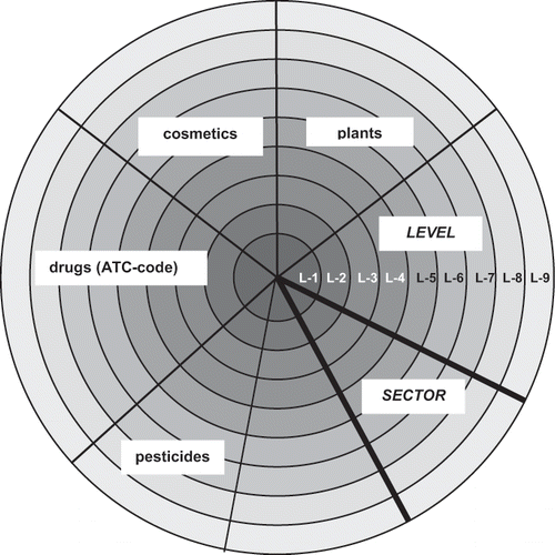
26 Prescription Opioid Abuse: Emerging Perspectives and Opportunities for Poison Centers
Dart RC. Rocky Mountain Poison and Drug Center, Denver Health, and University of Colorado, Denver, CO, USA.
Objective: Data from the Drug Abuse Warning Network suggest that the rates of prescription drug abuse are increasing in the United States. The RADARS® System was created in 2001 to study the misuse, abuse and diversion of prescription opioid drugs. The system was created by Purdue Pharma LP, but has since been transferred to the public sector. Due to the complexity of the issue, the system was constructed to provide four different perspectives on prescription opioid abuse. The Key Informant system includes over 300 clinicians, epidemiologists, treatment counselors, or other observers in a position to know about new and emerging drug problems. The Drug Diversion system includes over 300 law enforcement agencies nationwide reporting official seizures of prescription opioids. The Methadone Treatment clinic system includes 75 treatment centers. The Poison Center system includes 42 centers serving a population of over 222 million people. Poison centers were included because they: 1. provide widespread coverage of the US; 2. identify the specific drug product involved; 3. provide geographic specificity; 4. utilized trained staff and a standard reporting instrument and; 5. provide nearly real-time reporting. Methods: Seven drug groups are included in the system: buprenorphine, fentanyl, hydrocodone, hydromorphone, methadone, morphine and oxycodone. The four signal detection systems collect information from their informants and report quarterly the rates of cases involving a specific drug to the level of the 3 digit zip code. Like the Drug Abuse Warning Network, the concept of mentions is used. Every drug mentioned in a contact is counted as a mention. The event rate for each system is calculated for each geographic area and is trended over time. Two denominators are utilized in calculating rates: population of the area and unique recipients of dispensed drug (URDD). The use of population (e.g. cases per 100,000 population) gives a measure of the overall public health significance. In contrast, the use of URDD (cases per 10,000 recipients of drug) provides more information about the misuse and abuse of the drug or specific product in relation to other products. The pharmaceutical manufacturer is responsible for investigation of signals regarding their products. Results: The general trend in all systems has been an increase in rates of abuse over time for each opioid drug studied. In the Key Informant and Methadone clinic systems, oxycodone is the most common drug reported with hydrocodone second. In the Drug Diversion system, hydrocodone is the most common drug and oxycodone second. Poison centers receive the most calls and mentions regarding hydrocodone. However, the two denominators produced markedly different rates (). While hydrocodone and oxycodone products result in the greatest rate of mentions by population, methadone and buprenorphine product the highest rates in terms of individuals filling a prescription. Geographic trends in the signal detections systems indicate consistently high signal rates from the Appalachian region of the United States. Peak rates in the Appalachian region reach 128 mentions/100,000 population in contrast more typical rates of 11 mentions/100,000 in other areas. The use of pill identification (ID) has proven a useful. For example, the rate of pill IDs increased from 52 per 100,000 population to 210 pill IDs per 100,000 population in the weeks following the introduction of three new oxycodone controlled release products. Low population area tends to have higher rates than highly populated areas. The mean rate per 100,000 population for area with a population less than 100,000 is 20.3 mentions/100,000 compared to 4.4 mentions/100,000 for areas with a population greater than 1,000,000. Conclusions: Abuse of prescription opioid drugs appears to be increasing. Hydrocodone and oxycodone present the largest challenge in terms of the total amount of diversion and abuse occurring in the US When viewed by the number of patient receiving drug, however, methadone and buprenorphine appear to be disproportionately represented. Geographic differences are also important with rural areas contacting poison centers much more commonly than urban areas. References: Substance Abuse and Mental Health Services Administration, Office of Applied Studies. Emergency Department Trends From the Drug Abuse Warning Network, Final Estimates 1995–2002, DAWN Series: D-24, DHHS Publication No. (SMA) 03–3780, Rockville, MD, 2003.
TABLE 1 Comparison of poison center international exposure contact rates using population and URDDs denominators
27 Diversification in Activities of Poisons Centres
Kupferschmidt H. Swiss Toxicological Information Centre, Zuerich, Switzerland.
Objectives: Poisons Centre are institutions where information and experts on adverse and toxic effects of substances and products are available on a 24/7 basis. Answering emergency telephone inquiries from medical professionals and/or the public concerning toxic exposures is the core activity of Poison Centres. These prerequisites are the basis for the diversifications of a Poisons Centre's activities. Methods: Informations on various ways of diversification are collected from the own Poison Centre, from reports submitted as abstracts to the EAPCCT and NACCT congresses 1998–2005 and Congress Programs, and from the EAPCCT Poisons Centres Directory 2005–2006. Results: Currently Poisons Centres diversify their activities in various ways. The reasons for doing so are different (see ).
TABLE 1 Areas of diversification in poisons centres for different reasons
Toxicologists in Poisons Centres may want to broaden their scientific scope or to enlarge their professional field of activities, or there may be simply a need to find additional funds, as sources of funding are through direct patient revenue, academic affiliations, hospitals, industry and government. In the EAPCCT Poisons Centre Directory there are informations about particular services from 69% of the 81 Poisons Centres in 33 European countries. Of these 77% receive calls from the general public, 63% are involved in antidote or antivenin supply, 49% provide information about medications, 38% are involved in chemical incident control, 43% provide teratology information, 61% include information about herbal or traditional medications, and 51% anwser inquiries on veterinary toxicology. 36% of Poisons Centres are part of local or regional networks, mainly in France, Germany and Scandinavia. Other activities include the delivery of services outside its own geographical area, consultations in clinical, occupational and forensic medicine, in environmental toxicology, and allergology. Poisons Centre's databases may contribute to epidemiology, toxicovigilance and risk assessment. Some medical toxicologists deliver laboratoy services, mushroom identification, doping information and do consults in bio-statistics. Others provide clinical pharmacology services (information and consults on adverse drug reactions, drug interactions, therapeutic drug monitoring, tablet identification) and do reporting of adverse drug reactions. Some do toxicology research not only clinically but also in basic science and in this way contribute to bridge the gap between basic science and clini-cal medicine. Conclusion: There are many ways a Poisons Centre can diversify its activities, based on its core services. Diversifications can be useful from a scientific, professional or economical point of view and can provide a more broad and stable basis of a Poisons Centre's performance.
28 Improving Cooperation Between Poisons Centres and Industry – The Industry Perspective
Wilks MF. Syngenta Crop Protection AG, Basel, Switzerland.
Introduction: The majority of substances for which poison control centres (PCC) are consulted are produced by industry and fall broadly into two categories: those which are intended for human consumption or application (e.g. pharmaceuticals, cosmetics and personal care products), and those which are used in the home and the workplace but where exposure is not desired although it may be unavoidable (e.g. household and consumer products, industrial chemicals and agrochemicals). The respective industries are subject to different legislation and regulatory requirements, and their needs for PCC liaison and exchange of information are diverse but will always be related to one or both of the key functions of PCC, i.e. provision of expert advice and toxicovigilance. Pharmaceuticals: Most companies will have in-house staff and procedures to cover statutory pharmacovigilance activities including pre- and postmarketing surveillance, adverse event monitoring, and periodic safety update reports. Interaction with PCC is therefore often limited unless the centre has specific expertise in pharmacovigilance. Nevertheless, there are areas in which data from PCC can add useful information to drug safety assessments. For example, the safety of prescription medicines in overdose can be one of the factors to take into account when deciding whether a preparation should become available over the counter. Another area of potential collaboration is the provision of out-of-hour cover for clinical trials which is of particular interest for smaller companies. Cosmetics, personal care, household and consumer products: A characteristic of these product groups is that they are used directly by consumers, often without due consideration of safety precautions such as preventing access of children. This has the potential to generate frequent enquiries both to companies as well as to PCC. There is a need for good collaboration between the two, ideally on the basis of a formal agreement which ensures professional management of cases as well as access to product information when required. While the intrinsic hazards of many products are well understood there is a continuing debate between the industry and PCC about access to information. The desire of PCC to obtain comprehensive formulation details is understandable, however, from the companies’ point of view this appears often unnecessary as well as difficult to maintain. The number and concentration of hazardous ingredients may be limited and, given frequent formulation changes, there is the danger that a formulation database may be quickly out of date. A joint project by the EAPCCT and the European Cosmetics Toiletry and Perfumery Association (COLIPA) which has developed a set of ‘frame formulations’ specifying the type of ingredients and their maximum concentration in different cosmetics may well serve as an example for other industries (1). Industrial and agricultural chemicals: There is an increasing recognition in this industry sector that the professional management of adverse health incidents and toxicovigilance are an integral part of stewardship and responsible care activities. While basic toxicology information on products is often extensive, particularly for agrochemicals with their tight regulatory framework, in-house medical and product safety resources are often limited. This has prompted many companies to look for external service providers to deliver professional, high quality 24 h emergency response and management of incidents. In theory, PCC would be well placed to provide such services, however, for a number of reasons this has so far only happened on a limited scale. Many PCC are reluctant to enter into contracts with companies because they view a commercial relationship as a potential threat to their professional independence. It may be organisationally difficult for them to become service providers because they are part of local or national health services. There may also be concerns about medical data confidentiality. Financial contributions through industry contracts may also be viewed as subsidising essential services with the threat of further reduction of already very tight public financing of PCC activities. From an industry point of view, companies outside the pharmaceutical sector often feel that PCC do not fully appreciate the nature and uses of their products, or understand the different exposure scenarios and consequently tend to be rather conservative when assessing risks. In the area of toxicovigilance, regulatory bodies are asking for information on safety in use of products, particularly at the time of re-registration, however, this is often proving difficult to provide. Data which may be provided by different PCC are often not comparable due to a lack of harmonised data collection and interpretation. Conclusions: It is important that relationships between industry and PCC are established on the basis of openness, mutual trust and clarity of expectations. Any provision of services should be contractually defined. Different industry sectors have different needs for liaison with PCC but they all relate to the provision of advice in exposure situations and/or toxicovigilance activities. For various reasons many PCC are reluctant to enter into contractual agreements with commercial companies but, if handled correctly and sensitively, both partners can benefit from such relationships. Reference: 1. EAPCCT & COLIPA. Cosmetic Frame Formulations. Brussels, Belgium, European Cosmetics Toiletry and Perfumery Association, January 2000.
29 Enhancing Communication and Cooperation Between Chemical and Consumer Product Manufacturers and Poison Center: the Poison Center Perspective
Wruk KM, Bronstein AC. American Association of Poison Control Centers, USA.
Background: The first USA poison center (PC) opened in Chicago, IL in 1953. Currently, there are 61 US PCs. In 2004, the centers received more than 3.4 million calls from the public and health care professionals. Since 1983, these member centers of the American Association of Poison Control Centers (AAPCC) have contributed case information to a national database. Many of these involve exposures to specialty chemical and consumer products. Most exposures occur in the home, and some are occupationally related. Anomalous case clusters have been used to identify product problems. Product stewardship tenets require that both manufacturers and consumers understand health and environmental risk of product use. Poison centers play a vital role in this effort. Objective: Characterize the value of national and regional poison center data for chemical and consumer product manufacturers. Methods: PCs respond daily to questions providing standardized and accurate information for managing exposures through immediate case recommendations. Analysis of these calls provides insight into product formulation and use issues. Calls can be tracked and trended. Traditionally PC data were reported via annual or quarterly reports. These retrospective reports went to state and local health departments, funding, and governmental agencies. Development and implementation of real time toxicosurveillance heralds a major change in how PC data is used and valued. While the current recipients of these data are predominately public health entities, manufacturers could benefit from PC expertise and product surveillance activities. The AAPCC core data base application allows for near real-time product surveillance with the ability to generate company alerts to aid in-market icident support, evaluation and follow-up activities. Discussion: Poison center data can assist industry in post marketing surveillance; work in collaboration for new product launches, and public health issues of product related events. Rapid identification of unexpected outcomes of exposures to new or existing products helps minimize risk and liability. In addition to managing intentional and unintentional misuse issues, PCs have the specialized expertise necessary to recommend expert solutions for product health related events. This expertise assists the manufacturer in detection and event response solutions. Information collected and analyzed can be used for both in-house and governmental regulatory and reporting requirements. With the advent of near – real time mapping (GIS), companies can see problems develop as they occur. Centers can work with industry to monitor new product launches, or seasonal packaging problems that have the potential to increase exposure risk. While historically, industry was reluctant to use PC data, corporate poison center sponsorship initiatives can aid manufacturers in their product stewardship mission. Conclusion: Data collection and product surveillance are core competencies of PCs. Through a direct relationship PCs can work with manufacturers to provide incident reporting and toxicology support for product stewardship functions. In return corporate funding can provide assistance to PCs for operational needs. This collaboration results in a benefit to the public and corporation.
30 Should Poisons Centres Provide Veterinary Advice?
Campbell A. Veterinary Poisons Information Service, Medical Toxicology Unit, London, UK.
Introduction: In April 1992, the London Centre of the UK National Poisons Information Service, with the Leeds Centre as it was then formally started the UK Veterinary Poisons Information Service (VPIS). This subscription service was initiated, with support from the British Veterinary Association (BVA), in response to a noticeable increase in enquiries from veterinarians handled by the London centre. In addition our hospital management had demanded that this hitherto unfinanced activity was resourced appropriately from non-health service funds.
There were many initial concerns: a) Paying customers might expect a quality of service unachieveable with the limited veterinary toxicology data available and without significant, and potentially costly, input from qualified veterinarians; b) Some Poisons Centre staff wanted specific training in providing information about the management of animals and in locating species-specific information; c) The service might be costly to administer, particularly if the imposition of charges reduced overall veterinary activity – as occurred when the US Animal Poisons Control Centre introduced charges (1). Reference to the current EAPCCT directory of European Poisons Centres reveals that, of 81 centres listed, 41 (50.6%) regularly answer veterinary enquiries, whilst 15 (18.5%) do not, and for 25 centres such information was not available. Few centres are formally funded for such activities; but with over 13 years of experience the VPIS can offer insight into how these concerns were addressed, and how this initiative proved both rewarding and financially successful. Methods: Retrospective analysis of records from the VPIS database was undertaken to provide data on enquiry numbers and case details. Results/Discussion: The service and its usage: Practices take out annual subscriptions allowing them unlimited access to the VPIS on quotation of a membership number. The fees are dependent on the number of veterinarians in the practice. Initial uptake was remarkable with 333 practices registering in 2002–2003. Despite minimal advertising since, in 2005 over 1,250 were registered, with over 2,800 outlets. This represents over 80% of UK practices. To keep administrative costs low the subscription year is fixed (April to March each year). Enquiry numbers in 1992 did not fall as anticipated. Indeed, they have risen every year except 1999. Income has risen concomitantly (). The bulk of enquiries (95%) concern dogs and cats, and the agents to which each group are most commonly exposed follow readily identifiable patterns with Ibuprofen and Permethrin being the most frequent in dogs and cats respectively.
FIG. 1 Enquiry numbers and income of the VPIS (at 26.10.05)
Information Sources: Since the veterinary toxicology data available was perceived as limited, the VPIS adopted a systematic approach to data collection from the outset. The majority of referrals (about 80%) were, and are routinely followed up by, detailed postal questionnaire. Return rate has always exceeded 50%. Detailed data concerning the poisoning incidents, their clinical courses, analytical results and case outcomes can thus be documented. Initially these were summarised on paper, but in 1999 a tailored database was developed using Access™, which since 2000 been used to record all cases. In addition, all pre-2000 cases with follow-up have been transferred. The database automatically generates the questionnaires, tracks practice registrations, and permits useful statistical analysis. Summaries of past cases from the 50,000+ cases stored can be retrieved at the time of an enquiry. These summaries, together with data and reports from the published literature, from manufacturers, the internet and from other veterinary collaborators, are used to generate specific monographs on the management of specific poisons in specific species when sufficient in quantity and quality. Over 150 of these VPIS monographs are now in existence; all undergo regular review and update. Some have been published in a handbook (2). Regular scanning of veterinary and toxicology journals is undertaken to keep abreast of current practice. Training: As with handling human poisoning enquiries much of the necessary training is on-the-job. New staff members are given initial instruction into information sources, database usage, and the important factors that should be considered when handling animal cases (enzyme deficiencies, other metabolic differences, species and breed variations, the effects of animals grooming etc). All staff members are regularly informed of new monographs and changes. With growing experience the service has gradually improved. Very occasionally referrals for complex cases are needed and the VPIS has built a small network of veterinarians willing to assist in these rare instances. Perceptions of the service: On the follow-up questionnaires users are invited to comment on the service. Responses are usually detailed and positive, and adverse criticism is rare. VPIS activities are discussed on an annual basis at a meeting held by the BVA, at which most professional veterinary bodies, including industrial ones, are represented. In 2005, VPIS registration became a mandatory requirement of the Royal College of Veterinary Surgeons’ veterinary practice accreditation scheme – a decision made without any consultation with the VPIS! The service is now actively involved in the undergraduate training programmes of several veterinary colleges, and in various veterinary continuing education programmes too. In 2004, in association with the BVA's Animal Welfare Foundation, a “Pets and Poisons” leaflet was produced. Over one million copies have been distributed to practices for their clients. Further outreach campaigns are in planning. Conclusions: It is the VPIS experience that trained poisons centre staff can handle animal enquiries easily and well, as the skills required are similar to those needed for their routine human poisons calls. It is vital that systematic collection and processing of veterinary case data is conducted from the outset. It is possible to generate significant income from veterinarian users and bodies to more than cover the costs of service provision and development. The VPIS would strongly encourage other poisons centres to look at the potential opportunities of such activity. References: 1. Buck WB. A poison control center for animals: liability and standard of care. J Am Vet Med Assoc 1993; 203:1118–1120. 2. Campbell A & Chapman M, Handbook of Poisoning in Dogs and Cats, Oxford, UK: Blackwell Science, 2000.
31 Gluthatione – System Enzymes in the Liver and Erythrocytes in Acute Iron Intoxication in a Rat Model
Berkovitch M (1,2), Weinbaum S (2), Evans S (1), Zandback Y (2), Petrov I (1), Alkan Y (1), Siman-Tov Y (1), Rechavi M (2), Kozer E (1,2). 1. Clinical Pharmacology and Toxicology Unit, Pediatric Division, Assaf – Harofeh Medical Center, Zerif; 2. Sackler School of Medicine, Tel-Aviv University, Israel.
Objective: To study the effect of acute iron poisoning on the glutathione system in the liver, and to determine whether there is a correlation between glutathione system enzymes in the liver and erythrocytes in acute iron overdose. Methods: The study protocol was approved by the Animal Care Committee at Assaf Harofeh. Male Wistar rats weighing 150–250 gram were studied. They were double-housed in shoe box-type cages and were deprived food for 14 hours prior to iron administration. The rats were assigned to one of 3 groups. Group I rats (n = 20) received distilled water, group II (n = 30) received 400 mg/kg elemental iron and group III (n = 20), received 750 mg/kg elemental iron. Iron and water were administered orally by gavage. Groups were compared by one way analysis of variance or Kruskal-Wallis one way analysis of variance on ranks as appropriate. Results: Median serum iron level for groups 1–3 were 219 mg/l 735 mg/l and 979 mg/l, respectively (p < 0.001). Median time until sacrificing was longer in group 2 compared with group 3 (240 minutes versus 120 minutes, respectively, p < 0.001). Liver transaminases were higher in rats treated with iron. Reduced glutathione (GSH) in the liver in groups 2 (3.1 + 4.6 mmol/mg protein) and 3 (4.7 + 4.6 mmol/mg protein) was significantly lower than in group 1 (11.5 + 6.2 mmol/mg protein) (p < 0.001). Liver glutathione S-transferase (GST) was higher in group 3 (0.42 + 0.14 mmol/sec/mg protein) compared to groups 2 (0.3 + 0.07 mmol/sec/mg protein) and 1 (0.27 + 0.14 mmol/sec/mg protein) (p < 0.001). GSH levels in erythrocytes were higher in group 2 compared with group 3 (p < 0.05), there was no significant difference between groups 2 and 1 and between groups 1 and 3). There was a significant correlation between G6PD and GAD levels in the erythrocytes and in the liver (r = 0.52 p < 0.001 and r = 0.42 p < 0.001, respectively). There was no correlation between GSH, GSSG, GST, GR, GPX and Catalase levels in the erythrocytes and in the liver (p = 0.41, p = 0.48, p = 0.49, p = 0.53, p = 01.4 and p = 0.84 respectively). Conclusions: Iron intoxication is associated with depletion of reduced glutathione in the hepatocytes and with significant changes in the glutathione system enzymes. These changes are not reflected in the erythrocytes. These results suggest that oxidative stress contribute to the toxic effect of iron in hepatocytes. Whether these changes are in correlation with the severity of the poisoning has to be clarified.
32 Effects of Organophosphate Pesticide Poisoning on Time – Course of Cognitive Processing
Dassanayake WDMTL, Weerasinghe VS, Dangahadeniya U, Kularatne K, Senanayake N. South Asian Clinical Toxicology Research Collaboration, Faculty of Medicine, Peradeniya, Sri Lanka.
Objective: Recent evidence suggests that acute organophosphate poisoning could lead to delayed neuropsychological effects (1,2). However, consistent evidence is lacking on the effects of organophosphate poisoning on the time-course of cognitive processing of information. This study was conducted to determine the effect of organophosphate poisoning on more objective indicators of time-course of cognitive processing of visual and auditory stimuli. Methodology: This was a cross-sectional observational study. The study group comprising 25 patients who recovered from the cholinergic phase (on average, 14 days after poisoning) were compared with an age and sex matched control group. The tests of information processing used were simple visual reaction time, recognition visual reaction time, auditory event-related brain potentials (ERPs), visual evoked potentials and motor evoked potentials. Cognitive processing time of each visual reaction task was calculated by subtracting the afferent visual impulse duration and motor impulse duration from reaction time (cognitive processing time = Reaction time – P100 latency – total motor conduction time). Auditory stimulus evaluation time was measured with P300 wave latency of ERPs, recorded using oddball paradigm. Results: () P300 latency and cognitive processing time for recognition reactions were significantly prolonged in poisoned group compared to controls. There was no significant difference in cognitive processing times in simple visual reactions. Conclusions: Poisoned group showed a significant delay in cognitive processing in recognition visual reaction tasks and a significant delay in auditory stimulus evaluation in ERP experiments. However, cognitive processing in simple visual reaction task was similar in both groups. This suggests that organophosphate pesticide poisoning adversely affects higher order cognitive operations such as stimulus discrimination and stimulus evaluation, without affecting simpler cognitive processes operating in simple reaction tasks. Subsequent follow-up is necessary to observe any deterioration/improvement of these abnormalities. Being more objective indicators of time-course of cognitive processing, these measures may be useful in studying the effect of therapeutic interventions on cognitive processing of information. References: 1. Savage EP, et al. Chronic neurological sequelae of acute organophosphate pesticide poisoning. Arch Env Health 1988; 43:38–45. 2. Steenland K, et al. Chronic neurological sequelae to organophosphate pesticide poisoning. Am J Public Health 1994; 84:731–736.
TABLE 1 Measures of cognitive processing in test and control groups
33 1,4 Butanediol Pharmacokinetics and Clinical Effects in Humans
Haller CA (1), Thai D (2), Dyer JE (3), Benowitz NL (1). 1. Department of Medicine, University of California, San Francisco, San Francisco, CA USA; 2. Amgen, Thousand Oaks, CA, USA; 3. School of Pharmacy, University of California, San Francisco, CA, USA.
Objective: 1,4 Butanediol (BD) is a chemical precursor of gamma hydroxybutyrate (GHB) that is sometimes ingested to get “high.” The pharmacology of BD after oral ingestion has not been described in humans. The aim of this study was to characterize the pharmacokinetics and effects of BD on respiratory, cardiovascular, and cognitive function in healthy people. Methods: Eight healthy volunteers (5 men) were administered 25 mg/kg BD in a single oral dose after an overnight fast in a double-blinded, placebo-controlled, crossover study. Blood pressure, heart rate, skin temperature, electrocardiogram, and oxygen saturation (O2sat) were monitored. Blood samples were collected at 0, 5, 15, 30, 45, 60, 90 minutes, and 2, 3, 4, 5, 6, 12 and 24 hours, and analyzed by GC-MS for BD and GHB levels. Subjects completed visual analog scale (VAS) questionnaires about drug effects on 12 mood and physical symptom responses. Results: All subjects completed the study without significant adverse effects. Two subjects experienced drowsiness and nausea, which prevented completion of some cognitive testing but resolved without medical intervention. The plasma concentration-over-time profiles for BD and GHB are shown in . BD was rapidly converted to GHB with an elimination half-life (T½) of 14.5 minutes. A mean maximum GHB concentration of 44.7 mg/L was reached 30 minutes after BD ingestion. GHB T½ averaged 47 minutes. Compared to placebo, subjects reported feeling less awake and alert (p = 0.013), less able to concentrate (p = 0.045), and more lightheaded or dizzy (p = 0.0099) for as long as 4 hours after dosing, with maximal effects at approximately 60 to 90 minutes. Pulse oximetry readings were lower 45 minutes after BD dosing with a mean O2sat of 98.5% with BD, (95% C.I. 97.4–99.6%) versus 99.6% with PLC (95% C.I. 99.2–100.0%), (p = 0.031). Treatment-related changes in blood pressure, heart rate, and temperature were negligible. Conclusions: In vivo conversion of BD to GHB after oral administration is rapid. With modest BD doses, sedative effects and decreased oxygen saturation occurred in the first hour after dosing, but significant alterations in vital signs were not observed.
FIG. 1 Mean plasma concentrations over time for GHB and BD after oral administration of 25 mg/kg BD
34 Intoxications with Anticoagulants Cumatetralyl and Bromadiolone: Epidemiology, Case Reports and Toxicokinetic Analyses
Binscheck T, Grobosch T, Lampe D. Institute of Toxicology – Clinical Toxicology and Poison Information Centre, Berlin, Germany.
Objective: Ingestion of anticoagulant rodenticides are not a common feature in poison information service but can lead to severe disturbance of coagulation. To obtain epidemiological data about intoxications with anticoagulant rodenticides, ingestions reported to the Poison Information centre Berlin during the last five years were reviewed. In addition, two cases of severe adult intoxications with cumatetralyl and bromadiolone respectively were monitored and serum samples were analyzed for toxicokinetic parameters. Methods: 1. A number of 455 infant and 84 adult ingestions of seven most frequently asked anticoagulants were analyzed for circumstances and clinical presentation. 2. Serum samples of two adults intoxicated with cumatetralyl and bromadiolone, respectively, were analyzed using LC-MS with an internal standard method. Distribution and elimination half-life of cumatetralyl and bromadiolone were computed and compared to toxicokinetic data published elsewhere. Data were correlated to clinical findings and the time course of INR. Results: 1. Accidental ingestion of anticoagulant rodenticides occurs more frequently in children than in adults. In contrast, adults ingested larger amounts causing significant anticoagulation. 2. A 62-year-old male reported the intake of an unknown amount of a “rat poison” some days before. He had no symptoms, but INR was increased to 2,5 and treatment with vitamin K1 was immediately initiated. In LC-MS cumatetralyl was found at a serum concentration of 121 μg/L. The patient was treated with 3 × 20 mg of vitamin K1/day allowing a restored coagulation until discharge at day 11 with a cumatetralyl serum concentration below 10 μg/L. Computed elimination half-life was 60 hours indicating that cumatetralyl is a short-acting anticoagulant. In a second case, a 55-year-old man was admitted to the hospital with abdominal and muscular pain, had a bleeding lesion at his tongue and bloody expectorations but reported no ingestion of anticoagulants. At day 4, INR increased to 10 and intravenous treatment with vitamin K1 and PPSB was started. In toxicological analysis bromadiolone was quantitated at 440 μg/L serum. Subsequent measurements revealed a first order two-phase elimination of bromadiolone with an alpha-phase (25,5 hours) and a beta-phase of 140 hours resembling the terminal elimination half-life. Slow decline in bromadiolone serum concentration was paralleled by normalization of INR during the following 22 days with vitamin K1 p.o. Conclusion: Anticoagulation in children is a rare finding due to the low concentrations of anticoagulant in commercially available rodenticides and the little amount ingested. In adults with suicidal or criminal ingestions of larger doses, increase in INR and signs of bleeding occurrs more frequently. Toxicological analysis of anticoagulants with LC-MS allows identification and quantification of the anticoagulant involved in intoxication providing valuable information on prognosis and duration of treatment. The difference between short-acting cumatetralyl and long-acting bromadiolone is caused by the prolonged distribution phase and elimination half-life of the latter.
35 Poisoning by Topical Medications
Nelson LS. New York City Poison Control Center, New York, NY, USA.
Dermal exposures account for 8.0% of the calls and 1.3% of the fatalities reported to the AAPCC. The epidermis is the most important barrier to toxin bioavailability, requiring dissolution or permeation the ceramides, or skin lipids, in the stratum corneum in order to access the blood vessels of the dermis. Lipid solubility is thus the most important factor determining dermal absorption, although concentration, duration of exposure, molecular weight of the molecule, and specific skin characteristics (thickness, body folds, mucous membrane) are also important determinants. Illustrating this is mercury, Hg+, a metal with virtually no lipid solubility and, therefore, no skin penetration. The addition of two methyl groups, to form dimethylmercury, increases the systemic absorption sufficiently to produce fatal organic mercury poisoning following a single drop. The skin interface influences absorption, as is exemplified by patients with decreased levels of ceramide in the stratum corneum (e.g. psoriasis) who have increased toxin penetration. Similarly, the characteristics of the drug and its vehicle may influence transdermal absorption. Transdermal drug delivery systems were developed to alter the skin partition coefficient. For some agents, as with the fentanyl patch, a rate-limiting membrane is required to prevent rapid absorption due to the drug's high lipophilicity. Leakage of the patch has proven fatal. Still, with even fentanyl's penetrability characteristics, transdermal systems still require that large amounts of drug be present externally to maximize the transcutaneous gradient. For example, in order to deliver fentanyl at a constant rate for the three day life of the patch, a reservoir containing between 2.5 (25 mcg/hr patch) and 10 mg (100 mcg/hr patch) of fentanyl is needed. The fentanyl patch varies the rate of drug delivery simply by varying the skin contact surface area of the patch (10, 20, 30, and 40 cm2), for a release rate of 2.5 mcg/hr per cm2]. Dissolution of toxin in the stratum corneum may serve a depot function leading to continued systemic exposure despite apparent removal of the toxin. Fentanyl diffuses through to the dermis and is uptaken systemically. In adults, the time from application to minimal effective serum concentrations can range from 1.2 to 40 hours, and the time to reach maximum serum concentrations can range from 12 to 48 hours. When the patch is removed, fentanyl continues to be absorbed into the systemic circulation from the cutaneous depot. Other drugs of potential toxicologic interest that utilize transdermal drug delivery technology include clonidine (2.5–7.5 mg/patch, delivers 0.1–0.3 mg/d for 1 week), lidocaine (700 mg/patch,>650 mg remaining after 12 hours use), nicotine (approximately double the amount noted present, delivering 5–21 mg/day), and scopolamine (1.5 mg/patch, delivers 1 mg over 3 days). Despite the massive amount of drug in the patch, the serum concentrations obtained under most conditions are well below those associated with more conventional routes. In addition, morbidity and mortality are reported with the topical application of camphor, ethanol, organic phosphorus insecticides (including nerve agents), organochlorines (e.g. lindane), phenol, podophyllin, nitrates, and salicylic acid. Many of these are used themselves as medicinals or are representative of a class of therapeutic agents. Children are particularly at risk for toxicities from percutaneous absorption since their skin is more penetrable than that of adults. In addition, children may find the patches enjoyable for play, and since even a “used” patch has substantial quantities of drug remaining, poisoning in this circumstance is widely reported.
36 Educational Programmes in Australia
Dowsett RP. Clinical Pharmacology and Toxicology, Westmead Hospital, Sydney, Australia.
The University of Newcastle, Australia offers courses in post-graduate training in toxicology. The courses are available to those with a degree in Medicine, Pharmacy or a Health Science discipline from an Australian or overseas University. Four courses have been developed, each one semester in length, consisting of Core Principles, Clinical Toxicology of Pharmaceuticals, Clinical Toxicology of Plants and Animals and the Clinical Toxicology of Industrial and Chemical Agents. This presentation will demonstrate these topics in more detail and illustrate the teaching methods utilised. Undertaking these four courses fulfils the requirement for a Graduate Certificate in Clinical Toxicology. This can be extended to a Graduate Diploma in Clinical Epidemiology (Clinical Toxicology Specialisation) by undertaking four additional subjects in Epidemiology and Biostatistics. A Master of Medical Science (Clinical Toxicology Specialisation) can then be completed by additional course work in related fields or undertaking a thesis in clinical toxicology. The course is undertaken by distance education utilising a web-based interface for the discussion of clinical problems based on individual case histories. Approximately 30 case histories are presented for each course and students write their answers to specific questions about the assessment and management of each scenario. Discussion and clarification on each response can be posted by students or faculty. Contribution to this constitutes 40% of the overall assessment, the remainder coming from a mid-semester assignment (20%) and an end of assessment assignment (40%). The courses were developed by a faculty of Clinical Toxicologists based in Australia, Sri Lanka and Canada. For each topic there are written a summary, learning objectives, key readings and the clinical problems. Students are provided with books of these and reproductions of the key readings. HyperTox, a software program of information for the treatment of common and serious poisonings is also provided to students. The program began in 2004 and nine students have successfully completed the four toxicology courses. Further areas of development are being considered including additional courses to expand topics in the toxicology of pharmaceuticals and courses more focussed to the interests of pharmacy and health science graduates.
37 Educational Programmes in Clinical Toxicology in Western Europe
Locatelli C, Petrolini V, Lonati D, Butera R, Manzo L. Pavia Poison Center and Toxicology Unit, IRCCS Maugeri Foundation and University of Pavia, Italy.
Background: The birth of toxicology in Western Europe (WE) was in the 1955–1970s, mostly as a part of pharmacology. Gradually medical toxicology became an independent discipline. However, at the same time in the clinical field, poisonings have been the turning points on which clinicians from different medical specialties (e.g. anesthesia, intensive care, internal medicine, occupational medicine, pediatrics) developed clinical toxicology and the poison centers activities. Clinical toxicology has been in growth since the ‘70s, and parallelly, since education is one of the milestones for the improvement of the medical knowledge and the base for the characterization of a discipline and a specialty, as expected specific educational programmes in clinical toxicology (EPCT), distinct from the most general ones in medical toxicology, have been well established in WE. Nevertheless, EPCT are probably not sufficiently widespread in WE, and, if present, they may differ in the program and characteristic (years of training, skills, etc.) between the countries themselves and from those of the USs or Australasia. Objective: To evaluate the presence/absence in WE countries of a) a recognized educational degree of specialty in clinical or medical toxicology, and b) specific EPCT. Methods: A search and analysis of 1) web-reported sources (Google and Yahoo engines), and 2) published medical literature was conducted to find the educational degrees of specialty in clinical or medical toxicology and/or EPCT. The biggest western European countries (Austria, Belgium, Denmark, Finland, France, Germany, Greece, Ireland, Italy, The Netherlands, Norway, Sweden, Spain, Portugal, UK, Swiss) were included, whereas some other little countries (Andorra, Cyprus, Iceland, Liechtenstein, Luxembourg, Malta, Monaco, San Marino, Vatican City) were excluded because of loss of data or direct knowledge of absence or the studied objectives. The first 500 items (max) obtained from every web-search regarding EPCT were evaluated. EPCT were included if carried out by University, Scientific Society or official Medical Organizations. EPCT proposed by poison centers or other clinical department were excluded if not leading to a title recognized by the local Government or the European Union. Similarly, undergraduate courses and the not-well defined educational programmes were excluded, such as programmes of distance-learning for degrees (web-based programmes) and educational programmes comprehending courses on clinical toxicology as a limited part of general programmes of applied, forensic or environmental toxicology, ecotoxicology, occupational medicine, emergency medicine, intensive care, pediatrics, etc. Results: A degree of medical specialty in clinical or medical toxicology is not available in WE countries, except for Italy, where a specific 4-year postgraduate educational programme lead the European Union to recognize a title of specialist in medical toxicology. In other WE countries (1), the physicians treating poisoned patients are not formally educated in clinical toxicology, but in emergency or internal medicine, pharmacology, pediatrics, occupational medicine or other disciplines. The web-based search resulted in a diffuse availability of undergraduate, doctoral and postgraduate educational programmes in toxicology in almost all the WE countries, offering different types of degrees such as Ph.D., diploma, fellowships (e.g. France, 1 M.A. and more than 25 diplomas/Ph.D.; Ireland, 1 M.Sc.; Italy, more than 31 Ph.D.; UK, 6 or more M.Sc., more than 2 diplomas, several Ph.D.; Germany, 1 M.Sc., several Ph.D.; Netherland, 1 M.Sc., several Ph.D.; Austria, Belgium, Portugal, Spain, Swiss: several Ph.D.), but only a few of the postgraduate educational programmes (less than 3–4%) are specific for clinical toxicology. The medical literature shows more information about EPCT. Formerly, educational programmes in toxicology are operating in all the biggest WE countries, whereas EPCT result officially approved in France (2), Italy (3), UK (4). In Italy, for example, toxicology (and clinical toxicology) is an obligatory part of the course work in pharmacology as a part of undergraduate curricula in the Faculties of Medicine and Surgery. In the same Faculties, the postgraduate training in clinical toxicology includes 1) postgraduate schools of specialization (8 University), 2) Ph.D. programs (at least 3 in clinical toxicology and addiction; 3–4 years length), and 3) research fellowships (2–4 years). There is no evidence of specific EPCT in some WE countries (eg. Germany (1,5), Finland (6), Sweden (7). Conclusion: Toxicology is relatively young as a medical discipline in most WE countries, and in particular, clinical toxicology (the toxicological advice in the Poison Centers, the bed-side clinico-toxicological activity and consultancies) has achieved the clinical relevance needed to become an independent discipline only in the last two decades. The growing professional experience and knowledge in clinical toxicology need to be transmitted to young physicians that approach clinical toxicology and the treatment of poisoned patients. This preliminary evaluation show that is impossible to find and to compare EPCT in WE countries via web-sources or medical literature, and that this approach underestimates the real availability of the educational resources. To better evaluate the availability of EPCT in WE a different strategy of research has been recently started using a questionnaire sent to all WE poison centers, governmental institutions for education and university, and national scientific societies of toxicology. The EAPCCT website may be the better way to collect and store information regarding EPCT from all the European countries. This should be a way for EAPCCT to work, jointly with the academic institutions in all countries, with the aim to promote harmonization, mutual recognition and free movement in EU, and to help young physicians to approach the profession of clinical toxicologist choosing between the various European educational possibilities and capabilities. References: 1. http://www.iutox.org/newsletter_11_05 Full.asp 2. http://www.sftox.com 3. Preziosi P, Dracos A, Marcello I. Information resources in toxicology-Italy. Toxicology 2003; 190:35–54. 4. Pantry S. Toxicology digital sources produced and available in the UK (UK). Toxicology 2003; 190:75–91. 5. Kahl R, Desel H. Germany: toxicology information on the World Wide Web, 2003; 190:23–33. 6. Komulainen H. On-line information sources of toxicology in Finland. Toxicology, 2003; 190:15–21. 7. Heurgren-Calstrom G, Malmberg E. Online information resources of toxicology in Sweden. Toxicology 2003; 190:63–73.
38 Educational Programmes in Clinical Toxicology in Eastern Europe
Groszek B. Department of Clinical Toxicology, Jagiellonian University Medical College, Kraków, Poland.
Objective: The aim of the study was to evaluate educational programmes in clinical toxicology in Central and Eastern European countries. Material and methods: The questionnaire was worked out and distributed by e-mail to Poison Centres (PC) in Central and Eastern European countries (Bulgaria, Croatia, Czech Republic, Latvia, Lithuania, Macedonia, Poland, Romania, Russia, Slovakia, Slovenia, and Serbia). The questionnaire consisted of the following main points: name of the educational programme, scope and subject, target audience education level, background for postgraduate programmes, and role of Poison Centre. The data from questionnaires were analyzed. Results: In all countries, students of medical faculties are educated in clinical toxicology or management of acute poisonings in different time frame – from 4–30 hours (lectures, seminars, and clinical demonstrations). Toxicology is mainly a part of pharmacology education, only in a few universities (Poland, Latvia) is clinical toxicology a separate clinical discipline. Educational programmes are worked out by the universities. Postgraduate programmes: Most of the PC offer courses in postgraduate training in clinical toxicology for doctors specialized in emergency medicine, internal diseases, occupational medicine. Two weeks to one month of training in clinical toxicology in Poison Treatment Centre during specialization in emergency medicine is obligatory in Serbia, Latvia, and Poland. Educational programmes are accepted by the Ministry of Health. Specialisation in clinical toxicology for medical doctors is available in Serbia, Poland, Bulgaria, and Russia. The background required to achieve the specialisation in clinical toxicology is specialisation in emergency medicine, internal diseases, occupational medicine, anaesthesiology, paediatrics and two years training in the Centre, which has accreditation of the Ministry of Health. Educational programmes are prepared by experts in the clinical toxicology and accepted by the Ministry of Health. After two years of practical training, credit for courses and passing an examination, the degree diploma in clinical toxicology is conferred. Conclusions: There is no unification in students’ education in clinical toxicology, even in the same country. Postgraduate specialisation in clinical toxicology is available only in a few countries.
39 Clinical Toxicology Education in the United States
Mowry JB. Indiana Poison Center, Indianapolis, Indiana, USA.
Clinical toxicology education in the US occurs primarily in three distinct classes of health care professionals: physicians, pharmacists and nurses. Within each of these classes education in clinical toxicology can be divided into undergraduate and postgraduate offerings. Following formal training, education in clinical toxicology is offered by continuing education programs. Medical: Undergraduate medical education in clinical toxicology is limited. The Standards for Accreditation of Medical Education Programs address only pharmacology and therapeutics with no specific mention of toxicology. However, the Society of Academic Emergency Medicine (SAEM) has published an undergraduate Emergency Medicine model that addresses toxicology in the 2nd and 4th years. It is estimated that 2 to 4 hours of lecture time during pharmacology recitations is spent on clinical toxicology during undergraduate medical education. A clinical rotation with a medical toxicology service is sometimes available. Postgraduate medical education in clinical toxicology can be divided into residency and fellowship programs. Residency programs that specifically address clinical toxicology include Emergency Medicine, Pediatrics, and Preventative Medicine. Emergency Medicine Residency curriculum guidelines specifically require didactics and clinical information be provided on toxicology with 41 of the 49 toxicologic disorders in the Emergency Medicine Model for Clinical Practice being ranked Critical (e.g. life-threatening). SAEM has also published a model curriculum in for training emergency medicine residents in toxicology. Emergency Medicine residency programs contain a certain specified number of hours of didactic content with either the requirement (60% of programs) or the option (20% of programs) for a 4 week toxicology rotation. Pediatric Residency programs require poisonings and ingestion to be covered in the 4 months of emergency and acute illness experience. There is no requirement for subspecialty training in toxicology, although pediatric residents may an elective toxicology rotation. Preventative Medicine residency core content requirements specify competency in toxicology and hazardous outbreaks under the Occupational Medicine and Environmental knowledge content areas. Fellowship programs: The US Board of Medical Specialties recognizes Medical Toxicology as a subspecialty of Emergency Medicine, Preventative Medicine and Pediatrics. For subspecialty certification in Medical Toxicology, fellowship training is currently provided through 21 emergency medicine and 4 preventative medicine US Council on Graduate Medical Education (ACGME) accredited programs. There are no ACGME accredited pediatric medical toxicology training programs. ACGME maintains identical Medical Toxicology Fellowship requirements for Emergency Medicine and Preventative Medicine. Candidates must have completed an ACGME-approved residency. These 24-month programs must be affiliated with an ACGME-accredited residency program in Emergency Medicine, Preventative Medicine or Pediatrics, have faculty with certification in Medical Toxicology (or the equivalent) and be associated with a regional poison center. Key elements of the curriculum include planned educational conferences and clinical experience in both acute and occupational/environmental toxicology, education on pharmacokinetics and drug interactions, and progressive teaching responsibilities. Pharmacy: Undergraduate pharmacy education in clinical toxicology is a little more proscribed than for medicine. Training in toxicology is listed in the Pharmaceutical Sciences section of the ACPE Revised Standards and Guidelines, but not in the Clinical Sciences section. Preliminary survey data shows that 34% of the 91 colleges of pharmacy provide a standalone course in clinical toxicology, although it is required in only 42% of the 31 institutions that offer one. Courses are typically 2 semester hours in length for a total of 30 hours of instruction. Many pharmacy curriculums attempt to integrate clinical toxicology into their therapeutics or pharmacology courses, making it impossible to gauge the actual number of hours of clinical toxicology education resulting from those efforts. In addition, this usually results in non-clinical toxicologists teaching this material. Undergraduate pharmacy education also includes experiential practice rotations during the last year of the doctor of pharmacy curriculum. Preliminary data shows at least 29% of pharmacy schools offer a one month rotation in a poison center. While the vast majority of these are elective rotations, this rotation is required in at least 2 schools. Experience typically includes telephone poison management in the poison center and rounding with a medical toxicology team, if available. Postgraduate pharmacy education in clinical toxicology is provided in two ways: residencies in clinical toxicology or training in a poison center after employment as a specialist in poison information. There are currently 6 clinical toxicology residency programs available for pharmacists. There is currently no accreditation of clinical toxicology residencies / fellowships by the Board of Pharmaceutical Specialties due to the small numbers of programs and graduates. Doctoral trained pharmacists may become board certified in applied toxicology by examination through the US Board of Applied Toxicology after successfully completing a credentialing process following completion of a clinical toxicology residency or showing sustained involvement in the arena of clinical toxicology. Nursing: Undergraduate nursing education in clinical toxicology is not well developed. There are no specific courses in clinical toxicology in the nursing curriculum and whatever toxicology is taught is found in their pharmacology sections, similar to medical education. Some nursing schools as part of their training provide limited experiential time in a poison center. In our center, the experience is usually between 4 and 8 hours and intended to familiarize the nursing student with the services the poison center can provide to them once in practice. Postgraduate Nursing Education: There are no formal postgraduate training programs in clinical toxicology, however there are extensive training programs in poison control centers for nurses acting in the role of Specialists in Poison Information. The American Association of Poison Control Centers (AAPCC) Regional Certification Guidelines requires specialists in Poison Information to complete a training program approved by their medical director. AAPCC provides several resources for poison centers to aid in training their staff including an Orientation Manual provided as a resource for poison centers to extract and modify to their individual needs, a Staff Development Tool and a Toxicology References resource guide. Multidisciplinary continuing education in clinical toxicology is provided by individual poison centers servicing their catchment areas and nationally by AACT, ACMT and AAPCC through the North US Congress of Clinical Toxicology and other conferences.
40 Promoting Evidence-Based Clinical Toxicology
Dawson A. SACTRC, Univerity of Peradeniya, Kandy, Sri Lanka.
The promotion of evidence-based clinical toxicology requires both the identification of the areas of clinical toxicology that we wish to promote, and then assigning a level of evidence to those subject areas. This makes the information transparent to the recipients and confronting to the providers. There are a number of accepted systems for grading the level of evidence. In summary, the level of evidence in clinical toxicology is relatively poor compared to other specialties, although this situation is improving. Notably this has been enhanced by Cochrane and other systematic reviews and consensus statements. The identification of important areas where the evidence is weak is very important for justifying new clinical research and subsequent publications. However, it is clear that clinical toxicology needs to take a pragmatic approach and do the best it can with the existing evidence base it in order to address the needs of those treating acute and chronic poisoning. If we can not hasten the production of evidence we need to look carefully at the aspects of promotion. This requires us to recognise the target audience who require or need access to this information is more extensive that the immediate members of the associations and special societies. It obviously includes a broad range of people who are involved in the assessment and treatment of poisoning on a regular basis. Traditional promotion of clinical toxicology evidence includes publication in journals of original research and reviews. While this route provided a valuable resource the global access and utilisation of these journals is limited but may be improved by open access publication. A number of textbooks are available of varying standards. No textbook systematically categorizes the levels of evidence. There is considerable inter- and intra-textbook variation with any multi-authored textbook(1). Textbooks are expensive and often irrelevant to many areas of the world. Clinically bedside and small group teaching often from within specialty units is the most frequently cited activity. This may expand to short courses and workshops run by various clinical toxicology groups. Our capacity to deliver and promote by this method is limited by our numbers. Other groups have sought to develop more comprehensive postgraduate courses which can be delivered at a distance. Such course are expensive for students and this limits there accessibility. Self-study often in conjunction with other methods leading to an external postgraduate qualification such as board certification, is another common route in some countries. In this setting an appreciation of evidence is imposed by the marking system with the clear implication that the exam is evidence based. All of the previous approaches are really directed to acolytes or the converted. What is required and is most likely to give the greatest gains is a general increase in the standard of clinical toxicology. To access this broader audience requires the assistance of others to propagate the evidence base to an audience who may not be particularly seeking the information. Obvious avenues are the incorporation of clinical toxicology into undergraduate courses or into ongoing accreditation procedures delivered by a third party. A notable example of this is the inclusion of toxicology into the ACLS guidelines(2). To take this route requires the identification of content, the level of audience it is directed to and supporting material to enable it to be taught. This has been recently achieved for agrochemical with IPCS Multilevel Training Resource for agrochemical poisoning. Work has been done to identify a curriculum for clinical toxicology(3). What is now required identifying the components of such a curriculum that need to be delivered to different audiences and a linking of that curriculum to resource materials whose level of evidence is identified to provide teachers and trainers a standardised resource for promoting evidence based toxicology. Models of development and distribution of such course material exist in the opensource software development. Much of the resources already exist on out desks. It is time to develop and promote a global core curriculum in clinical toxicology. References: 1.Eddleston M, Buckley NA, Checketts H, Senarathna L, Mohamed F, Sheriff MH, Dawson A. Speed of initial atropinisation in significant organophosphorus pesticide poisoning – a systematic comparison of recommended regimens. J Toxicol Clin Toxicol. 2004; 42:865–875. 2. Albertson TE, Dawson A, de Latorre F, et al. TOX-ACLS: toxicologic-oriented advanced cardiac life support. Ann Emerg Med 2001; 37:S78–S90. 3. Medical Toxicology Core Content Task Force. The Core Content of Medical Toxicology. Ann Emerg Med 2004; 43:209–214.
41 The Effect of a Short Tutorial on the Incidence of Prescribing Errors in Pediatric Emergency Care – A Prospective Cohort Study
Kozer E (1,2,3), Scolnik D (1,2), Macpherson A (4), Rauchwerger D (1), Koren G (2). 1. Division of Emergency Services, The Hospital for Sick Children, Toronto, ON, Canada; 2. Division of Clinical Pharmacology and Toxicology, Hospital for Sick Children, Toronto, ON, Canada; 3. Pediatric Emergency Services, Assaf Harofeh Medical Center, Zerifin, Israel; 4. Institute for Clinical Evaluative Sciences, Toronto, Ontario, Canada.
Objective: To determine whether a short educational intervention reduces the incidence of prescribing errors among trainees in a pediatric emergency department (ED). Methods: A prospective cohort study at the ED of a tertiary pediatric hospital. Participants: Fellows and residents arriving at the emergency department at the beginning of the academic year. Interventions: All fellows and residents arriving at the ED were invited to participate in a 30-minute tutorial focusing on appropriate methods for prescribing medications, followed by a written test. Eighteen days were selected randomly during July 2001. All the charts from these days were reviewed for medication errors. Two reviewers, blinded to whether or not a particular physician attended the tutorial, independently decided whether or not an error had occurred. Main outcome measures: Number of medication errors. Results: Twenty-two trainees worked in the ED during July 2001. Of these, 13 trainees attended the tutorial. During the study dates there were 2157 visits to the ED. 2058 (95.4%) charts were available for review. Eight hundred and ninety nine orders given by trainees were evaluated. We identified 66 (12.4%) errors in 533 orders given by those who attended tutorial, and 46 (12.7%) errors in 363 orders given by those who did not attend tutorial. The adjusted odds of a medication error was not significantly different between those who attended the tutorial and those who did not (OR: 1.03 95% CI: 0.66–1.73). Conclusions: A short tutorial, followed by a written test, administered to trainees before entering their rotation in the pediatric ED, did not appear to reduce prescribing errors.
42 Medical Toxicology – Regulations, Quality of Care and Licensing Practitioners Across Europe
Manzo L. Department of Internal Medicine, National Toxicology Information Center, University of Pavia, Italy.
Introduction: Interest of physicians practicing medical toxicology is primarily linked to caring for overdose patients in the emergency department. Other areas, such as drug and alcohol misuse, side effects of medicines, diagnostic services, regulatory affairs, workplace and legal issues, risk assessment, and biochemical terrorism, have recently become core contents of the discipline (1). Thus, the dimension of medical toxicology is evolving and this process will certainly impact on regulations, training programs and practice. Qualifications and recognition of diplomas: There are significant differences in regulations for specialist training and qualification in Europe. The Directive 93/16 represents the legal framework promulgated to guarantee the free mobility of practitioners and specialist doctors, and mutual recognition of medical credentials throughout the EU. It is specified that each member state shall recognize the diplomas, certificates and other evidence of formal qualifications in specialized medicine awarded to nationals of member states by the other member states, by giving the same effect in its territory as those which the member state itself awards. However, the majority of countries have not yet adopted the European legislation this resulting in remarkable differences in the national systems for accreditation of medical specialists. Specialties recently awarded in one country are not listed among the specialties recognized at the community level. Although minimum training periods are specified for each listed specialty, many of these are shorter than those indicated by the EU legislation or have a totally different structure in individual countries. In certain fields, the desired mutual recognition of certificates is not possible without further training, leading to mismatches in the practical experience of specialists with apparent equivalent qualifications. Medical toxicology is not recognized by the European Union of Medical Specialists (UEMS) as an established medical specialty. However, it is officially recognized in Italy as a specialty meeting the European regulations. The postgraduate school of medical toxicology (PGSMT) is open to students who have completed basic medical training, leading to formal qualification of the physician as a specialist, in accordance with the Directive 93/16. Based on a Decree issued by the Ministry of Health, the school is recognized as one of the postgraduate schools meeting the objectives of the National Health Service. Recently, the core curriculum of PGSMT has been revised, in accordance with UEMS recommendations, in the framework of a national reform of postgraduate specialist training programs. The specialist in medical toxicology must possess knowledge and skills that enable him or her to be competent and appropriately manage situations in the entire field of medical toxicology, including acute poisoning, drug addiction, and effects of workplace chemical exposure. A five-year, full-time training program must be completed to be qualified as a specialist. The curriculum is structured into two cycles: 1. a two-year common trunk training program, shared with postgraduate schools of anesthesiology, physical medicine & rehabilitation, and audiology. During this period, the trainees rotate through various basic specialties including internal medicine, emergency medicine, pediatrics, neurology, pathology and radiology. 2. A three-year cycle of elective instruction, covering the areas of knowledge essential for the practice of medical toxicology. Teaching programs are coordinated by university, i.e. a medical school, which must be officially linked to a network of affiliated hospitals, health institutions and specialized centers in which faculty instruct physicians-in-training. The Ministry of Education, in cooperation with the Ministry of Health and regional health authorities, is responsible for coordinating on a regular basis the manpower planning procedures, which aim at balancing demand and training for specialists. Other diplomas in medical toxicology are offered in Europe to physicians and other qualified professionals as part of approved programs for continuing medical education and continuous professional development (CME/CPD). This field is characterized by an extreme variety of regulatory situations, with differences between and within the individual countries regarding educational providers, forms of delivery (formal courses, seminars and conferences, distance learning, self-study, etc.) and the coordinating bodies which may be under control of the Ministry of University, the Ministry of Health or professional organizations. Recognition of teachers and teaching institutions: The Directive 93/13 specifies that Member States shall designate the authorities and bodies competent to issue the diplomas, certificates and other evidence of formal qualification. The process is regulated locally by national governmental authorities. An important instrument, recommended by UEMS, is the accreditation of teaching institutions coupled with the certification of teachers at national level in accordance with national rules. In UK, the Postgraduate Medical Education and Training Board (PMETB) assumed statutory powers on 30 September 2005, as the official body responsible for coordinating postgraduate medical education and training activities for all specialties, including general practice. In Italy, the National Authority responsible for the accreditation of postgraduate medical schools is the Osservatorio Nazionale della Formazione Medica Specialistica (ONFMS) which is nominated by the Ministry of University. Credentials are recognized in accordance with national rules and EU legislation as well as considering the UEMS recommendations. Accreditation is recognized to schools that possess a certified training staff and are connected with a network of teaching hospitals and training institutions appropriate for the purpose. For example, the medical toxicology network must include emergency and intensive care medicine, poison center, and a service for management of drug dependence. Impact on quality of care: Harmonized rules for qualifications of specialists may have significant impact on quality of health care in medical toxicology. The toxicologist is undoubtedly the physician most indicated for using scientific knowledge, expert judgement and personal experience to provide proper care to patients. Benefits may include adequate treatment of severe and unusual cases, optimizing use of referrals and strategies for generalist-specialist co-management as well as limiting excessive admission rates, medical errors and overuse of diagnostic procedures. Reference: 1. Wax PM, Ford MD, Bond GR, et al. The core content of medical toxicology, Ann Emerg Med 2000; 43:209–214.
43 Experience with a Novel Internet – Based Tool for the Prevention of Poisoning in Pre – School Children
Rauber-Lüthy C (1), Otto M (2), Kupferschmidt H (1). 1. Swiss Toxicological Information Centre, Zuerich, Switzerland; 2. pnn Pharma Nation Network AG, Technopark, Zuerich, Switzerland.
Objectives: Children under the age of 5 are at greatest risk for accidental self-poisoning. Poison Centres not only give advice to parents and physicians in case of intoxication but also make efforts to prevent such accidents by promotion of childproof containers or by providing information materials to the parents. The aim of this project was to create an online game that matches 3–5-years-old children's skills and teaches about toxic risks in house and garden. Methods: We developed the game “Toxli” with a flash movie serving as the user interface in a client-server application. The games logic, the generation of individualised learning scenarios as well as the tracking of user behaviour were implemented on a server where server linux, apache, mySQL and PHP were used. All graphics are integrated in Flash. In a virtual household a child encounters everyday objects, dangerous or harmless, and has to decide via mouse http://www.toxi.chclick whether or not it may touch them and play with; the child is immediately told whether the choice was correct. Once the tour is completed, the child receives a summary evaluation basing on an arbitrary score. With every new tour other items appear, so that the game can be played repeatedly. In addition, the game offers up-to-date information for parents and caregivers about important toxins in the house and garden. The game can be played in Swiss German, French and Italian and is accessed by clicking on the Toxli logo on the website of the Swiss Toxicological Information Centre (http://www.hpa.org.uk/research/office.htm. 5. Pirkle JL, Osterloh J, Needham LL, Sampson EJ. National exposure measurements for decisions to protect public health from environmental exposures. International Journal of Hygiene & Environmental Health 2005; 208:1–5. 6. National Report on Human Exposure to Environmental Chemicals 3rd Report Jul 2005 http://www.cdc.gov/exposurereport/7. The European Environment & Health Action Plan 2004–2010 http://europa.eu.int/comm/environment/health/pdf/com2004416.pdf. 8. National Blood Service for UK & North Wales http://www.blood.co.uk/.
66 Increased Photic Driving in EEG – An Early Sign of the Neurotoxic Effect of Mercury ?
Urban P (1), Krajča V (2), Nerudová J (1), Čábelková Z (1), Cikrt M (1). 1. National Institute of Public Health, Prague, Czech Republic; 2. Department of Neurology, Teaching Hospital Bulovka, Prague, Czech Republic.
Objective: To assess the potential of EEG photic driving (PD) as a marker of an early neurotoxic effect of long-term, low-level exposure to mercury vapor. Methods: Twenty-four chloralkali workers (aged 42 ± 2.6 yrs) exposed to mercury vapor; 24 age-and gender-matched controls. Indices of exposure: the workplace air-borne Hg0 concentration (mean 59 micrograms/m3), the duration of exposure (median 12.5 yrs), and urinary mercury (median 43 micrograms/24 hours). Photic stimulation with frequencies of 1–30 Hz was performed as a part of a standardized EEG examination. Automatic computerized evaluation of PD was performed using five quantitative parameters. Results: The median of the number of stimulation frequencies eliciting PD in the exposed and in the control group was 17 and 10, respectively (p < 0.001). The median of the maximum value of PD in the exposed and control groups was 24.6 z-units and 9.4 z-units, respectively (p < 0.001). The median of the stimulation frequency eliciting the maximum PD was 15 Hz in the controls and 20 Hz in the exposed (p < 0.01). The median of the sum of PD in the exposed and control groups was 153.5 z-units and 47.7 z-units, respectively (p < 0.001). The median of the PD index in the exposed and control groups was 9.4 z-units and 4.4 z-units, respectively (p < 0.001). There was no significant association between the parameters of PD and the indices of exposure (1). Conclusion: In the group of workers exposed to mercury vapor, we observed a significantly increased PD in comparison with the controls. Although we were unable to demonstrate a dose-response relationship, we nevertheless consider a causal connection between exposure to mercury and PD changes to be conceivable. The hypothesis is supported by the following facts: Our observation is consistent with that of other authors (2), it was reproduced experimentally on laboratory rats, and it is biologically plausible. The increased PD may be interpreted as an electrophysiological marker of CNS hyperexcitability induced by an early neurotoxic effect of mercury, the clinical manifestation of which is erethism. However, the issue of whether or not the intergroup differences in PD were mercury-related could not be definitely decided on the basis of our results, and further research is needed. References: 1. Urban P, Nerudová J, Čábelková Z, et al. EEG Photic Driving in Workers Exposed to Mercury Vapors. Neurotoxicol 2003; 24:23–33. 2. Rousková V, Stýblová V. Die Lichtreizantwort im EEG bei Arbeitern die Quecksilberdämpfen ausgesetzt sind. In: Proceedings of the 2nd International Congress of Industrial Neurology. Praha, 1974:377–381.
67 Combined Conventional and Hazardous Materials Mass Casualty (CCHMMC) Preparedness Drills in a 800 Bed Level I Hospital
Mordel A (1), Sadovsky G (2), Bar Chaim S (1), Davidson B (3). 1. Department of Emergency Medicine, Assaf Harofeh Medical Center, Israel; 2. Department of Disaster & Emergency, Assaf Harofeh Medical Center, Israel; 3. Departmnet of Hospital Management, Assaf Harofeh Medical Center, Israel.
Objective: Following current world and local events, CCHMMC is no longer a hypothetical scenario and is considered a major threat. There is no real “hands on” experience dealing with such event in the western world. Detailed plans and continuous drills are only way of preparedness. Methods: We present experience gathered in 5 CCHMMC drills conducted during a period of 14 months. Our hospital CCHMMC emergency plan is for receiving 160 casualties. Each drill started as a conventional mass casualty alarm, following delayed detection of a responsible toxicological agent and transformation to a full response to a CCHMMC, opening the in-hospital mass decontamination site (IHMD) after several contaminated casualties were inside an unprepared ED. Each drill lasted 2–3 hours, with 60 “patients” (actors and dummies). The communications equipment used in drills was: line phones, cellular phones with digital walkie-talkie service, intercoms, SMS, hospital announcement system and megaphones. Results: Numerous conclusions and lessons, unpredictable in advance were gathered, the most important: 1. For the use of the chemical warfare protective suits (CWPS) and decontamination tasks pre-assignment and specific training of the administrative and maintenance personnel is a necessity. Several dozens of personnel in chemical warfare protective suits (CWPS) are needed: stretcher bearers, non-ambulatory decontamination personnel, bag/mask ventilation assistants, supervisors, security, physicians (triage and intubators), nurses – mostly not suited to work in CWPS. On the initial CCHMC drill (January 2003), the IHMD became operative after 45 minutes. There was observed heat fatigue over time of the personnel in CWPS. Careful selection by predetermined criteria, mainly from the administrative and maintenance staff, repeated training of over 300 persons in putting on the CWPS and repeated drills shortened the response time of the IHMD to under 25 minutes. There are plans and equipment for replacement of the fatigued decontamination teams. 2. Communications: Proper treatment of contaminated casualties necessitates coordination of newly assembled teams not used to work together in sites (IHMD) that are not used otherwise is a communications nightmare. Communicating while in CWPS is an additional hardship. The main lessons learned were: (A) Advantage of the cellular walkie-talkie service over the cellular phones during the first minutes after alarm because of the cellular network collapse due to overload. (B) The need for special phone/cell phone list of the various personnel in easy possession/sight. (C) Powerful megaphones/public announcement system at the decontamination area. (D) Hands-free headsets for the cellular phones for the personnel in CWPS. (E) Communications assistants for the key-personnel. 3. Location of separate triage and intubation teams in CWPS next to the casualty's arrival site. 4. Decontaminating while treating contaminated casualties during the transformation into the CCHMMC mode. Conclusion: Advanced planning and frequent revisions following drills are necessary in order to prepare a hospital for a possible CCHMMC. Proper selection of the personnel and training of the decontamination, proper communication equipment and frequent field tests are essential to assure maintenance of the chain of command and information flow in a CCHMMC.
68 US Brimonidine Exposures 1997–2004: The Value of Postmarketing Toxicosurveillance
Woolf AD (1), Lai M (2). 1. Children's Hospital, Boston, Massachusetts, USA; 2. American Association of Poison Control Centers, USA.
Objective: Brimonidine tartrate (Alphagan®, Alphagan-P®) is a centrally-acting selective alpha-2-adrenergic agonist introduced in the US in 1996 for use as a topical ophthalmic agent to lower intraocular pressure in patients with glaucoma and following optic surgery. The pharmacology and potency of brimonidine are often compared to clonidine. There are previous case reports of severe pediatric exposures (1,2), but no large epidemiologic studies of the drug since its release. We investigated the trends and patterns of poisoning with brimonidine in the United States, with a particular interest in pediatric exposures and implications for prevention. Methods: All exposures to brimonidine in the categories: “unintentional general” or “therapeutic error” were retrieved from the American Association of Poison Control Center's Toxic Exposure Surveillance System (TESS) database from 1997–2004. Results: There were 287 unintentional exposures/therapeutic errors involving brimonidine over the 9-year period. Brimonidine poisonings increased five-fold within the 1st 5 years post-marketing and have remained at 43–50 exposures per year since 2000. One hundred fifty (52%) inadvertent exposures to brimonidine occurred in children ≤5 years old, including 131 (87%) ingestions. One hundred eighteen (41%) exposures occurred in patients>50 years old (77 (65%) reporting an ocular adverse reaction). One hundred sixty eight (59%) were treated at home; 75 (26%) were treated and released from a health care facility (HCF); 27 (9.4%) were admitted; 22 (7.6%) received charcoal; 16 (5.6%) required naloxone; one atropine. Toxic effects: bradycardia (N = 9), hypotension (N = 9), vertigo (N = 7), drowsiness (N = 60). There were no reported deaths. Conclusion: This is the first national study of a new centrally acting, alpha-adrenergic agonist agent: brimonidine tartrate, available to the public by prescription. The study demonstrates the value of poison center-originated post-marketing surveillance. Brimonidine toxic exposures have a bimodal age distribution: unintentional ingestions in the very young and therapeutic errors among older adults. It is packaged in a screw top squeeze bottle without child resistant features, so that inadvertent exposures among children are possible. Since the drug's neurological and cardiovascular toxicity, potency and possible need for intensive therapies have been compared to those of clonidine, a more aggressive triage to an HCF may be prudent. Further studies of the appropriate triage of cases of brimonidine exposure and how to prevent inadvertent childhood exposures are warranted. References: 1. Prok L, Hall D. A 24-day old with episodic lethargy, hypotonia, and apnea: the eyes have it. Curr Op Pediatr 2003; 15:226. 2. Berlin RJ, et al. Ophthalmic drops causing coma in an infant. J Pediatr 2001; 138:441.
69 Mortality from Poisoning by Chemicals in the Spanish Toxic Event Surveillance System
Ferrer Dufol A (1), Nogué Xarau S (2), Royo Hernandez R (1), Menao Guillén S (1), Civeira Murillo E (1), Vargas Marcos F (3), Castillo Soria O (3). 1. Toxicology Unit, Clinical University Hospital, Zaragoza, Spain; 2. Toxicology Unit, Clinic Hospital, Barcelona, Spain; 3. Ministry of Health, Madrid, Spain.
Objective: To study the mortality of incidents caused by chemical products within the frame of the Spanish Toxic Event Surveillance System. This system has been operating since 1999 following the profile of these incidents admitted in the Emergency Departments of Spanish Hospitals and accordingly, 3,358 cases have been collected between 1999 and 2004 from 20 hospitals. Methods: Data are submitted by members of the staff of the emergency department of the hospitals involved in the study. The clinical data for each patient include: sex, age, symptoms, treatment and outcome and product identification, exposure cause, location of exposure and exposure route. Chemical substances have been classified in seven families: toxic gases, caustics, solvents, detergents, pesticides, metals and others. Results: We present the main characteristics of the 56 cases with a fatal outcome reported by 14 out of the 20 participating hospitals in 6 years. Average age is 59 years. Males represent 67.8% and females 32.2%. Reason for the exposure has been suicide in 40 cases, domestic accidents in 13 cases, occupational 1 and unknown in 2 cases. The route of exposure has been oral in 47 cases and respiratory in 9 cases. The chemicals causing the fatal cases have been: CO (8 cases), Solvents (9 cases: 7 methanol; 1 perchloroethylene and 1 unknown), caustics (16 cases: 13 HCl; 1 H2SO4, 1 NaOH and 1 unknown) pesticides (22 cases: 15 paraquat, 3 organophosphates insecticides, 3 carbamates and 1 gliphosate) and detergents (1 case). Mortality rates for the main agents are: CO 1.11%, Methanol 17.95%, Paraquat 51.72% and HCl 16.05%; and global mortality rate is 1.66% higher than the mortality rates of the total acute poisoning that is under 0.5% in Spanish hospitals. Conclusion: 1. Mortality rate in the toxic incidents caused by chemical agents is higher than in the total poisoning cases; 2. It affects an older population in comparison with the general toxic cases and the age is also over the average of the total chemical cases; 3. The main exposure reason is suicidal cases, but this also happens in a few domestic accidents; 4. The most dangerous agents are methanol, paraquat, HCl and CO.
70 Intoxications with Occupational Toxicants in the Czech Republic
Pelclová D (1), Fenclová Z (1), Kucera I (2). 1. Department of Occupational Medicine, Toxicological Information Centre, Prague, Czech Republic; 2. Regional Hygienic Station of the Kralovehradecky Region, Hradec Králové, Czech Republic.
Objective: To demonstrate the evolution of poisonings with occupational toxicants in the Czech Republic during past decades. The first Department of Occupational Medicine in the Czech Republic was founded in 1956 by Professor Jaroslav Teisinger, first Vice-President of the EAPCC. This department used to treat the poisonings with industrial chemicals, and in 1962 the Toxicological Information Centre was started here. We describe the trends in the number of both occupational intoxications in the Czech Republic and all hospitalisations due to occupational toxicants. Another objective is to present severe non-occupational intoxications with lead, mercury, organic solvents and dioxins, treated at the Department of Occupational Medicine in Prague. Methods: Data concerning intoxications, acknowledged as occupational diseases, have been gathered from the National Register of Occupational Diseases of the Czech Republic. Other diseases, such as occupational asthma caused by chemicals, and contact dermatitis including corrosives damage, have not been included. Data, concerning intoxications in the Czech republic, available since 1981 according to the 9th and 10th International Classification of Diseases, were obtained from the Institute of the Health Care Statistics of the Ministry of Health. Numbers of workers, currently at risk of highly toxic chemical substances have been found in the Categorization of Work Operations of the Public Health Offices database, which provides data on the extent and level of exposure to occupational risk factors. Results: In the Czech population of 10 million, 68,625 workers are exposed to chemicals, classified as toxic (lead, organic solvents, mercury, carbon monoxide, etc.); among them, 414 subjects are working with chemicals classified as highly toxic (aromatic nitro and amino-derivates, hydrogen disulfide, cyanides, organic solvents, etc.). Only about a half of them (30,562 workers) are at risk of chemicals at a level exceeding 30% of the maximum allowed concentration. The counts of workers, recently working with chemicals, that caused most frequent occupational intoxications, are shown in .
TABLE 1 The number of workers exposed to selected chemicals in the year 2005
presents the numbers of occupational intoxications. Besides the cases shown in the table, 15 further intoxications were acknowledged in the year 2004: 7 chronic intoxications by 2,3,7,8-tetrachlorodibenzo-p-dioxin, two by phosphorus compounds, and sulphur dioxide, one by zinc oxide, isocyanates, ether, and by pyrethroids. shows total hospitalisations due to poisonings in the Czech population caused by the same groups of chemicals. In addition to that chemicals, corrosives caused 121 hospitalisations in the year 1996, and 132 in the year 2004. Conclusion: The decrease of the total count of occupational intoxications during the time interval 1956–2004 is about 22fold, during the past 10 years 3.75 fold. The decrease of the count of non-occupational intoxications, caused by the same chemicals in the population in the past decade, is only 1.37 fold. Prevention of intoxications in industry, especially in the large-scale enterprises, seems to be easier than in the general population. Carbon monoxide was the leading cause here, followed by corrosives. In contrast to all other chemicals, carbon monoxide and chlorine compounds show an increasing trend. More information oriented to both physicians and to the public about the danger and symptoms of poisonings with chemicals is necessary to prevent these intoxications. References: 1. Pelclová D, Fenclová Z, Lebedová J. Occupational diseases in the Czech republic in the year 1998. The need for unifying European standards/criteria for all occupational diseases. Cent Eur J Publ Health 2000; 8:49–52. 2. Hinnen U, Hotz P, Gossweiler B, et al. Surveillance of occupational illness through a national poison control center: an approach to reach small-scale enterprises? Int Arch Occup Environ Health 1994; 66:117–123. 3. Woolf A, Alpert HR, Garg A, et al. Adolescent occupational toxic exposures: a national study. Arch Pediatr Adolesc Med 2001; 155:704–710.
TABLE 2 Acute and chronic occupational intoxications with chemicals; most common causes and total number of intoxications
TABLE 3 The number of all hospitalisations due to intoxications with chemicals
71 Biological Terrorism – What Poison Center Staff Need to Know
Krenzelok EP. Pittsburgh Poison Center, University of Pittsburgh Schools of Pharmacy and Medicine, Pittsburgh, USA.
Contemporary biological terrorism has its roots in World War 1. The world was shocked by the use of chemical warfare in WW1 and the fact that biological weapons were on the verge of being used. This led to the development of an international treaty (1925 Geneva Protocol for the Prohibition of the Use in War of Asphyxiating, Poisonous or other Gases and of Bacteriological Methods of Warfare) that banned the use of these agents. However, the treaty was weak and lacked enforcement sanctions and during WW2 the Axis and Allied forces experimented with biologicals and weaponized some agents. Biological weapon research and development continued to crescendo through the ‘Cold War’ and interest re-emerged as these unconventional weapons of warfare were transformed into what we now refer to as biological terrorism agents, or more commonly as bioterrorism agents. The mention of uncommon diseases such as anthrax, tularemia, plague, smallpox and viral hemorrhagic fever produced anxiety and forced the medical community, public health providers and government agencies to address the stark reality of a perceived doomsday scenario that might occur should a terrorism event involve an exotic biological pathogen. The media amplified and embellished the questions of uncertainty: Were these contagious diseases? How many people would perish? Were antibiotics effective? How do you know if you have been infected? These questions overwhelmed health care professionals and public health officials. The anthrax terrorism events of October and November, 2001 in the United States further heightened concern and ushered in the current era of preparedness for bioterrorism. The anthrax-related terrorism incidents and the associated increased media awareness thrust poison centers worldwide into the unfamiliar territory of having to respond to questions about infectious disease. Poison center specialists were often forced into the position of being reactive rather than proactive in response to the surge of inquiries about anthrax and other biological toxins and pathogens. Clear lessons were learned from the 2001 anthrax incidents. There was an expectation from the public, health care professionals and government officials that poison centers had expertise about bioterrorism agents and were prepared to respond to the volume of inquiries—this was not true universally. While poison centers may have had access to information about biological terrorism agents, there was little familiarity or expertise with the agents. As the events unfolded in the media, the surge capacity of poison centers was often compromised. What do poison centers need to know about biological terrorism agents? First and most importantly, they need to be aware of the myriad of biological pathogens and toxins that may be used in a bioterrorism event and have a perspective on the degree of morbidity and mortality that might be associated with each agent. Therefore, initial and continuing education on an array of potential bioterrorism agents is critical and poison specialists must be more informed than those who customarily contact poison centers for information. Some of the diseases, pathogens and toxins that specialists in poison information must be aware of and familiar with include but are not limited to: anthrax, botulism, brucellosis, cholera, ebola, Marburg, mycotoxins, plague, ricin, saxitoxin, smallpox, staphylotoxins, tularemia. The focus of staff education should be on the most probable and deadly bioterrorism agents and should include awareness about key pathognomonic features such as contagious potential, disease incubation times, laboratory data, x-ray findings that can assist clinicians in the differential diagnosis of various disease states (e.g. mediastinal widening [anthrax] vs. pneumonia [plague]), physical and chemical properties (e.g. it is unlikely that ricin would be a viable inhalation hazard due to particle size), etc. Poison centers must be knowledgeable about antidotal, antibiotic and antiviral therapy as well as preferred prophylactic therapies and the appropriate management of systemic disease caused by bioterrorism agents. Understanding the agents, their associated pathophysiology and management are critical to poison center preparedness. With this background knowledge poison centers can develop toxicosurveillance systems that may help to identify symptom outliers and result in the identification of a sentinel bioterrorism event that may otherwise go unrecognized until the problem has intensified. It is useful to develop basic fact sheets on bioterrorism agents that will aid specialists in poison information with rapid reorientation to the most vital aspects of a particular agent. Each center should have a basic disaster response plan that addresses staffing plans to meet the expected surge of calls that may occur in a bioterrorism event. Critical to the success of responding to a bioterrorism event is to conduct realtime testing of the poison center disaster plan. Other than responding to an actual bioterrorism incident, this is the only way to determine if the poison center staff are able to recognize a bioterrorism event and communicate relevant information to the public, medical professionals, public health officials and government representatives. Some poison centers have distanced themselves from the responsibility of providing service in a bioterrorism event by indicating that this type of information dissemination is beyond the scope of the traditional poison center. However, the future of clinical toxicology and poison centers is to accept new challenges and to expand the traditional role. This also has the advantage of creating greater need for poison center services and often decreases the financial vulnerability of poison centers since some government entities have recognized poison center expertise and are willing to reward the centers financially for bioterrorism preparedness. References: 1. Krenzelok EP. Biological and chemical terrorism: a pharmacy preparedness guide. Bethesda, Maryland; US Society of Health-System Pharmacists, 2003. 2. Krenzelok EP. The critical role of the poison center in the recognition, mitigation and management of biological and chemical terrorism. Przeglad Lekarski 2001; 58:177–181. 3. Mrvos R, Piposzar JD, Stein TM, Locasto D, Krenzelok EP. Regional pharmaceutical preparation for biological and chemical terrorism. J Toxicol Clin Toxicol 2003; 41:17–21. 4. Mrvos R, Krenzelok EP. Realtime testing of a regional poison information center's disaster plan. J Homeland Security Emerg Management 2005; 2:1–3.
72 Poisoning with Exotic Animals: A Real Hazard of Keeping Venomous House Pets
de Haro L. Centre Antipoison, Hôpital Salvator, Marseille, France.
Introduction: The first cases of envenomation by exotic pets in Europe were reported at the beginning of the 1980s, about 10 years after similar observations in the US(1,2). At that time such accidents were uncommon events (for example one snakebite every 2 years in France during the 1980s), involving eccentric individuals or professional collectors. Since 1990, the number of people keeping exotic house pets has been grown steadily in Western Europe, resulting in both a greater range of imported venomous species and an increasing number of envenomations in private homes (2). Today, a wide variety of dangerous pets including not only snakes, but also tarantulas, scorpions and poisonous fish, are readily available for purchase (2). Discussion: Snakes are by far the most dangerous animals for private owners. Clinical features of envenomations vary considerably depending on the snake species. Viper and rattlesnake bites produce extensive swelling and coagulopathy while elapid venoms induce neurotoxicity and respiratory distress. Antivenoms made in other continents are not easily available in Europe, inducing treatment difficulties in case of severe systemic disturbances after exotic snakebites (2–4). Tarantulas have recently gained rapid popular as pets because they require little care and can be easily kept by people with little training. In the early 1990s, the first pet spider-related accidents reported in the Europe involved species imported from South US. Although the venom of these spiders is often mildly toxic for humans (local signs and isolated non-infectious fever), some species have urticating hairs that can cause serious eye lesions (keratitis that may require several months of treatment) (1). Experienced tarantula keepers are now often interested in other arachnids which can induce more serious poisonings, such as black widows (Latrodectus sp.), funnel web spiders (Atrax robustus or Hadronyche sp.) or dangerous scorpions (Androctonus sp. are really “trendy” in France …). Stings by tropical fish species are common since the development of marine aquarium in the 1990s, concerning lion-fish, scorpion-fish or stingray aggressions. Recent advances in aquarium technology now enable home hobbyists to simulate mini-coral reef systems in which numerous invertebrate species can develop. Reef aquariums usually contain few or no fish. The main interest is to keep various colorful coral, shellfish, starfish, sponge and anemone species. Unlike their inoffensive kin living in temperate waters, several invertebrate species from tropical waters can cause skin reactions after the slightest contact (2). Pet dealers have responded to the growing demand for exotic animals by selling a wide range of dangerous species. This fact explains that personnel working in pet stores constitute a high-risk population for envenomation just after the private collectors. Most pet store personnel receive little specific training and consequently are at risk especially when handling these creatures during delivery operations and sales transactions. Conclusion: The experience reported here corroborates the non-negligible hazards associated with the currently popular practice of keeping exotic pets in private homes. Physicians and health care personnel should be trained to manage these accidents. Bites involving snakes and some species of spiders and scorpions can lead rapidly to invalidating and life-threatening complications. These accidents associated with imported species that do not occur naturally in Europe and North US also raise an open question as to the responsibility of society for covering treatment costs. References: 1. de Haro L, Jouglard J. Dangers of pet tarantulas: experience of the Marseilles Poison Centre. J Toxicol Clin Toxicol 1998; 36:51–53. 2. de Haro L, Pommier P. Envenomation: a real risk of keeping exotic house pets. Vet Hum Toxicol 2003; 45:214–216. 3. Lonati D, Butera R, Cima M, et al. Serpents exotiques en Europe: un cas de morsure par mocassin du Mexique (Agkistrodon bilineatus). Presse Med 2004; 33:1582–1584. 4. Schneemann M, Cathomas R, Laidlaw ST, et al. Life-threatening envenoming by the Saharan horned viper (Cerastes cerastes) causing micro-angiopathic haemolysis, coagulopathy and acute renal failure: clinical cases and review. QJM 2004; 97:717–727.
73 Scorpionism Versus Latrodectism: A Comparative Study to Aid in Distinguishing Between the Two Syndromes
Müller GJ, Hoffman BA, Wium CA. Tygerberg Poison Information Centre, Tygerberg Hospital, Cape Town, South Africa.
Objective: Scorpionism and latrodectism share similar clinical features and it is often difficult to distinguish between the two syndromes where the culprit has not been identified. This study was conducted to highlight the distinguishing features of each of the syndromes with the purpose of facilitating diagnosis. Methods: Ninety-nine cases of scorpionism and 96 cases of latrodectism were reviewed and compared. Patients were treated either in the local teaching hospital, or elsewhere in close consultation with the authors. Similarities and differences in the clinical features were recorded. Results: The following clinical symptoms and signs, although they differed in quality and degree, were relatively common to both latrodectism and scorpionism: The bite or sting mark was obvious in 67% of latrodectism versus 36% of scorpionism cases. Local pain was present in 80% of both syndromes. Systemic muscle pain and cramps were observed in 63% of spider bites versus 34% of scorpion stings. General weakness, ataxia and inability to stand or sit were present in 62% of scorpion sting versus 31% of spider bite cases. Profuse sweating was noted in 46% of spider bites, versus 29% of scorpion stings. Hyperactivity, restlessness and anxiety were prominent in 57% of cases of scorpionism (especially in children) versus 34% of latrodectism cases. Raised blood pressure and increased pulse rate were noted in 33% of both syndromes. The following represent distinguishing features unique to each of the two syndromes: Dysphagia, dysarthria and absent gag reflex were prominent features in 63% of scorpionism cases, but absent in latrodectism. Increased salivation was present in 60% of scorpionism cases, but was not a feature in latrodectism. 40% of scorpionism patients presented with difficulty in breathing, 24% of whom required intubation and or ventilation. Only 1% of latrodectism cases had difficulty in breathing. Voluntary muscle spasm of the abdomen was present in 38% of latrodectism cases, but not in scorpionism. Regional lymphadenitis was present in 38% of cases of latrodectism, but absent in scorpionism. Ptosis, a feature not seen in latrodectism, was observed in 30% of scorpionism cases. Generalized paraesthesia and hyperaesthesia were noted in 58% of scorpionism cases. Although paraesthesia was also present in latrodectism (16%), it was restricted to the hands and feet, especially soles of the feet. Muscle fasciculation, trembling and involuntary movements were prominent in scorpionism (58%), but uncommon in latrodectism (5%). Conclusion: While some patients may present only with features common to both syndromes, the above list of unique features should aid in distinguishing between latrodectism and scorpionism.
74 Neurotoxic Effects After Viper Envenomations in Italy
Lonati D, Giampreti A, Petrolini V, Butera R, Locatelli C, Manzo L. Pavia Poison Center, IRCCS Maugeri Foundation and University of Pavia, Pavia, Italy.
Introduction: Systemic effects after European Vipera species envenomation include mostly gastrointestinal, cardiovascular and coagulation disorders. Recently, cases involving neurological symptoms, sometimes with permanent sequelae, have been reported both in Italy and South-Eastern France. Neurotoxic findings include bilateral ptosis, ophthalmoplegia, bilateral symmetric facial nerve palsy, dysphonia, dysphagia, dyspnea, deficit of masticatory, sternocleidomastoid and nucal muscles; these effects have been always observed in patients with moderate to severe signs of non-neurological systemic toxicity. Objective: To describe neurotoxic effects observed in patients referred to Pavia Poison Center after viper bite envenomation. Methods: Medical records of patients observed over a two years period were reviewed, in order to identify cases with neurological signs after Vipera envenomation. Cases were assessed for the severity of local effects, presence of non-neurological associated systemic signs, time course of neurological effects, overall management, and outcome. Results: Six adult patients (30 to 75-years-old) with evident neurological signs were studied. The bite site was the lower limb in four cases and the upper limb in two cases. All patients showed local signs with a various degree of severity (mild perilesional edema in 2 cases, edema involving the distal part of the limb in 3 cases, massive limb edema in 1 patient). Systemic non-neurotoxic effects occurred in 4/6 patients (vomiting, diarrhea); two patients showed local and neurological toxic effects, in absence of any other systemic manifestation. Neurological signs were observed 4–30 hours after the bite, while the other systemic effects occurred earlier (40 minutes – 17 hours). Neurotoxic effects included bilateral (3/6 patients) or monolateral (1/6) ptosis, diplopia (3/6), dysphagia (1/6) and bilateral deficit of masseter muscles (1/6). Antivenom was administered in 3 patients, as soon as neurological signs were observed. In treated patients neurological signs started improving and eventually resolved 6–11 and 24–26 hours after the onset respectively. In untreated patients with spontaneous evolution, improvement and resolution of neurotoxic effects was observed 7–44 and 55–64 hours after the onset. Conclusion: These observations suggest that neurotoxic effects can occur even in patients without other systemic signs of toxicity, with only local effects. This, together with the delayed onset of neurological manifestations, implies the need of a thorough clinical evaluation also for patients with local minor effects. Antivenom administration seems to be beneficial in shortening the duration of neurological effects, but its efficacy needs to be further investigated in a larger series.
75 Hepatotoxicity of Plants
Jones AL, Dargan PI. Guy's and St Thomas Poisons Unit, London, UK.
Hepatic impairment from use of therapeutic drugs is widely recognised but there is far less awareness of the potential hepatotoxicity of herbal preparations. Plants and herbs are widely believed to be harmless and are commonly used for self-medication without supervision. However, many plants and herbs can cause severe acute and chronic hepatotoxicity, and even cirrhotic transformation and acute liver failure. Germander (teucrium chamaedrys) – the diterpenoids of this plant are transformed by cytochrome P450 3A into hepatotoxic epoxide metabolites and this can resultin mixed hepatocellular and cholestatic or acute cholestatic hepatitis.[1] Use of plants that contain unsaturated pyrrolizidine alkaloids (PAs) such as symphytum officinale (comfrey), tussilago farfara, heliotroprium, senecio, t’u-san-ci’I (compositae) is associated with hepatotoxicity due to veno-occlusive disease (1). This is due to the conversion of PAs to cytotoxic pyrroles which damage both hepatic sinusoidal and endothelial cells and can result in ischaemic damage and centrilobular necrosis. Liver failure can occur in the acute phase with mortality of 20–40%, but complete recovery has also been reported. Chronic PA veno-occlusive disease has a poor prognosis (1). Pennyroyal oil contains pulegone which depletes hepatic glutathione, and is metabolized to menthofuran which is directly toxic to hepatocytes. Both pulegone and menthofuran are metabolized via CYP-2E1. Administration of N-acetylcysteine is recommended in all cases of ingestion of more than 10 mL of pennyroyal oil (1). Skullcap has a diterpenoid containing metabolite which causes hepatotoxicity (1). Teucrium polium has been associated with a case of acute liver failure requiring liver transplantation (1). Chaparral leave ingestions (creosote bush, larrea tridentate) have resulted in 19 reported cases of hepatotoxicity (mixed-cholestatic hepatitis), occurring 3–52 weeks after exposure with resolution over 1–17 weeks in most patients. However, there have been 2 cases of acute liver failure with successful liver transplantation and four cases of chronic liver disease progressing to cirrhosis (1). Kava (piper methysticum) exposure has resulted in at least 22 cases of acute hepatitis including 18 of acute liver failure. Usually the hepatotoxicity settles within 8 weeks, but 2 of the patients with ALF required liver transplantation and the explanted livers showed extensive hepatocellular necrosis; additional cholestasis was identified in two cases. In one report, 2 patients with kava hepatoxicity were poor metabolisers of debrisoquinine and so it is possible that CYP2D6 deficiency may increase the risk of kava hepatotoxicity (1). The fruit of the cycad tree, contains a potent hepatotoxin (1). Senecio, Heliotropium, Crotolaria contain more than 100 alkaloids and these cause centrizonal necrosis and veno-occlusive disease. The clinical picture is of relatively acute or subacute hepatic failure, with ascites, jaundice and a mortality rate of 20% (1). Camphor causes hepatitis. Mediterranean glue thistle (Actractylis gummifora) can cause hepatic necrosis, possibly due to intereference with hepatic ADP and ATP transport inhibiting oxidative phosphorylation and induction of the mitochondrial membrane permeability transition pore resulting in apoptosis (1). The carboxyatractyloside component of Impila (caleopsis laureola) produces acute abdominal pain, vomiting, convulsions and acute renal and liver failure (centrizonal necrosis) with profound hypoglycaemia. Up to 63% patients die within 24 hours, with an overall mortality of 91% at 5 days (1). Cascara sagrada causes cholestatic hepatitis (1). Venencapsan (horse chestnut leaf) has been associated with steatosis (1). Prostata has been associated with hepatic fibrosis (1). Ma Huang (ephedra spp) ingestion has resulted in two case reports of severe acute hepatitis in patients using this substance for weight loss. However, the contents of the products were not formally analysed to confirm botanical identity (1). Jin Bu Huan – there have been ten reported cases of acute hepatitis related to jin bu huang use and one case of chronic hepatitis. Levo-tetrahydropalmatine is the active agent and probably responsible for the hepatotoxicity which developed at a mean of 20 weeks (range 6 days to 52 weeks) in the reported cases. Liver biopsy in one case showed eosinophilic infiltrates and cholestasis (1). Greater celandine (chelidonium majus) ingestion has resulted in 10 reported cases of reversible mixed cholestasis-hepatitis within 3 months, one un-intentional rechallenge resulted in a recurrence of hepatotoxicity. Commercial extracts of greater celandine contain more than 20 alkaloids and the toxic component has not been identified (1). This list above probably represents the tip of an emerging iceberg. The diagnosis of herbal hepatotoxicity is often delayed as patients may not readily give a history of their use and so it is important that a detailed history is taken of exposure to drugs, chemicals, plants and traditional-medicines in all patients with hepatotoxicity. There have been a number of reports of hepatotoxicity related to Chinese herbal medicines (CHM) including at least three cases of acute liver failure (1). It can often be difficult to identify the specific component of the CHM responsible for hepatotoxicity, but the genus Paeonia has been present in at least four cases of severe hepatotoxicity (2–4). Two studies have looked at the incidence of hepatotoxicity in patients taking such agents (5–6). In a review of 1265 patients taking CHM, one developed acute hepatitis and 106 (8.4%) developed a rise in ALT up to 3x normal, this returned to normal in 95% patients (5). In another study of 1507 patients using CHM for chronic pain, 14 had a reversible rise in ALT> 2x normal, the risk was greater with use of glycyrrhizae radix and atractylodis macrophalae (7). Prescriptions of CHM often contain up to 25 different ingredients and so, if hepatotoxicity develops, it can often be difficult to identify which agent(s) is responsible. Rarely, a toxin may damage sinusoids and endothelial cells directly, for example monocrotaline, a plant alkaloid from the crocus, which is metabolized to a reactive molecule causing damage and blockage of the venous return to the liver and secondary ischaemic death of hepatocytes (1). Toxin-induced liver toxicity can mimic almost any type of liver disease and a high index of suspicion is required in making the diagnosis. The degree and extent of liver injury should be monitored by serial PTRS or INRs, liver function tests, including bilirubin, aminotransferases, alkaline phosphatase and albumin. Liver biopsy should be considered only if the extent of liver damage or etiology is in doubt. Accurate clinical assessment of renal function – that is, more than simply monitoring plasma urea and electrolytes, is also required. Patients who develop hepatotoxicity must be advised to stop exposure to the toxin. The role of liver transplantation in acute liver failure is controversial and emotive. In the UK, the association with two of the following 4 features are considered an indicator for emergency liver transplantation (8); jaundice to encephalopathy developing over more than one week, age below 10 years or above 40 years, serum bilirubin> 300 micromol/L, prothrombin time> 50 secs. Contraindications to transplantation may vary between transplant centres but in the UK include HIV positive status, patients in delirium tremens, serious sepsis, history of IV drug abuse, serious chronic psychiatric illness associated with high risk of repeat suicide attempts and extrahepatic malignancy (9). References: 1. Jones AL, Dargan PI. Hepatotoxicity. Haddad and Winchester, Eds Shannon, 1996 2. Yoshida EM, McLean CA, Cheng ES. Chinese herbal medicine, fulminant hepatitis and liver transplantation. Am J Gastroenterol 1996; 91:2647–2648. 3. Gorey JD, Wahlqvist ML, Boyce NW. Adverse reaction to a Chinese herbal remedy. Med J Aust 1992; 157:484–486. 4. Kane JA, Kane SP, Jain S. Hepatitis induced by traditional Chinese herbs; possible toxic components. Gut 1995; 36:146–147. 5. Al-Khafaji M. Monitoring of liver enzymes in patients on Chinese medicines. JCM 2000; 62:6–10. 6. McRae CA, Agarwal K, Mutimer D, Bassedine MF. Hepatitis associated with Chinese herbs. Eur J Gastroenterol Hepatol 2002; 14:559–562. 7. Melchart D, Linde K, Hager S. Monitoring of liver enzymes in patients treated with traditional Chinese drugs. Compl Ther Med 1999; 7:208–216. 8. O’Grady JG, Alexander GJM, Hayllar KM et al. Early indicators of prognosis in fulminant hepatic failure. Gastroenterology 1989; 97:439–445. 9. Jones AL. Recent advances in the management of late paracetamol poisoning. Emerg Med 2000; 12:14–21.
76 Toxicological Hazards of Molds and Algae
Martin TG. University of Washington, Seattle, WA, USA.
Toxic Molds: Molds are visible fungi, that digest their food externally in the environment before absorption. Mycotoxins are produced by molds to reduce competition for the food source by other molds and bacteria. Mycotoxins are small molecular weight compounds with low volatility. Credible evidence of adverse health effects from “toxic molds” stems from reported cases of animal or human ingestion or high dose inhalation and from laboratory animal studies. The most important mycotoxins in toxic human ingestions are: aflatoxins, tichothecenes, ergots, ochratoxins and patulin. Aflatoxins caused the death of 100,000 turkeys due to moldy Brazilian peanuts in the poisonous turkey feed. Many types of food and feed contain aflatoxin. Crops and foods contaminated by fusarium species may contain toxic amounts of trichothecenes. Stachybotrys chartarum may produce a trichothecene called satratoxin. Trichothecenes inhibit protein and DNA synthesis and inhibit mitochondrial electron transport. Manifestations of trichothecenes poisoning may include GI upset, bone marrow suppression, seizures and death. Alimentary toxic aleukia (ATA) killed hundreds of thousands after WWII and was due to ingestion of moldly grain contaminated with the trichothecene T‐2. Poisoning from heavy exposures to inhaled stachybotrys has been reported in Russian farm workers. St. Anthony's Fire was due to ergot mycotoxins from the mold, Claviceps purpurea. Ochratoxin A (OTA) is the prototype of ochratoxins, produced by Aspergillus and Penicillium species. OTA inhibits protein, DNA and RNA synthesis, induces lipid peroxidation and impairs hepatic microsomal function. OTA has also been implicated as the cause of Balkan nephropathy. Patulin is produced by molds growing on fruit such as Penicillium and Aspergillus. Patulin toxicity manifests by vascular congestion and edema of the lungs, liver and mesentery. Substantial concern has arisen over the purported adverse effects of “toxic mold” growth in indoor environments. These fears and concerns were magnified and strengthened by a reported association between pediatric acute pulmonary hemosiderosis and stachybotrys. A multidisciplinary review concluded that the available evidence was not sufficient to establish this association and the findings of this association were officially withdrawn. Many still believe in this association and innumerable claims of personal injury and property damage have been filed worldwide based upon exposure to toxic molds. Toxic Algae: Algae produce cysts that may lie dormant for years on the sea bottom. When conditions are optimal, exponential growth occurs and up to 8,000 cells may be produced from one cell within a week. When suitable conditions such exponential growth results in algae blooms. Algae toxins may be produced as secondary metabolites by the algae itself or may result from metabolism of protoxins by shell fish or fish. Release of algae toxins and protoxins requires lysis of cells, often a result of storms. Once released, algae toxins generally pass quickly up the food chain and shellfish may accumulate toxic levels within 24 h. As concentrations of algae toxins and protoxins drop, detoxification of contaminated shellfish and fish may require weeks to months depending upon the toxin and the contaminated host. The number of toxic or harmful algae blooms appear to be increasing in frequency, intensity and geographical distribution. Manifestations of ciguatera poisoning include temperature reversal sensation, GI upset and myalgia. The onset is usually rapid, but may be delay up to 30 h. Symptoms may last for weeks to years and are aggravated by stress, alcohol and fish, which contain ciguatoxin. Ciguatera poisoning affects 50,000 people annually. Ciguatoxins are concentrated in by tropical coral reef fish. Ciguaterra outbreaks have occurred after disturbances of coral reefs by hurricanes or human activity. Ciguatoxins bind sodium channels increasing intracellular sodium influx. Paralytic shellfish poisoning (PSP) can be life-threatening with an rapid onset. The PSP toxin, saxitoxin, blocks sodium channels in nerve and muscle. PSP manifestations include perioral numbness, which may spread to the face and neck, fingertip paresthesia, headache, fever and GI upset. Death due to respiratory paralysis has been reported. PSP toxins are distributed worldwide and found in many types of shellfish. Neurotoxic shellfish poisoning (NSP) are similar to ciguatera poisoning, but less severe with complete recovery usually within days. NSP may also be caused not only by inhalation of aerosolized toxin. NSP has a rapid onset with perioral and extremity numbness and GI upset. In severe cases, shock, respiratory failure and seizures may occur. NSP toxins are brevetoxins, which act on the voltage-sensitive sodium channels. NSP blooms occur off the coast of Florida and the Gulf of Mexico. Amnesic shellfish poisoning (ASP) occurred in 1987 in Canada with over 100 poisoned, 14 suffered severe neurological injury and 3 died. ASP manifestations include GI upset within 24 h, dizziness, headache, confusion, short-term memory deficits, dyspnea and coma. The onset of neurological symptoms is usually under 48 h and short-term memory deficits may be permanent. The prototypical ASP toxin is domoic acid, which is absorbed resulting in delayed symptoms. Domoic acid is a glutamate receptor agonist, which increases calcium influx leading to cell death. ASP toxins have been detected wide geographical distribution including the coasts of Canada, US, Portugal, Spain, France and UK. In the UK, the highest concentrations of domoic acid has been reported in king scallops. Diarrhetic shellfish poisoning (DSP) first occured in 1976 in Northeastern Japan. DSP manifestations include GI upset with rapid onset of diarrhea. Complete recovery usually occurs within 3 days. Okadaic acid is the prototype DSP toxin with potent protein phosphatase inhibition. DSP toxins are found in a variety of shellfish with a global distribution. The first outbreak of azaspiracid poisoning (AZP) occurred in 1995 in the Netherlands. Other outbreaks have occured in Italy, France and UK from Irish mussels. Azaspiracids cause severe GI upset with an onset of 6–18 h and duration up to 5 d. The main source of azaspiracids is Protoperidinium, a genus with distribution throughout northern Europe.
77 Acute Intentional Self-Poisoning with Zinc Phosphide
Roberts DM (1,2), Ranganath H (1), Buckley NA (2). 1. South Asian Clinical Toxicology Research Collaboration, N Central Province Clinical Units, Sri Lanka; 2. Medical School, Australian National University, Canberra, Australia.
Metal phosphide pesticides are widely used, the most common being aluminium phosphide (AlP) which is extremely toxic, leading to severe toxicity and death in up to 90% of patients with self-poisoning(1). Zinc phosphide (ZnP) is generally considered to have a toxicity profile similar to that of AlP (2,3) although there are few reports on which to base this. Objective: To describe the clinical effects following acute intentional self-poisoning with ZnP. Case series: Clinical details were prospectively recorded from patients with acute poisoning as part of a large, multicentre cohort study in Sri Lanka. 43 patients presented to study hospitals with a history of acute ZnP poisoning. All patients received supportive care. One death was noted, corresponding to a case fatality ratio of 2.3%. This patient presented with a GCS of 15/15, vomiting, abdominal pain and hypotension (BP 70/50 mmHg) that could not be corrected, leading to death at 31h post-ingestion. Of the remaining patients, while 42% had spontaneous vomiting post-ingestion, this had resolved in all but two patients (5%) at the time of hospital admission (median time to admission 6h). At this time, 84% of patients were asymptomatic and remained so until discharge (median duration 37 h) with the exception of three patients who developed self-resolving abdominal pain beyond 6 h. Conclusion: Outcomes were favourable in this case series of ZnP poisoning, with only one death. Features similar to AlP poisoning were observed, although severity was markedly reduced. This reason for this is unknown, but may relate to 1. lower intrinsic toxicity of ZnP compared to AlP (4,5) and 2. smaller exposures to ZnP (formulation as a powder, low ZnP content in some products or poor manufacturing standards causing undetectable ZnP in others) (5,6), 3. importance of diet in ZnP toxicity, or 4. the co-formulated metal. References: 1. Banjaj R, Wasir HS. Epidemic aluminium phosphide poisoning in Northern India. Lancet 1988; 1:820–821. 2. Chugh SN, Dushyant, Ram S, et al. Incidence and outcome of aluminium phosphide poisoning in a hospital study. Indian J of Med Res [B] 1991; 94:323–325. 3. Stephenson JB. Zinc phosphide poisoning. Archives Environ Health 1967; 15:83–88. 4. International Programme on Chemical Safety: “Phosphine”, Poisons Information Monograph 865, Jul 1999. 5. Casteel SW, Bailey EM. A review of zinc phosphide poisoning. Vet Hum Toxicol 1986; 28:151–154. 6. Bruggers RL, Griffin DL, Haque ME. Analysis of commercially available zinc phosphide from Bangladesh – Implications for rodent control. International Biodeterioration and Biodegradation 1995; 36:25–33.
78 Drug – Induced Encephalopathy Caused by an Alkylating Agent – A Case for Treatment with Methylene Blue
Liebelt EL. University of Alabama at Birmingham School of Medicine, Birmingham, Alabama, USA.
Introduction: Drug-induced encephalopathy has numerous etiologies, the majority of which can be treated with supportive care alone. Chemotherapeutic agents may cause central neurotoxicity, yet rarely can these serious adverse effects be reversed by a specific drug(1). Case: 29-year-old male with Non-Hodgkin's lymphoma, resistant to past chemotherapy, and acute renal failure began treatment with RICE therapy (Rituxan Iosfamide, Carboplatin, Etopside). During his second infusion of ifosfamide, the patient developed confusion, disorientation, and combativeness. Forty-eight hours after his first infusion, he was mute and lethargic which progressed to obtundation and coma. Full evaluation for altered mental status was completed and included a negative Head CT scan, normal electrolytes and blood sugar, sepsis evaluation and initiation of antibiotics. Intravenous methylene blue was started at a dose of 50 mg q 6 hours and was continued for 5 days. His mental status eventually returned to baseline over the course of this time period. Blood culture was positive for Klebsiella pneumoniae on Day 4. The patient received intravenous ifosfamide 1 month later and became disorientated and combative during his first treatment. He was started on IV methylene blue, 50 mg every 4 hours during his 3 day course of treatment and his mental status reversed to normal baseline. Discussion: Ifosfamide is an alkylating agent used in the treatment of many solid tumors. Ifosfamide-induced acute encephalopathy represents a dose-limiting adverse effect, affecting up to 50% of patients treated orally and a smaller percentage of patients treated with intravenous regimens of this drug. The onset of ifosfamide (IFOS)-related central nervous system (CNS) toxic effects have been reported between 12 and 146 hours after the start of administration and in general, spontaneously reverse within 48–72 hours of its discontinuation. However, cases of progressive encephalopathy leading to coma and death have been reported. Clinical signs include confusion and disorientation, decreased level of arousal, stupor and mutism, seizures, hallucinations and personality changes, blurred vision, extrapyramidal symptoms, cerebellar signs, and urinary incontinence. Various predisposing factors have been reported for IFOS encephalopathy including renal failure, female sex with bulky disease, and hypoalbuminemia(2). A greater incidence of neurotoxicity has been reported at higher doses of IFOS and for short duration intravenous infusion. The neurotoxic effects are more common and severe when oral regimens are used. Several mechanisms of IFOS neurotoxicity have been proposed and focus on the mitochondrial toxicity (3,4). It is hypothesized that both chloroacetaldehyde and chloroethylamine are the toxic metabolites responsible for neurotoxicity. The production of chloroacetaldehyde, structurally related to the metabolites of ethanol and chloral hydrate, has been demonstrated to cause glutathione depletion and inhibit long-chain fatty oxidation, both contributing to toxicity. Excessive urinary excretion of glutaric acid and sarcosine, similar to what is seen in congenital glutaric aciduria, is also observed in patients with IFOS-induced encephalopathy. Chloroethylamine conjugates with cysteine, forming thialysine, which can be metabolized to thialysine ketimine which can inhibit the electron-binding flavoproteins in the mitochondrial respiratory chain. This inhibition can lead to a disturbance of the intracellular NAD/NADH balance with the accumulation of NADH which, in turn, prevents the dehydrogenation of chloracetaldehyde. The NAD/NADH ratio influences hepatic gluconeogensis, which is impaired at low NAD concentrations, and is a necessary cofactor for oxidation of aldehydes such chloroacetaldehyde, and is markedly impaired in patients treated with IFOS. Several investigators have reported the efficacy of methylene blue in both preventing and reversing IFOS-induced encephalopathy (2,4,5). It acts as an electron acceptor and can stimulate long-chain fatty acid oxidation, thus counteracting some of the metabolic pathways by replacing inhibited flavoproteins and restoring the mitochondrial respiratory chain. It may also oxidize NADH, allowing dehydrogenation of the aldehydes and inhibit the plasma and extrahepatic monoamine oxidases In the literature to date, 31 patients have been reported to be treated successfully with methylene blue for IFOS-induced encephalopathy. Recommended doses are 50 mg/dose intravenous 6 times a day for treatment of IFOS-induced encephalopathy and 4 times a day either IV or orally for secondary prophylaxis. Conclusion: IFOS-encephalopathy is a known complication of chemotherapy and can be both prevented and treated with methylene blue. References: 1. Verstappen CC, Heimans JJ, Hoekman K et al. Neurotoxic complications of chemotherapy I patients with cancer: clinical signs and optimal management. Drugs 2003; 63:1549–1563. 2. David KA, Picus J. Evaluating risk factors for the development of ifosfamide encephalopathy. Am J Clin Oncol 2005; 28:277–280. 3. Nicolao P, Giometto B. Neurological toxicity of ifosfamide. Oncology 2003; 65:11–16. 4. Kupfer A, Aeschlimann C, Cerny T. Methylene blue and the neurotoxic mechanisms of ifosfamide encephalopathy. Eur J Clin Pharmacol 1996; 50:249–252. 5. Pelgrims J, De Vos F, Van den Brande J, et al. Methylene blue in the treatment and prevention of ifosfamide-induced encephalopathy: report of 12 cases and a review of the literature. British J Cancer 2000; 82:291–294.
79 Hypoglycaemia-Induced by Sulfonylurea Agents: Glucose, Diazoxide or Octreotide?
Lheureux PER. Department of Emergency Medicine, Erasme University Hospital, Brussels, Belgium.
Although the number of oral medications available to treat diabetes mellitus has increased, sulfonylurea agents (SUA) remain a mainstay of therapy for hyperglycaemia in type 2 diabetes. The major potential adverse effect of use of SUA is a hyperinsulinaemic state that causes hypoglycaemia. It may be observed during chronic therapeutic dosing, even with very low doses of a SUA, and especially in older patients. The annual incidence of SUA-induced hypoglycaemia is probably about 1–2% of SUA-treated patients. Case fatality rates of up to 10% have been reported, and 5% of survivors may have permanent neurological impairment. Hypoglycaemia may also result from accidental or intentional poisoning in both diabetic and non-diabetic patients. Severe hypoglycaemia, leading to hospital admission and sometimes fatal outcome, has mainly been reported in the settings of overdose and type 2 diabetes managed with long-acting rather than short-acting SUA. Poor nutritional status or calorie restriction, sustained physical exercise, acute systemic illnesses, alcohol consumption, and renal, hepatic and cardiovascular disease are other risk factors for development of severe hypoglycaemia. The traditional approach to SUA overdose includes repeated measurement of blood glucose levels, every 20–60 min, and infusion of hypertonic glucose as needed. Indeed, hypoglycaemia can be prolonged and may recur during a period of more than 24–48 hours despite glucose supplementation. Hypertonic glucose infusion rapidly corrects hypoglycaemia, but it then acts as a potent secretagogue for SUA-sensitized cells; insulin secretion is stimulated, and so the hypoglycaemia recurs. This effect is particularly important in nondiabetic persons, non-insulin-dependent diabetic patients and those not previously treated with SUA. Therefore, central venous access is often required for continuous and prolonged infusion of hypertonic glucose, and frequently repeated measurement of blood glucose level is mandatory; strict euglycaemia should be the goal and hyperglycaemia, as well as hypoglycaemia, should be avoided. Apart from glucose administration, two other antidotal approaches to SUA overdose have been employed in the past, one using glucagon and the other diazoxide. Glucagon has been shown to produce only transient beneficial effects on glycaemia. Indeed, it also dramatically stimulates the release of endogenous insulin and thereby contributes to subsequent hypoglycaemia. Diazoxide, an antihypertensive agent, acts as a potassium channel opener and has been used to reduce insulin release and limit rebound hypoglycaemia: however, its efficacy appears limited, it must be administered by intravenous infusion and its use could be associated with hypotension, reflex tachycardia, nausea and vomiting. Such adverse effects may be especially problematic in elderly patients. Other measures that have been proposed include corticosteroids and urinary alkalinization to enhance urinary elimination of the SUA (e.g. chlorpropamide), but their usefulness has not clearly been established. Octreotide is a synthetic analogue of the natural hormone somatostatin that is able to bind to the same receptors with effects similar but with greater potency and longer duration of action (up to 12 hours). Furthermore, octreotide overcomes some of the shortcomings of exogenous somatostatin, namely a need for intravenous administration, a short duration of action, and a postinfusion rebound of hormonal secretion. It inhibits the secretion of several neuropeptides, including insulin, and has successfully been used to control life-threatening hypoglycaemia caused by insulinoma or persistent hyperinsulinaemic hypoglycaemia of infancy. Therefore, this agent should in theory also be useful to decrease glucose requirements and the number of hypoglycaemic episodes in patients with SUA-induced hypoglycaemia – effects that are related to the hyperstimulation of endogenous insulin production. This hypothesis seems confirmed by few animal studies and one controlled randomized crossover study in 8 human volunteers using a model of simulated glipizide overdose. The clinical experience, that consists of one small retrospective observational series based on chart review, and several anecdotal case reports, suggests that octreotide is effective in treating prolonged or refractory hypoglycaemia induced by SUA, as well as in preventing rebound hypoglycaemia by breaking the vicious circle that can result from glucose supplements and consequent insulin release. Octreotide has been used in adults as well as in children. Treatment with octreotide appears to be safe and is usually well tolerated. Rare reported adverse effects include injection site pain, nausea, vomiting, dose-related transient abdominal pain and diarrhoea. The most commonly used regimen consists of an initial 50 μg dose in adults, which is repeated two to three times a day. Octreotide administration may be required for several days, especially for long-acting or sustained release SUA. However, in the majority of reported cases, a treatment course limited to 12–72 hours was needed to resolve the hypoglycaemia. Because some patients experience delayed hypoglycaemia after cessation of octreotide therapy, they should be observed for rebound hypoglycaemia for at least 12–24 hours after the last dose. Octreotide is not currently licenced for the indication of SUA induced hypoglycaemia. Depending on local regulations, this may prevent reimbursement of its cost by social insurance agencies. Although it would not be expected to reduce mortality and long-term morbidity rates markedly compared with a carefully monitored glucose infusion, octreotide renders the management of SUA poisoned patients easier. The treatment is also economical because such a short term treatment with low doses is relatively inexpensive and it potentially reduces the need for frequent glucose measurements, insertion of central line access or intensive care unit admission. However, the hospital stay is not likely to be shortened, whatever the treatment option. Indeed, both intravenous glucose supplements and subcutaneous octreotide administration may be required for several days. It is thus unwise to discharge patients with SUA-induced hypoglycaemia after a satisfactory initial response. From available data, however, octreotide appears to be highly efficacious and safe in the management of SUA-induced hypoglycaemia. It should be considered, along with glucose supplementation and gastrointestinal decontamination, for first-line therapy in any patient with SUA-induced hypoglycaemia. There is, however, obviously a need for continuing research to clearly establish the optimal route and dosing guidelines, dosing interval, duration of treatment and inpatient monitoring requirements.
80 Place of Extracorporeal Life Support (ECLS) in the Treatment of Acute Chloroquine Poisoning
Résière D (1), Mégarbane B (1,2), Guerrier G (1), Deye N (1), Théodore J (1), Karyo S (1), Rettab S (1), Ekhérian JM (1), Baud F (1,2). 1. Réanimation Médicale et Toxicologique, Hôpital Lariboisière, Paris, France; 2. INSERM U705, Hôpital Fernand Widal, Paris, France.
Objectives: Chloroquine is responsible for severe poisonings with membrane stabilizing effects in which early treatment is extremely urgent. The mortality remains high despite of prognostic factor understanding, early pre-hospital management, and optimal medical treatment in intensive care unit. ECLS represents a therapeutic hope to reduce the mortality of this intoxication, but its benefit is not yet established. The objective of this present study is to report a series of patients with severe chloroquine poisoning, treated by ECLS. Methods: Prospective study of patients admitted with severe chloroquine poisoning refractory to conventional medical treatment defined by: persistent cardiac arrest or refractory cardiogenic shock unresponsive to intravenous fluid repletion (>1000 ml in <8 h), administration of molar sodium bicarbonate (>375 ml in <8 h), and epinephrine infusion (>3 mg/h), associated with acute renal failure (oliguria or serum creatinine concentration>90 μM (F) or 120 μM (M)) or respiratory failure (PaO2/FiO2, ratio <150 mmHg). ECLS was performed using cardiovascular assistance device with a modified circuit adapted to ICU patients (BEHQV 50600 – Jostra-Maquet®). ECLS was surgically implanted using femoral arterial and venous percutaneous canula, with retro-perfusion of the superficial femoral artery. Results: Six patients (1male and 5 females, age: 39 years [22–50] (median[extreme]), and SAPS II: 74 [56–92]) were included during a 3 year-period. All patients were depressive and 4/6 had already attempted suicide. These patients had ingested multiple drugs with a supposed ingested dose of chloroquine of 7.0 g [2.8–10.0]. On admission, systolic blood pressure was 80 mmHg [70–130], heart rate 101/min [75–128], Glasgow Coma Score 5 [3–15], and QRS duration 160 msec [100–200]. Plasma lactate concentration on admission was 6.5 mmol/l [4.4–24.5], serum creatinine concentration 94 μmol/l [70–114], serum potassium concentration 2.3 mM [1.6–4.1], PaO2/FiO2 ratio 107 mmHg [57–422], and chloroquine blood concentration 40.3 μmol/l [9.8–92.5] (therapeutic zone: 1–6 μM). All patients had a cardiac arrest, either pre-hospital (2/6) or within hospital (4/6). The cardiac arrest was refractory to external cardiac massage in 2/6 patients. ECLS allowed successful outcome in 3/6 patients without significant neurological sequelae. ECLS complications were hemorrhages requiring massive blood transfusions (3/6), including 1 hemorrhage in the canulation site, crush syndrome due to lower limb acute ischemia (1/6), and hospital-acquired infections (3/6). Three patients died. Death occurred following brain death (2/6), capillary leaking syndrome (1/6), and acute respiratory distress syndrome (1/6). Conclusions: These data suggest that ECLS should be considered as the only therapeutic alternative in the treatment of patients with severe chloroquine poisoning complicated with a refractory cardiogenic shock.
81 Is There Value in Pharmacokinetic-Pharmacodynamic Modeling of Overdose Patients? The Example of Citalopram and QT Prolongation
Isbister GK (1,2), Friberg LE (3), Duffull SB (3). 1. Clinical Toxicology, Newcastle Mater Hospital, Newcastle, Australia; 2. Tropical Toxinology Unit, Menzies School of Health Research, Darwin, Australia; 3. School of Pharmacy, University of Queensland, Brisbane, Australia.
Objectives: Management of overdose patients is often based on anecdotal evidence and case reports. The relationship between drug concentration and clinical effects is poorly understood for most drugs in overdose. There is often uncertainty in the exact dose and time of ingestion and blood sampling is sparse with few samples in the absorption phase. We aimed to develop guidelines for the management of citalopram overdose, including decontamination and cardiac monitoring, based on a pharmacokinetic-pharmacodynamic (PKPD) model. Methods: Clinical data for the PKPD-modelling was collected prospectively from 62 citalopram overdoses. Demographics, dose history, co-ingested drugs, administration of single dose activated charcoal (SDAC) and serial ECGs and blood samples were collected from patients. A PKPD model was developed by analysing plasma concentration and ECG data in WinBUGS using a fully Bayesian approach. The primary outcome was QT, RR combinations above an abnormal threshold as a surrogate predictor of Torsades de Pointes (TdP). The model was used to address the following questions: 1. Above what dose should patients be decontaminated?; 2. Above what dose should patients have cardiac monitoring?; 3. For what period of time should patients be monitored? Simulations from the final PKPD model were performed using MATLAB to answer these questions. Results: The pharmacokinetics of citalopram were linear over the dose range and uncertainty in the dose and dose time was accounted for by the clinical investigator rating the estimated uncertainty on a 5-point scale. The estimated t1/2 of elimination was 40 hours in patients not administered charcoal and patients administered charcoal were estimated to have a 22% lower fraction absorbed and 72% higher clearance of citalopram. The heart rate corrected QT interval was linearly dependent on predicted citalopram concentrations in a hypothetical effect-compartment (t1/2 of effect-delay was 1.4 hours). Charcoal resulted in a pronounced reduction in QT prolongation. Simulations showed that for overdoses above 600 mg it was advisable to give SDAC and above 1000 mg the patient should receive SDAC and be monitored. In patients with a normal QT at 13 hours, the risk of later having an increasing QT was less than 1% so the minimum monitoring time for overdoses over 1000 mg should be 13 hours. Conclusions: Simple guidelines for the management of citalopram overdose were developed from a PKPD model, using the dose ingested and the ECG. The model will help clinicians to decide which patients to decontaminate and to monitor. Further work is required to characterize the predictive ability of the QT interval for TdP.
82 Should Sodium Calcium Edetate Remain the Chelating Agent of Choice in the Management of Lead Poisoning?
Bradberry SM. National Poisons Information Service (Birmingham Centre), City Hospital, Birmingham, UK.
Introduction: Worldwide sodium calcium edetate (edetate calcium disodium) has been the antidote of choice in the treatment of lead poisoning since it was introduced in 1954. However, the dimercaprol derivative, succimer (DMSA [dimercaptosuccinic acid]), which was first used as an antidote for heavy metal poisoning in 1965 in China, has been considered increasingly as a potential alternative. Are there sufficient clinical data to attribute antidotal superiority to either agent in the treatment of inorganic lead poisoning? In considering all available data, it must be recognized that because of their different molecular weights when both agents are administered in equal mass doses, the molar amount of DMSA is approximately double that of sodium calcium edetate. Sodium calcium edetate versus DMSA. Published studies: Wang et al. (1) were the first to suggest that intravenous DMSA was as effective as intravenous sodium calcium edetate in occupational lead poisoning. Four other studies have compared the efficacy of oral DMSA with intravenous sodium calcium edetate. However, these studies have used different therapeutic regimens, they often involved only small numbers of patients and reported the impact of chelation as grouped data rather than each patient acting as his own control. Friedheim et al. (2) treated five lead poisoned smelter workers with DMSA (8.4 g over 5 days, the daily dose ranging from approximately 8–42 mg/kg/day) while four other patients received a five day course of intravenous sodium calcium edetate 1 g/day (approximately 12–16 mg/kg/day). Mean daily urine lead excretion was significantly (p < 0.001) greater in response to sodium calcium edetate (4620 ± (SE) 540 μg/day) than DMSA (2680 ± 220 μg/day). However, sodium calcium edetate treated patients had a significantly (p < 0.01) higher mean pre-chelation blood lead concentration (116 ± (SE) 9 μg/dL) than those who received DMSA (97 ± (SE) 6 μg/dL). Sodium calcium edetate also caused a significantly (p < 0.01) increased urine excretion of zinc compared to control while DMSA did not, though DMSA significantly (p < 0.05) enhanced copper elimination compared to controls. In a paediatric study (3), six lead poisoned children (mean blood lead concentration 39.7 μg/dL) administered intravenous sodium calcium edetate 1050 mg/m2/day (30 mg/kg/day) for five days had higher urinary lead excretion (though numerical data were not given) than in five others (mean blood lead 36.0 μg/dL) given oral DMSA 1050 mg/m2/day (30 mg/kg/day) for the same time, despite the advantage in molar terms of DMSA. Urine lead excretion was estimated from spot urine collections (μg lead per mg creatinine), since complete urine collections could not be obtained from the study population. Graziano et al. (4) undertook a further comparative study of intravenous sodium calcium edetate 30 mg/kg/day (approximately) for five days (n = 4) and oral DMSA 30 mg/kg/day (approximately) for five days (n = 19). This study suggested that the impact on urine lead excretion following the first dose was similar with both agents. Thereafter mean daily urine lead elimination declined in both groups, the decline being significantly (p < 0.0001) more rapid in the DMSA group so that over five days more lead was eliminated in the urine of the sodium calcium edetate-treated patients, even though larger amounts of DMSA were administered in molar terms. Interestingly, however, the blood lead concentration fell significantly (p < 0.0007) further over five days in the DMSA group. A possible explanation for this apparent discrepancy was that sodium calcium edetate mobilized more lead from tissue stores than did DMSA. Lee et al. (5) compared the effect of low dose chelation therapy (oral DMSA 10 mg/kg and intravenous sodium calcium edetate 1 g [approximately 14.3 mg/kg]) on mean urine lead excretion of 34 Korean lead workers. Seventeen workers received DMSA only while the other 17 workers received both chelating agents with a two-week ‘wash-out’ period between treatments. In the second group nine workers received sodium calcium edetate first and eight received DMSA first. In the first eight hours after antidote administration, mean urine lead excretion in sodium calcium edetate treated patients (n = 17) was approximately four times greater than in the 17 patients who received DMSA alone. Interestingly, urine lead excretion after DMSA was significantly (p < 0.0002) greater in men who had received sodium calcium edetate prior to DMSA than in those who received DMSA alone. This again might imply mobilisation of lead by sodium calcium edetate from tissues stores not accessible to DMSA. Unpublished data: In our own studies, using individual pre-chelation data as control, intravenous sodium calcium edetate 75 mg/kg/day (n = 10) increased urine lead excretion by a mean factor of 24.7, compared to a mean factor of 15.3 following oral DMSA 30 mg/kg (n = 20); (p = 0.037). However, it should be noted that DMSA 30 mg/kg is equivalent to 0.16 mmols/kg while sodium calcium edetate 75 mg/kg is 0.2 mmols/kg, so that, in molar terms these doses slightly bias comparison in favour of the parenteral antidote. Moreover, sodium calcium edetate-treated patients had a significantly (p = 0.03) higher mean pre-chelation blood lead concentration (90.9 ± (SD) 38.0 μg/dL) than those who received DMSA (69.3 ± (SD) 14.6 μg/dL) which may also have favoured greater urine lead elimination with the parenteral antidote. Blood lead concentrations also fell significantly (p = 0.01) further after sodium calcium edetate than DMSA. DMSA did not cause increased zinc excretion in our patients, but sodium calcium edetate caused a mean 27 fold increase in daily urine zinc excretion above baseline with a fall in the serum zinc concentration to below the normal range in half the chelation courses. Mild reversible impairment of renal function occurred in approximately one quarter of sodium calcium edetate chelations with a mild reversible increase in transaminase activity in a similar proportion of DMSA courses. Oral DMSA significantly (p < 0.0001) enhanced urine copper concentrations but without any significant effect on serum copper concentrations. Conclusions: Intravenous sodium calcium edetate is more effective than oral DMSA in increasing lead excretion, though it produces greater adverse effects. DMSA is well tolerated and can be administered orally, but is three times more expensive than sodium calcium edetate. Sodium calcium edetate should therefore remain the treatment of choice in severe inorganic lead poisoning. If available, DMSA may be considered in patients who are less severely poisoned or in those patients where an oral antidote is preferable. References: 1. Wang S-C, Ting K-S, Wu C-C. Chelating therapy with Na-DMS in occupational lead and mercury intoxications. Chin Med J (Engl) 1965; 84:437–439. 2. Friedheim E, Graziano J, Popovac D et al. Treatment of lead poisoning by 2,3-dimercaptosuccinic acid. Lancet 1978; 2:1234–1236. 3. Graziano JH, Lolacono NJ, Meyer P. Dose-response study of oral 2,3-dimercaptosuccinic acid in children with elevated blood lead concentrations. J Pediatr 1988; 113:751–757. 4. Graziano JH, LoIacono NJ, Moulton T et al. Controlled study of meso-2,3-dimercaptosuccinic acid for the management of childhood lead intoxication. J Pediatr 1992; 120:133–139. 5. Lee B-K, Schwartz BS, Stewart W et al. Provocative chelation with DMSA and EDTA: evidence for differential access to lead storage sites. Occup Environ Med 1995; 52:13–19.
83 Does Urine Alkalinization Prevent or Reduce the Severity of Non – Traumatic Rhabdomyolysis-Induced Renal Failure?
Vale JA. National Poisons Information Service (Birmingham Centre) and West Midlands Poisons Unit, City Hospital, Birmingham, UK.
Introduction: In 1944, Bywaters (1) recommended the use of “alkaline diuresis” to prevent renal failure in patients with crush syndrome. Since then, urine alkalinization has often been incorporated in treatment regimens designed for the prevention or amelioration of renal damage in patients with rhabdomyolysis, irrespective of the cause. Is this management rational? Pathogenesis of rhabdomyolysis-induced renal failure: Three main mechanisms are involved (2). First, tubular necrosis occurs by free-radical mediated lipid peroxidation. This involves oxidation of haem in myoglobin from Fe2+ (ferrous) to Fe3+ (ferric), that is, via production of metmyoglobin (3). The ferric iron of metmyoglobin enters a cycle of redox reactions in which lipid hydroperoxides (LOOH) are oxidized to form lipid peroxide radicals (LOO−), with concomitant formation of a ferryl (Fe4+) species. Cyclical free radical formation drives progressive tubular damage by lipid peroxidation. Secondly, renal vasoconstriction occurs due to activation of the sympathetic nervous system and the renin-angiotensin system in response to reduced effective circulating blood volume, scavenging of the vasodilator nitric oxide (NO) by myoglobin and release of isoprostane, a renal vasoconstrictor, formed as a result of free radical damage to phospholipid membranes. Thirdly, tubular obstruction occurs due to the formation of tubular casts formed by binding of free myoglobin to Tamm-Horsfall protein, a renal glycoprotein (4). Rationale for urine alkalinization: experimentally, urine alkalinization has been shown to suppress the rate of conversion of ferryl (Fe4+) myoglobin to ferric (Fe3+) myoglobin particularly at a urine pH>7.0. Thus, alkalinization inhibits the cyclical formation of lipid peroxide radicals and limits lipid peroxidation (5), so reducing tubular damage. The associated reduced isoprostane release lessens vasoconstriction. Consistent with this, in isolated perfused kidneys, myoglobin induces vasoconstriction only at acid pH (6). In addition, binding of myoglobin to Tamm-Horsfall protein is reduced under alkaline conditions (4). However, Heyman et al. (6) have shown that although acidosis exacerbates myoglobin toxicity in isolated perfused kidneys, acute or chronic exogenous acid loads prevent renal damage in vivo. The authors suggested that this observation may reflect a beneficial effect of any volume replacement or solute load. These findings are consistent with Zager's report (4) that the administration of a neutral non-reabsorbed solute prevented renal retention of myoglobin and renal damage to the same extent as urine alkalinization (pH =8). Clinical studies: There are no adequately controlled studies and two of the three studies cited here involve traumatic rhabdomyolysis and the concomitant administration of mannitol. Eneas et al. (7) performed a retrospective review of 20 patients with myoglobinuria (the majority were due to non-traumatic causes) treated within 48 hours of admission with intravenous mannitol 25 g and sodium bicarbonate 100 mEq in 5% dextrose 1 L at an infusion rate of 250 mL/hr for 4 hr (17 cases); two patients received intermittent injections of mannitol and sodium bicarbonate and one patient received mannitol alone. Nine of 20 patients responded with a higher urine output; none of this group required dialysis and all survived. The remaining 11 patients did not respond to mannitol and sodium bicarbonate and 10 required dialysis; one patient died. The non-responders had significantly higher peak serum creatine kinase (CK) activities, serum phosphate concentrations and haematocrit (suggesting a more severe degree of haemoconcentration) than the responders. Homsi et al. (8) undertook a retrospective analysis of 24 patients admitted to an ITU with a diagnosis of traumatic rhabdomyolysis (CK>500 IU/L [normal range <50 IU/L]; muscle injury <48 hr previously; serum creatinine concentration <272 μmol/L [<3 mg/dL]). Fifteen patients were treated with saline (mean 204 mL/hr over 60 hr), mannitol (mean 56 g/day) and sodium bicarbonate (mean 225 mEq/day for a mean of 4.7 days). Nine patients received only saline (mean 206 mL/hr over 60 hr). The initial CK activity was significantly higher in the group receiving mannitol and sodium bicarbonate. Four patients died in the mannitol and sodium bicarbonate group and two patients died in the saline only group (p>0.05). The authors claimed that progression to established renal failure could be avoided with prophylactic treatment and that once saline expansion was provided, the addition of mannitol and bicarbonate was unnecessary. Brown et al. (9) reviewed the clinical course of 2,083 trauma admissions to an ICU of whom 85% had abnormal CK activities (CK>520 U/L); renal failure (plasma creatinine>182 μmol/L [>2 mg/dL] occurred in 10% of cases and a CK activity of 5,000 u/L was the lowest activity associated with renal failure. 382 patients had CK activities>5,000 I/L; 228 of these 382 patients did not receive mannitol/sodium bicarbonate, whereas the remainder (154 patients) received a bolus of mannitol 0.5 g/kg and sodium bicarbonate 100 mEq diluted in 1 L 0.45 normal saline. This was followed by an infusion of mannitol 0.1 g/kg/hr and an infusion of sodium bicarbonate 100 mEq diluted in 0.45 normal saline 1 L at a rate of 2–10 mL/kg/hr. The administration of mannitol and sodium bicarbonate did not prevent renal failure, dialysis or mortality in patients with CK activities greater than 5,000 U/L. Conclusions: Experimental data suggest that both the administration of sodium bicarbonate to produce urine alkalinization and volume replacement can reduce the likelihood of rhabdomyolysis-induced renal failure. Limited clinical data suggest that early volume replacement is more important than urine alkalinization in preventing rhabdomyolysis-induced renal failure. References: 1. Bywaters EGL. Ischemic muscle necrosis. Crushing injury, traumatic edema, the crush syndrome, traumatic anuria, compression syndrome: a type of injury seen in air raid casualties following burial beneath débris. JAMA 1944; 124:1103–1109. 2. Holt SG, Moore KP. Pathogenesis and treatment of renal dysfunction in rhabdomyolysis. Intensive Care Med 2001; 27:803–811. 3. Holt S, Moore K. Pathogenesis of renal failure in rhabdomyolysis: the role of myoglobin. Exp Nephrol 2000; 8:72–76. 4. Zager RA. Studies of mechanisms and protective maneuvers in myoglobinuric acute renal injury. Lab Invest 1989; 60:619–629. 5. Moore KP, Holt SG, Patel RP et al. A causative role for redox cycling of myoglobin and its inhibition by alkalinization in the pathogenesis and treatment of rhabdomyolysis-induced renal failure. J Biol Chem 1998; 273:31731–31737. 6. Heyman SN, Greenbaum R, Shina A, Rosen S, Brezis M. Myoglobinuric acute renal failure in the rat: a role for acidosis? Exp Nephrol 1997; 5:210–216. 7. Eneas JF, Schoenfeld PY, Humphreys MH. The effect of infusion of mannitol-sodium bicarbonate on the clinical course of myoglobinuria. Arch Intern Med 1979; 139:801–805. 8. Homsi E, Leme Barreiro MFF, Orlando JMC, Higa EM. Prophylaxis of acute renal failure in patients with rhabdomyolysis. Ren Fail 1997; 19:283–288. 9. Brown CVR, Rhee P, Chan L, Evans K, Demetriades D, Velmahos GC. Preventing renal failure in patients with rhabdomyolysis: do bicarbonate and mannitol make a difference? J Trauma Injury Infect Crit Care 2004; 56:1191–1196.
84 Treatment of Drug-Induced Hypotension
Baud FJ, Mégarbane B, Deye N, Résière D. 1Réanimation Médicale et Toxicologique, Hôpital Lariboisière, Université Paris VII, Paris, France.
Objectives: Hypotension is a common feature of acutely poisoned patients admitted in ICU. Different drugs are susceptible to induce hypotension through various toxic mechanisms. Our objective was to discuss the mechanisms and treatments of drug-induced hypotension. Methods: A critical review of published articles regarding the management of acute poisoning-related hypotension. Results: According to Poiseuille's law, ΔP = Q × SVR where ΔP denotes the perfusion pressure or the mean arterial pressure, Q the cardiac output, and SVR the systemic vascular resistance. Furthermore, Q = VS × HR, where VS denotes the stroke volume and HR the heart rate. Finally, ΔP = Vs × HR × SVR. Perfusion pressure is thus determined by three main factors. However, in healthy humans, rapidly acting compensation mechanisms precludes any significant modification of the perfusion pressure due to the variation of one parameter. A decrease in HR caused by digitalis will be associated with an increase in VS and in SVR. A decrease in Vs caused by drug-induced hypovolemia will be associated with an increase in HR and SVR. Finally, a decrease in SVR caused by arterial vasodilating agents will be initially associated with an increase in HR and VS. Thus, the occurrence of hypotension is always associated with some degree of failure of the compensation mechanisms. Does it mean that hypotension consistently requires treatment? To address this point requires looking at the definition of hypotension. Dorland et al (28th Ed) states that hypotension is an abnormally low blood pressure seen in shock but not necessarily indicative of it. This definition highlights the need to look at what is going on behind hypotension. Hypotension may occur in a large number of conditions. Orthostatic hypotension is a drop in blood pressure upon standing or after standing motionless in a static position. A large number of drugs may cause orthostatic hypotension. This side-effect should be looked for after initiation of treatment. However, rather than to immediately treat orthostatic hypotension, the physician should inform the patient of this side-effect and possibly related signs and symptoms. More than the degree of hypotension, treatment of drug-induced orthostatic hypotension would be considered according to the occurrence of related symptoms. Eventually, the severity of the symptoms may result in the withdrawal/change of the causative drug. Hypotension only describes some modification of macrocirculation. However, the decision of treatment should merely take into account for the cellular consequences of hypotension which reflect the degree of impairment of microcirculation related to hypotension. This example emphasizes that more than consideration on macrocirculation assessed by a decrease in mean arterial pressure; it is of utmost importance to look at the consequences at the microcirculation level. Alteration of microcirculation may be evidenced by signs and symptoms including dizziness, transitory loss of consciousness and collapse, discoloration of the skin, or even chest pain. Alteration of microcirculation may also be evidenced by more sophisticated signs requiring a close and repeated assessment of any change in the mental status, low urine output and routine clinical chemistry including plasma lactate, serum creatinine concentrations and liver function tests. Accordingly, controlled hypotension has been used in a number of surgical procedures using various agents including sodium nitroprusside. Very low levels of mean arterial pressure were accepted during periods of surgical hours providing that no impairment of closely checked microcirculation was observed. Similarly, severe hypothermia may result in both bradycardia and hypotension without signs of cell dysfunction whose basic treatment include progressive rewarming and avoidance of the use of cardiotropic drugs till microcirculation is preserved. It is our common experience that a number of patients poisoned with arterial vasodilating agents will experience significant hypotension with no evidence of failure of microcirculation in spite of repeated and frequent check of cellular dysfunction providing that the patient remains at rest in the supine position and receives moderate intravenous fluid repletion. Eventually drug-induced hypotension may result in failure of the microcirculation. To address this point requires considering our capability of assessing microcirculation in critical care units (CCU) that remains still limited in comparison with that of macrocirculation. Now, macrocirculation can be assessed using a large number of devices including clinical evaluation, echocardiography coupled with Doppler which remains operator-dependent, right heart catheterization is performed in the majority of CCU but must be completed by the simultaneous measurement of arterial and mixed venous blood gases providing insights on oxygen transfer, delivery and consumption as well as the ability of macrocirculation to meet metabolic cellular demand. Of course, in some conditions there is evidence of failure of microcirculation including cardiopulmonary arrest, a pulse less tachycardic unconscious female with flecaine or chloroquine nearby. There is no question about the need for life supports. These have been clarified in the TOX-ACLS: toxicological oriented advance cardiac life support (Ann Emerg Med 2001; 37:S78–S90) that remains our standards of treatment. However, regular updating of these recommendations would be worthwhile. In other conditions it appears quite more difficult to assess whether failure of macrocirculation induces failure in microcirculation. The most common cases we have to cope with are patients with a past history of significant hypertension and patients with advanced cardiac disease. In the former, apparent normal value of macrocirculation may be associated with progressive deterioration of microcirculation that would be not evidenced or too late if the past history of hypertension is unknown. In the latter, abnormal signs of macrocirculation may be still associated with abnormal signs of microcirculation as plasma lactate at the upper level of the normal range, low urine output, increased serum creatinine. However, the concern of the attending physician is to know whether deterioration is progressing due to the effect of the suspected poisoning or the patient being in his stable poor condition. We have to assess in the next future new devices allowing the non invasive or poor invasive assessment of microcirculation including continuous SVO2 measurement and Near Infrared Spectroscopy. Conclusion: Drug-induced hypotension is common in acute poisoned patients. A large number of drugs acting on the cardiovascular system have been used and claimed efficient to treat drug-induced hypotension in medical reports. Medical literature has always promoted publication of single or limited case reports with no control study. To our knowledge, only one published (chloroquine) and the other pending publication (extracorporeal life support in membrane stabilizing poisoning) fill out the recommendation of controlled groups. The scientific community should become aware that, though medical literature, they promote the common accepted definition of antidote: “any drug which administered to a poisoned patient provides a publication.” References: Riou B, et al. N Engl J Med 1988; 318:1–6; Clemessy JL, et al. Crit Care Med 1996; 24:1189–1195. Clemessy JL, et al. Intensive Care Med 1996; 22:1400–1405. Bailey B. J Toxicol Clin Toxicol 2003; 4:595–602. Mégarbane B, et al. Toxicol Rev 2004; 23:215–222. Albertson TE, et al. Ann Emerg Med 2001; 37:S78–90. Bradberry SM, et al. Toxicol Rev 2005; 24:195–204. Maclaren G, et al. Anaesth Intensive Care 2005; 33:120–123.
85 Is Foodborne Botulism Really the Deadly Disease we Think?
Zilker T. Department of Toxicology, Klinikum rechts der Isar, Munich, Germany.
Introduction: In 1820, the Swabian poet and physician Justinus Kerner described 36 cases of food-poisoning caused by smoked sausages of whom 15 (41%) died. Since then, botulism was thought to be a deadly disease though van Ermengen in 1896 studied 39 cases of whom only 3 died. He isolated the germ responsible. Objective: As we had the impression that most of the botulism cases in Germany had a mild course and the equine antitoxin was useless and sometimes harmful, we looked into our cases treated at our department during the last 30 years. Methods: In a retrospective study we looked at 49 cases of botulism treated by us between 1975 and 2005. Fifteen patients had eaten self canned food, 25 smoked food and in 9 cases the origin became not clear. Results: 41/49 patients had dry mouth (84%), 36/49 showed mydriasis (73%), 33/49 had double vision (67%), 22/47 had ptosis (45%) and 21/49 had severe constipation (43%). Severity grading was done according to PSS. 35/49 (71%) were classified as mild, 9/49 (18%) as moderate, 3/49 (6%) as severe (need for mechanical ventilation), 2/49 fatal (4%). One patient of the fatal cases died of lung embolism when he was off the respirator. Botulism antitoxin was administered in 14 of the mild cases and in all moderate, severe and fatal cases. In 7/28 cases (25%) adverse effects due to the antitoxin were seen (3 times immediate rise in temperature, 4 times allergic skin reaction; in 23 patients Neostigmine and in 4 patients Guanidine was given. The mean time to the onset of symptoms was 72.6 hours in the mild group, 34.1 hours in the moderate group and 18 hours in the severe plus fatal group. There was no obvious immediate benefit of any pharmaceutical treatment. The one person who died and one patient on the respirator for 1 month had received the antitoxin within the first 12 hours. Conclusion: Over 70% of all botulism cases are mild and could be treated as out-patients. The more severe the course of botulism is, the earlier the onset of the symptoms ensues. No treatment besides symptomatic care with few patients on the respirator (10%) seems to be necessary though controlled studies for the efficacy of the antitoxin treatment are lacking.
86 Mysteries of the Mysterious Metal – When and if Mercury Should be Chelated
Brent J. Toxicology Associates/NewFields, University of Colorado Health Sciences Center, Denver, Colorado, USA.
The assessment of the utility of chelation for heavy metals must consider two fundamental questions: 1. Does the potential chelator enhance the excretion of the metal?, and 2. Does this enhancement of excretion alter the clinical course of the metal-poisoned patient? As will be discussed in this presentation, the answer to the first question is yes. However, there are no firm data allowing a similar affirmation of the second. Numerous chelators have been utilized to enhance mercury excretion, most prominently dimercaprol, penicillamine, and more recently 2,3-dimercaptosuccinic acid (proper generic designation succimer) and 2,3-dimercaptopropane-1-sulfonate (DMPS), for which there is no analogous generic designation. This presentation focuses on the latter two agents. These are the primary chelators of mercury utilized over the last approximately 2 decades. Succimer was originally developed in the 1950s and was first used clinically in China during that decade. More recently it has been developed as a treatment of childhood lead poisoning. However succimer also enhances the excretion of arsenic and mercury. It is distributed primarily to the extracellular compartment. Adverse events associated with succimer are reversible elevation of serum transaminases, rashes, and gastrointestinal symptoms. It appears to be excreted primarily via the kidney. DMPS was originally developed by Petrunkin in the Soviet Union as Unithiol in the 1950s. Its first well-described use however was during an episode of poisoning of seed grain by methylmercury in Iraq (1). DMPS was subsequently developed by Heil as Dimaval in Germany, where it is widely used. DMPS appears to have advantages over succimer, the two most prominent being its availability for parenteral administration and its apparent ability to distribute into intracellular compartments. There are data indicating that DMPS disulfide metabolites are the species that actually distribute intracellularly. It is unknown to what extent, if any, these act as chelators. Importantly, it appears that in the kidneys DMPS disulfides may be reduced to parent chelator and thus much of the mercury mobilized by this molecule derives from renal stores. The kidney is a major site of mercury storage. DMPS administration causes a significant increase in copper, selenium, zinc, and magnesium but does not affect manganese or cobalt stores (2). It has been associated with a number of adverse reactions, primarily rashes (3) and hypersensitivity reactions (4). DMPS appears to be excreted in both the urine and the bile. Multiple studies on rodent models have shown that both succimer (5–7) and DMPS (5–9) increase urinary mercury excretion. The medical literature is replete with case reports and series of patients with possible or confirmed mercury toxicity who have been treated by chelation. However this data is highly anecdotal and uncontrolled and thus the amount of information regarding the clinical efficacy of chelation in altering patient outcome that may be gleaned from these reports is very limited. Succimer or DMPS, and less commonly other chelators, have been utilized in a number of instances in attempts to assess clinical efficacy in the treatment of mercury toxicity. Many of these studies derive from the Iraqi seed grain episodes (1, 10–17). None of these studies, however, were designed to assess clinical efficacy and none provide sufficient data to draw any firm conclusions regarding an effect on outcome. Several studies have attempted to evaluate the effects of chelation on patients with dental amalgams but these have failed to show a beneficial effect. For example, Sandborg-Englund (4) reported a clinical trial of 20 patients who had symptoms which the patients attributed to dental amalgams. Treatment consisted of 20-mg of succimer/kg/day versus placebo in three divided doses. This study, like others, showed a significant correlation between pre-treatment amalgam burden and urinary mercury. The initial mercury excretion in these patients averaged 6.2 ug/day and increased 1.65-fold following chelation. However, out of 10 symptom indices that were assessed, chelation had a statistically significant effect on only one (fatigue/inertia), a result that could easily be attributed to chance given the multiple comparisons in this study. Importantly this study originally was going to randomize 23 subjects, but the trial was interrupted because of three cases of generalized hypersensitivity reactions, consisting of two cases of urticaria and one case of angioneurotic edema with fever. Similarly other trials have failed to find a beneficial effect of chelation on patients bearing dental amalgams (e.g. 18). Attempts have been made to assess the validity of “chelation challenge tests” for the determination of mercury body burden. Such exercises have been hampered by the lack of a referent “gold standard” for the determination of this parameter and thus, despite multiple attempts, there is no valid way of making this assessment. Despite these limitations, there have been a number of studies attempting to validate a challenge test using succimer. Roels et al. (19) demonstrated that 2 grams of oral succimer in non-occupationally exposed individuals resulted in an approximately 2-fold increase in urine mercury concentration over a 24-hour period. Similarly, Hibbard (20), publishing in an alternative medicine journal, administered 30-mg succimer/kg to patients who attributed various symptoms to their dental amalgams and reported a 163% increase in total urinary mercury excretion, similar to that reported for unexposed patients by Roels (19). No conclusions can be drawn from the Hibbard study because of the lack of a control group. Bradstreet et al. (21) published a retrospective analysis of 221 patients evaluated at a clinic promoting chelation therapy for autism. The mean urinary mercury concentration after three days of 10-mg succimer/kg/dose given three times a day was 4.06-ug/g creatinine (range 0–58.6). His comparison group consisted of 18 non-autistic controls that were evaluated at their clinic because of concerns regarding environmental mercury exposure. Their mean urinary mercury excretion after 9 doses of succimer was 1.29-ug/g creatinine (range 0–6.2). The authors state that this was a statistically significant difference between the two groups. However, attempts to reproduce their statistics by the method they describe in their paper suggest a P value of 0.17. Similar statistical inconsistencies occur in their attempted subgroup analyses. In the study by Bradstreet there were no pre-chelation urine determinations and hence it is impossible to reach any conclusions about the results of chelation; nor was there control for dietary mercury ingestion, which is likely an important confounder in this study. Frumkin et al. (22) attempted to assess the validity of a succimer challenge test among workers exposed chronically to mercury at a chloralkali plant. They compared 119 workers that had been previously exposed to mercury to 101 unexposed workers and assessed the enhancement of urinary excretion after 2 10-mg succimer/kg doses. Of note the mean time since last working in a mercury-exposed environment was 6.1 years. There was no significant difference in urinary mercury excretion by various parameters between the exposed and unexposed subjects. Similar to other attempts at challenge studies, urine mercury concentrations tended to go up approximately 2–1/2 times following chelation; this was similar in both groups. Given that the normal range may extend up to 2 standard deviations above the mean Frumkin et al. suggest that normative 24‐hour urinary mercury excretion may increase to approximately 20-ug after a succimer challenge. Most recently Archibald et al. (23) assessed chelation challenge with succimer in 14 asymptomatic, healthy volunteers and showed, consistent with other studies, that there was an increase in urinary mercury excretion from an average of 2.2-ug/L to 12.7 following succimer. Here too there was one patient who a developed significant hypersensitivity reaction to this agent. Thus it appears that a succimer challenge test has not been shown to correlate with mercury body burden and there are no data to suggest that it is of clinical utility. There is a similar parallel body of data with DMPS. In summary, there is little question that chelation therapy enhances urinary mercury excretion. However it is unknown whether this affects patient outcome. Chelation does not appear to benefit patients with symptoms they attribute to their dental amalgams There is no support at the present time for chelation challenge tests to assess either mercury toxicity or body burden. Adverse reactions to chelators are common. References: 1. Clarkson T, et al. Tests of efficacy of antidotes for removal of methylmercury inhuman poisoning during the Iraq outbreak. J Pharmacol Exp Ther 1981; 218:74–83. 2. Torres-Alanis O, et al. Urinary excretion of trace elements in humans after sodium 2,3-dimercaptopropane-1-sulfonate challenge test. J Toxicol Clin Toxicol 2000; 38:697–700. 3. Risher JF, et al. Mercury exposure: evaluation and intervention. The inappropriate use of chelating agents in the diagnosis and treatment of putative mercury poisoning. Neurotoxicol 2005; 26:691–699. 4. Sandborgh Englung GS, et al. DMSA administration to patients with alleged mercury poisoning from dental amalgams: a placebo-controlled study. J Dent Res 1994; 73:620–628. 5. Aaseth J, et al. Treatment of mercuric chloride poisoning with dimercaptosuccinic acid and diuretics: preliminary studies. J ToxicolClin Toxicol 1982; 19:173–186. 6. Planas-Bohne F, et al. The effect of 2,3-dimercaptopropane-1-sulfonate and dimercaptosuccinic acid on the distribution and excretion of mercuric chloride in rats. Toxicol 1981; 19:275–278. 7. Buchet J, et al. Influence of 2,3-dimercaptopropane-1-sulfonate and dimercaptosuccinic acid on the mobilization of mercury from tissues of rats pretreated with mercuric chloride, phenylmercury acetate or mercury vapors. Toxicol 1989; 54:323–333. 8. Pingree S, et al.: Effects of 2,3-dimercapto-1-propanesulfonic acid (DMPS) on tissue and urine mercury levels following prolonged methylmercury exposure in rats. Toxicol Sci 2001; 61:224–233. 9. Goyer RA, et al. Role of chelating agents for prevention, intervention, and treatment of exposures to toxic metals. Environ Health Persp 1995; 103:1048–1052. 10. Amin-Zaki L, et al. Studies of infants postnatally exposed to methylmercury. J Peds 1974; 85:81–84. 11. Amin-Zaki L, et al. Perinatal methylmercury poisoning in Iraq. Am J Dis Child 1976; 130:1070–1076. 12. Amin-Zaki L, et al. Methylmercury poisoning in Iraqi children: clinical observations over two years. Br Med J 1978; 1:613–616. 13. Amin-Zaki L, et al. Methylmercury poisoning in mothers and their suckling infants. Dev Toxicol Environ Sci 1980; 8:75–78. 14. Amin-Zaki L, et al. Methylmercury poisoning in the Iraqi suckling infant: A longitudinal study over five years. J App Toxicol 1981; 1:210–214. 15. Bakir F, et al. Clinical observations on treatment of alkylmercury poisoning in hospital patients. Bull World Health Organ 1976; 53:87–92. 16. Clarkson TW, et al. The three modern faces of mercury. Environ Health Perspect 2002; 110:11–23. 17. Clarkson TW, et al. Human exposure to mercury: the three modern dilemmas. J Trace Elements Experimental Med 2003; 16:321–343. 18. Grandjean P, et al. Cognitive deficit in 7-year-old children with prenatal exposure to methylmercury. Neurotox & Teratol 1997; 19:417–428. 19. Roels H, et al. Urinary excretion of mercury after occupational exposure to mercury vapour and influence of the chelating agent meso-2–3-dimercaptosuccinic acid. Br J Ind Med 1991; 48:247–253. 20. Hibbard AR, et al. Mercury from dental amalgam fillings: studies on oral chelating agents for assessing and reducing mercury burdens in humans. J Nutr & Enviro Med 1998; 8:219–231. 21. Bradstreet J, et al. A case-control study of mercury burden in children with autistic spectrum disorders. J Am Phys & Surgeons 2003; 8:76–79. 22. Frumkin H, et al. Diagnostic chelation challenge with DMSA: a biomarker of long-term mercury exposure? Environ Health Persp 2001; 109:167–171. 23. Archibold GP, et al. Dimercaptosuccinic acid loading test for assessing mercury burden in healthy individuals. Ann Clin Biochem 2004; 41:421–423.
87 Use of Beta – Adrenergic Antagonists in Acute Clenbuterol Poisoning
Kirrane BM (1), Johnson-Arbor K (2), Halcomb SE (1), Chan GM (1), Ford M (3), Ginsburg BY (1), Marcus S (4), Hoffman RS (1). 1. New York City Poison Control Center, NY, USA; 2. Dept. of Emergency Medicine, University of Connecticut/Hartford Hospital, Hartford, USA; 3. Carolinas Poison Center, Carolinas Medical Center, Charlotte, USA; 4. Dept. of Preventative Medicine/Pediatrics, New Jersey Medical School, Newark, USA.
Objective: Clenbuterol is a potent, long-acting beta-2-adrenergic agonist. Clinical toxicity follows consumption of contaminated live-stock, illicit use for body-building, and exposure to adulterated heroin. Acute toxicity resembles other beta-2-adrenergic agonists including palpitations, tachycardia, tremor, anxiety, agitation, and vomiting. Additionally, hyperglycemia, hypokalemia and lactic acidosis are well described. Clenbuterol's positive inotropic and chronotropic effects also raise concerns for potential myocardial injury. Currently, there are a paucity of clinical data to suggest the optimal treatment of clenbuterol poisoning. Specifically, the use of beta-adrenergic antagonists is poorly evaluated. Beta-adrenergic antagonists have been used successfully to treat tachycardia and palpitations in isolated cases of clenbuterol poisoning. In addition, a patient on atenolol who consumed clenbuterol-contaminated meat never developed symptoms (1). Beta-adrenergic antagonists are contraindicated in acute cocaine toxicity, which is a concern in substance users. An outbreak of clenbuterol-tainted heroin provided a unique opportunity to observe treatment effects in acutely poisoned patients. We report the use of beta-adrenergic antagonists in 10 patients presenting with clenbuterol toxicity after exposure to tainted heroin. Methods: During January-October 2005, cases of potential exposure to tainted heroin reported to regional poison centers (RPC) were collected. Patients were identified based on a provisional case definition as described in the MMWR(2). In each case, care was determined by the primary physicians in consultation with the RPC. Only cases where patients received beta-adrenergic antagonists are included in this analysis. Results: Five US RPC (CT, NC, NJ, NY, SC) reported a total of 34 patients who met the initial case definition for heroin-related clenbuterol poisoning. Ten patients received beta-adrenergic antagonists. All were male, aged 21–56 years. Presenting signs and symptoms included: chest pain (5); palpitations (5); abdominal pain (2); vomiting (4); and unresponsiveness (1). In 5 patients, ECGs were reported as suspicious for ischemic changes. All patients were initially tachycardic (mean heart rate 122/min). Although the mean blood pressure was 124/66 mm Hg, both hypotension and hypertension were reported. 6/10 had urine or blood confirmation of clenbuterol use. 2/6 also had evidence of recent cocaine use. All 10 patients had clinical resolution of symptoms after administration of beta-adrenergic antagonists, and no adverse drug effects were reported. Conclusion: The use of beta-adrenergic antagonists appears to be safe and effective in acute clenbuterol poisoning. However, we continue to advise caution when there is evidence of recent cocaine use. References: 1. Maistro S, Chiesa E, Angeletti R, et al. Beta blockers to prevent clenbuterol poisoning. Lancet 1995; 346:180. 2. CDC. Atypical reactions associated with heroin use - Five states, January– April 2005. MMWR 2005; 54:793–796.
88 Appropriate Use of Drugs in the Management of Confusion Caused by Poisoning
Shannon M. Children's Hospital Boston, Harvard Medical School, MA/RI Poison Control System, USA.
Introduction: This presentation will use a case history to illustrate some if the issues that need to be considered in the management of confusion caused by poisoning. Case history: A 15-year-old male with a history of substance abuse is brought to the emergency department after reportedly ingesting 3.5 grams (60 mg/kg) of diphenhydramine and unknown amounts of diazepam. Witnesses report that the patient had a brief generalized convulsion before ambulance personnel arrived. On arrival, he has a blood pressure of 148/95 with a pulse of 158, respiratory rate 24 and rectal temperature 38.2. Him physical examination is notable for a severe agitated delirium; he is thrashing and mumbling incoherently. He is extremely combative, requiring 3 security personnel to restrain him. An intravenous line is established with considerable difficulty. Attempts to obtain an electrocardiogram are unsuccessful. What therapeutic or antidotal interventions, if any, would you provide at this point? Relevant Poisoning Epidemiology: 1. Six of the 10 most common ingestions among children and 8 of the top 10 most common ingestions among adults are of agents capable of producing acute confusion. 2. Among cases of drug-associated confusion, the cause is unintentional in 84%, intentional in 12%, and the result of adverse reaction in 2.5%. 3. Among US exposures in 2004, antidotal therapy for acute confusion was uncommon: among 2,438,00 exposures, naloxone was provided in 12,618 cases, flumazenil was administered in 2,148 cases and physostigmine was administered in 157 cases (1). Differential Diagnosis of Drug-Induced Confusion (DRINC): The differential diagnosis of DRINC is extremely long. The most common etiologies are outlined in . Given the increasing use of behavior-altering drugs in clinical medicine, it is likely that anxiolytics, antidepressants and anti-psychotics will remain the most common causes of DRINC. Substances of abuse, including toxic alcohols and opioids, form another common category. Finally, natural toxins, derived from plants and animals, form a third general category of confusion-producing drugs. Agents and Antidotes Used for the Treatment of DRINC: Many pharmacological interventions are available for the treatment of drug-engendered confusion (). Many of these are true antidotes, generally acting through competitive receptor antagonism of the offending agent. True antidotes possess both diagnostic and therapeutic value in DRINC. Other agents are non-specific calmatives, having little diagnostic value. Controversial Treatments for DRINC: Droperidol, Physostigmine, and Flumazenil: Droperidol: Pharmacology/Pharmacokinetics: A member of the butyrophenone class, droperidol has potent sedative, anxiolytic and antiemetic effects. It can rapidly produce a state of mental detachment and indifference with relative maintenance of alertness. Its onset of action after intramuscular or intravenous administration is 3–10 minutes; peak effects occur within 30 minutes. The duration of its tranquilizing effects is typically 2–4 hours but may persist for as long as 12 hours. Indications, Contraindications, Adverse Effects – What is the Evidence?: Droperidol is indicated for induction of tranquilization. Dose: 2.5–10 mg IM or IV (child: 0.1–0.15 mg/kg). An ECG should be obtained prior to its administration. It is contraindicated in those with a prolonged QT interval. FDA added a “Black Box” in December 2001, based on European studies by Lischke et al and Reilly et al, warning that the drug was associated with fatal cardiac arrhythmias; FDA also removed most drug indications. Like all buyrophenones, droperidol can produce alpha-adrenergic blockade and peripheral vascular dilatation, leading to hypotension (2–11). Physostigmine: Pharmacology/Pharmacokinetics: Physostigmine is a reversible anticholinesterase inhibitor which exaggerates and prolongs the effects of acetylcholine at sites of cholinergic transmission. A tertiary amine, physostigmine readily penetrates the blood brain barrier, unlike quaternary amines such as neostigmine. Physostigmine reverses both central and peripheral anticholinergic effects. Central anticholinergic manifestations such as delirium, disorientation, hallucinations, and hyperactivity can be promptly terminated by physostigmine. Physostigmine reverses central anticholinergic toxicity within minutes of intravenous administration; it may be ineffective after mixed drug exposures. Physostigmine's duration of action is relatively short – approximately 45 to 60 minutes. However, single doses are sometimes capable of producing sustained reversal of a central anticholinergic syndrome. Indications, Contraindications, Hazards – What is the Evidence?: Physostigmine should be used to reverse the adverse CNS effects produced by anticholinergic agents; it is superior to benzodiazepines for treatment of DRINC. DOSE: 2–5 mg IV or IM @ 1 mg/min (child: 0.02 mg/kg intravenous @ 0.5 mg/min). Ingestion of tricyclic antidepressants or other proconvulsant agents is a relative contraindication to its use; Newton reported that physostigmine produced seizures in 2 of 21 patients. The drug is also relatively contraindicated in those with cardiac arrhythmias; a baseline ECG is recommended prior to its administration. Other adverse effects include hypersecretion, bradycardia, and hypotension (12–19). The weight of evidence indicates that physostigmine has a very favorable safety profile when given properly (13,19). Flumazenil: Pharmacology/Pharmacokinetics: Flumazenil antagonizes the actions of benzodiazepines on the CNS by competitively inhibiting the activity of the benzodiazepine recognition site on the GABA/benzodiazepine receptor complex. Flumazenil does not antagonize the CNS effects of drugs affecting GABA-ergic neurons by means other than the benzodiazepine receptor (including ethanol, barbiturates, or general anesthetics) and does not reverse the effects of opioids. Flumazenil antagonizes the sedation, recall, and psychomotor impairment produced by benzodiazepines. After intravenous administration, the onset of reversal occurs within 1–2 minutes; peak effects within 6–10 minutes (20–27). Indications, Contraindications, Hazards – What is the Evidence?: Flumazenil is indicated for the reversal of known or suspected benzodiazepine overdose. Dose: 0.2 mg IV (child:0.01 mg/kg, max 0.2 mg), administered over 30 seconds. Escalating doses of 0.3 mg then 0.5 mg can be administered if the desired level of consciousness is not achieved, to a cumulative dose of 5 mg. Flumazenil is contraindicated in those who have taken tricyclic antidepressants, cocaine, lithium, methylxanthines, isoniazid, propoxyphene, monoamine oxidase inhibitors, or bupropion (26). Reversal of sedation by flumazenil is associated with an increased frequency of CNS excitation; 1% to 3% may require treatment for agitation or anxiety. Serious side effects are uncommon but include seizures. In one series, 6 of 446 patients (1.3%) treated with flumazenil developed seizures; 4 of these 6 patients had ingested a cyclic antidepressant. In animal models, flumazenil is associated with increased mortality in mixed cocaine-benzodiazepine intoxication (Derlet et al.). Conclusions: Drug-induced confusion is extremely common and may occur intentionally or unintentionally. An long list of agents is capable of producing this specific type of altered mental status. Both non-specific agents and targeted reversal agents (antidotes) are available to treat DRINC. However, there is considerable controversy around the use of three such agents: droperidol, physostigmine, and flumazenil. The literature is useful in providing guidelines for the appropriate use of these chemicals, although considerable controversy remains around when and how to use them in clinical practice. References: 1. Watson WA, Litovitz TL, Rodgers GC, et al. 2004 Annual report of the American Association of Poison Control Centers Toxic Exposure Surveillance System. Am J Emerg Med 2005; 23:589–666. 2. Anonymous. Droperidol to be discontinued in the UK. Inpharma Weekly 2001; 1271:20. 3. Coon TP, Patel MM, Wright DW, Miller M. Derailing the myth of the droperidol dangers: Ann Emerg Med 2004; 44:S90–S91. 3. Gan TJ. “Black Box” warning on droperidol: A report of the FDA convened expert panel. Anesthesia & Analgesia 2004; 98:1809. 4. Jacoby JL, Fulton J, Cesta M, Heller M. After the black box warning: dramatic changes in ED use of droperidol. Am J Emerg Med 2005; 23:196. 5. Kao L, Kirk M, Evers S, Rosenfeld S. Droperidol, QT prolongation, and sudden death: What is the evidence? Ann Emerg Med 2003; 41:546–558. 6. Knott J, Taylor D, Castle D. A randomised double-blind clinical trial of droperidol vs. midazolam for sedation of the acutely agitated patient in the emergency department. Emergency Medicine Australasia 2004; 16:A37. 7. Lischke V, Behne M, Doelken P, et al. Droperidol causes a dose-dependent prolongation of the QT interval. Anesthesia & Analgesia. 1994; 79:983–986. 8. Mullins M, van Zwieten, K, Blunt JR. Unexpected cardiovascular deaths are rare with therapeutic doses of droperidol. Am J Emerg Med 2004; 22:27–28. 9. Reilly JG, Ayis SA, Ferrier IN et al. QTc-interval abnormalities and psychotropic drug therapy in psychiatric patients. Lancet. 2000; 355:1048–1052. 10. Shafer SL. Safety of patients reason for FDA black box warning on droperidol. Anesthesia & Analgesia 2004; 98:551–555. 11. van Zwieten K; Mullins ME, Jang T. Droperidol and the black box warning. Ann Emerg Med 2004; 43:139–140. 12. Bigger JT, Kantor SJ, Glassman AH, Perel JM. Is physostigmine effective for cardiac toxicity of Tricyclic Antidepressant drugs? JAMA 1977; 237:1311. 13. Burns MJ, Linden CH, Graudins A, Brown RM, Fletcher KE. A comparison of physostigmine and benzodiazepines for the treatment of anticholinergic poisoning. Ann Emerg Med 2000; 35:374–381. 14. Hershey LA. The use and abuse of physostigmine. Drug Therapy 1980; 10:143–148. 15. Maister AH. Atrial fibrillation following physostigmine. Can Anaesth Soc J 1983; 30:419–421. 16. Newton RW. Physostigmine salicylate in the treatment of Tricylic Antidepressant overdosage. JAMA 1975; 231:941–943. 17. Pentel P, Peterson CD. Asystole complicating physostigmine treatment of Tricyclic Antidepressant overdose. Ann Emerg Med 1980; 9:588–590. 18. Rumack BH. Anticholinergic poisoning: treatment with physostigmine. Pediatr 1973; 52:449. 19. Schneir AB, Offerman SR, Ly BT, Davis JM, Baldwin RT, Williams SR, Clark RF. Complications of diagnostic physostigmine administration to emergency department patients. Ann Emerg Med 2003; 42:14–19. 20. Anonymous. Promethazine: Coma due to a medication error in an elderly patient treated with flumazenil: case report. Reactions Weekly 1994; 532:13. 21. Barnett R, Fanzca GM, Boothe P, Latozek K, Neal C, Legatt D, Finegan B. Flumazenil in drug overdose: randomized, placebo-controlled study to assess cost effectiveness. Crit Care Med 1999; 27:78–81. 22. Derlet RW, Albertson TE. Flumazenil Induces Seizures and Death in Mixed Cocaine-Diazepam Intoxications. Ann Emerg Med 1994; 23:494–498. 23. Lassaletta A, Martino R, Gonzalez-Santiago P, Torrijos C, Cebrero M, Garcia-Frias E. Reversal of an antihistamine-induced coma With flumazenil. Ped Emerg Care 2004; 20:319–320. 24. Paloucek F. Antidotal flumazenil use-the protamine of the 90s. Crit Care Med 1999; 7:10–11. 25. Rubio F, Quintero S, Hernandez A, Fernandez S, Cozar L, Lobato IM, Pantoja S. Flumazenil for coma reversal in children after cannabis. Lancet 1993; 341:1028–1029. 26. Spivey WH. Flumazenil and Seizures: Analysis of 43 Cases. Clin Ther 1992; 14:292–305.27. Weinbroum A, Rudick V, Sorkine P, Nevo Y, Halpern P, Geller E, Niv D. Use of flumazenil in the treatment of drug overdose: A double-blind and open clinical study in 110 patients. Crit Care Med 1996; 24:199–206.
TABLE 1 Common causes of acute, drug-induced confusion
TABLE 2 Drugs useful in the management of drug-induced confusion
89 Beyond the Kidney: Calcium Oxalate Cytotoxicity is Responsible for Endothelial Damage from Ethylene Glycol
McMartin KE, Crenshaw BL. Department of Pharmacology, Toxicology & Neuroscience, LSU Health Science Center, Shreveport, USA.
Objective: Ethylene glycol (EG) intoxication begins with CNS effects and severe acidosis, followed by cardiopulmonary effects and acute renal failure. EG is metabolized to glycolic acid, which is responsible for the metabolic acidosis, and to oxalic acid, which complexes with calcium to form calcium oxalate monohydrate (COM). The mechanism of the cardiovascular collapse, edema, and perivascular inflammation after EG has been presumed to result from cytotoxicity of an aldehyde metabolite. Autopsy studies in cases of EG poisoning have revealed marked accumulation of COM crystals in cerebral vessels, with associated edema and inflammation. Such COM accumulation could induce endothelial damage, leading to edema, by producing endothelial cell death. These studies have evaluated the cytotoxicity of COM in an endothelial cell model, human umbilical vein endothelial cells (HUVEC). Methods: Confluent cultures of HUVEC were treated with COM suspensions in physiologic buffer at concentrations from 50–1470 mg/mL for up to 8 h at 37°C. Results: COM produced cell death in HUVEC in a dose and time dependent manner. Although sodium oxalate (at 10 mM) in calcium-free buffer did not induce cell death, COM (at an oxalate equivalence of 5 mM) in the calcium-free buffer produced 60% cell death. Using multiple fluorescent dyes to detect reactive oxygen species (ROS), no significant increase in ROS was elicited by COM-treated HUVEC. However, cell death decreased when HUVEC were co-treated with COM and the antioxidant MnTBAP (SOD mimetic), but not when treated with COM and N-acetyl-L-cysteine. Conclusion: These studies have demonstrated that COM, not the oxalate ion, produces endothelial cytotoxicity, apparently by increasing intracellular superoxide anions, but not by decreasing intracellular glutathione. COM accumulation at endothelial cells could induce the cardiovascular or cerebral complications associated with EG exposure by causing leakage of vascular contents into the interstitium, leading to edema and inflammatory responses.
90 Is There Still an Indication for Ethanol and/or Use of Dialysis in Ethylene Glycol and Methanol Poisoning?
Jacobsen D. Department of Acute Medicine, Ullevaal University Hospital, Oslo, Norway.
Background: Methanol and ethylene glycol (EG) poisonings are challenging medical emergencies where the clinical features may be explained by well defined pathophysiological mechanisms. Both conditions share clinical characteristics such as developing metabolic acidosis and treatment principles are much the same: correction of metabolic acidosis, antidotal inhibition of production of toxic metabolites and removal of toxic alcohols and their metabolites by hemodialysis. Studies on the new antidote fomepizole have so far been encouraging and its use may also limit the need for dialysis in these poisonings. The future role of ethanol and/or dialysis in these situations should therefore be reconsidered, especially after clinical research during the recent methanol outbreak in Norway where fomepizole was the prefered antidote (1). Although clinical toxicologists have awaited the development of new antidotes, such as fomepizole, the commercial use of some new antidotes have been hampered by economic implications. One problem in evaluating the cost-benefit ratio in countries with a public health care system is that the bill from the pharmacy is very visible while the obvious savings in intensive care given to these patients are much harder to spot. Such comparisions are difficult to perform on a scientific basis. Because ethanol also effectively blocks methanol and EG metabolism, the advantages of fomepizole treatment are mainly reflected in soft and surrogate endpoints such as less morbidity, less side effects, ease of administration and no need for therapeutic drug monitoring. This differenece was clearly demonstrated when comparing the recent Norwegian outbreak with the mass poisoning in Estonia where ethanol was the antidote used (1,2). In terms of evidence-based medicine the recommendations for fomepizole before ethanol therefore will therefore be in the D or E category (A best, E worst). As a clinician treating many of such patients in a public hospital first with ethanol as the antidote – and later manily with fomepizole—the savings in intensive care resources are easy to spot but difficult to prove. Methanol: one major difference between the two toxic alcohols is that the intrinsic elimination of methanol (T/2 40–70 hrs) is by far slower that that of EG (T/2 10–15 hrs) when their metabolism is inhibited by an antidote (3,4). If patients are not dialysed, this means that antidotal therapy must be continued for about 10 and 2 days in order to eliminate the toxic alcohol endogenously in methanol and EG exposure, respectively (5 half-lives). As a consequence, monotherapy with ethanol (without dialysis) may not be appropriate as most patients do not tolerate several days with a therapeutic S-ethanol concentration around 22 mmol/L (100 mg/dL) which is usually recommended. If ethanol is the preferred antidote, methanol poisoned patients should therefore be dialysed even if there is no metabolic acidosis. If fomepizole is the preferred antidote, hemodialysis may not be necessary unless there is pronounced metabolic acidosis (base deficit> 25 mM) and/or visual disturbances. The efficacy of fomepizole and its lack of CNS-side effects allow for monotherapy with antidote even over days if dialysis is not available as recently demonstrated (1). In the event of mass poisoning, which is often the case in methanol poisoning (2), fomepizole treatment allows patients to be dialysed electively when resources are available (1). Ethylene glycol (EG): Early French case studies demonstrated that patients with EG poisoning might be treated sucessfully with fomepizole and bicarbonate alone, without dialysis, even with pronounced acidosis if renal function was normal (3). These findings have later been confirmed by several authors and EG poisoning is now in most places treated with fomepizole witout dialysis (3,5). There are few studies on the use of ethanol without dialysis, probably because the additional CNS-depression caused by ethanol may lead to respiratory depression and respiratory arrest. Although this may not effect outcome using hard end-point such as mortality, the morbidity associated with ethanol treatment in these poisonings may be considerable and resource demanding, especially when it comes to intensive care. Conclusion: Based on clinical experience and clinical data, there are few indications for ethanol as an antidote in methanol or EG poisoning except for places where economy limits antidote supplies. The advantages of fomepizole are maily in less morbidity, easier administration and less use of intensive care resources. The use of dialysis depends on when in the course of the poisoning the patients are admitted. In patients admitted late with pronounced acidosis (base deficit> 25 mM) and organ complications (visual disturbances and renal failure), dialysis may still be indicated if readily available. Improved early diagnosis in these patients may also limit the need for dialysis as treatment with antidote and bicarbonate may be intitiated early before complications develop. References: 1. Hovda KE, Hunderi OH, Tafjord AB, Dunlop O, Rudberg N, Jacobsen D. Methanol outbreak in Norway 2003–2004. Epidemiology, clinical features, treatment and prognostic signs. J Int Med 2005; 258:181–190. 2. Paasma R, Hovda KE, Tikkerberi A, Jacobsen D. Methanol mass poisoning in Estonia. Outbreak in 154 patients. Clin Toxicol 2006, in press. 3. Megarbane B, Borron SW, Trout H, Hantson P, Jaeger A, Krencker E, Bismuth C, Baud FJ. Treatment of acute methanol poisoning with fomepizole. Intensive Care Med 2001; 27:1370–1378. 4. Hovda KE, Andersson KS, Urdal P, Jacobsen D. Methanol and formate kinetics during treatment with fomepizole. Clin Toxicol 2006; 43:221–227. 5. Brent J, McMartin KE, Phillips S, Burkhart KK, Donovan JW, Wells M, Kulig K. Fomepizole treatment for ethylene glycol poisoning. N Eng J Med 1999; 340:832–838.
91 Fomepizole or Ethanol in Ethylene Glycol Poisoning: Discussion of Aspects in a Female Patient Admitted Extremely Frequently
Hovda KE, Heyerdahl F, Froyshov S, Jacobsen D. Department of Acute Medicine, Ullevaal University Hospital, Oslo, Norway.
Objective: Fomepizole is now the antidote of choice in ethylene glycol and methanol poisonings. There are few phase-IV data available, and frequent use in young patients may therefore be questioned; what is the safety when fomepizole is given frequently to the same patient? Does ethanol represent an alternative even if fomepizole is available? Methods: A 26-year old woman with a dissociative disorder and Ehlers-Danlos syndrome was admitted to multiple hospitals in Norway, more than 60 times with ethylene glycol poisonings, whereas 14 times to our hospital over the last five years. In most cases, she was admitted between 0.5 and 4 hrs after ingestion, with a moderate to severe acidosis, and usually with no renal impairment. Due to her psychatric diagnosis, she had to be sedated with propofol, and hence she was consequently treated in the ICU. During the later admissions, she was given ethanol instead of fomepizole because of unknown side-effects of fomepizole when used frequently. Retrospectively, we compared the intial laboratory data, clinical course and outcome with the two antidotal regimens. Results: She responded well to treatment with antidote and bicarbonate. Hemodialysis was only performed in 2 of 14 cases. She received fomepizole 6, and ethanol 8 of the 14 times she was hospitalized, and there were no significant differences in outcome (). Only one episode resulted in elevated S-creatinine and this was the most acidotic episode with pH 7.11. Fomepizole was given, and S-creatinine was normalized after two weeks. Conclusion: Fomepizole is effective and easier to administer than ethanol. However, due to limited knowledge about repeated use over long periods, potential long-term side-effects cannot be excluded. In addition, a high frequency of poisonings in a single subject has economic implications. Treatment with ethanol was uncomplicated in this patient. Due to her psychiatric disease, sedation was necessary; in this case the CNS-depressive side-effect of ethanol could be seen as a therapeutic effect.
TABLE 1 Outcome after frequent ethylene glycol (EG) poisonings
92 Is There a Place for Non – Invasive Ventilation in Poisoning?
Bédry R. Polyvalent and Toxicological Intensive Care Unit, Clinique Mutualiste, Pessac, France.
“Noninvasive ventilation” (NIV) allows respiratory support in conscious patients without the use of an access to the lower airway routes (intubation or tracheotomy canula). The interface between the ventilator and the patient is usually a facial mask, which covers the nose and the mouth. The aim of the technique is to reverse respiratory failure and to avoid intubation. If tracheal intubation remains the most employed technique for the treatment of respiratory failure, NIV has proved efficacy in case of different types of acute respiratory failure including severe exacerbations of COPD patients (2) and cardiogenic pulmonary oedema (3). This technique, first used in intensive care units, is now more and more used in pulmonary units, medical wards, emergency departments, and in pre-hospital care. NIV is not usually employed in case of poisoning because most of the intoxicated patients present at least one contra indication to NIV: a Glagow coma scale under 10, a failure of at least one organ other than respiratory, or an immediate need for intubation (3). We did not find any reference in the literature indicating the use of this technique in poisoned patients. Nevertheless, in our opinion, NIV could be employed in selected situations, and having in mind the strict respect of contraindications: 1) persistant hypercapnic respiratory failure in COPD patients intoxicated with benzodiazepines in spite of flumazenil treatment, 2) “mild” ARDS following pneumonitis, and 3) initial management of respiratory failure in the first 24–72 hours following extubation. In our experience with nonintoxicated patients, this first session lasts one hour, and the following sessions 30 to 45 minutes every three to four hours. NIV is a useful technique if used with an experimented team, and need close surveillance of conscious state, absence of vomiting, respiratory frequency, vital signs, and gas exchanges. Evolution of the clinical situation is based upon improvement of oxygenation, respiratory rate and feeling of dyspnea at the end of the first session. At this time, we must decide: 1) to stop NIV (usually in case of cardiogenic pulmonary oedema), 2) to continue NIV sessions (usually in COPD patients who improve their respiratory condition), or 3) to intubate the patient, in the absence of improvement. In conclusion, NIV is NOT the first technique to be used in case of respiratory failure in intoxicated patients, but, performed by experimented teams, could be an alternative technique in selected cases. References: 1. International Consensus Conferences in Intensive Care Medicine: noninvasive positive pressure ventilation in acute Respiratory failure. Am J Respir Crit Care Med 2001; 163:283–291. 2. Lightowler JV, Wedzicha JA, Elliott MW, Ram FSF. Non-invasive positive pressure ventilation to treat respiratory failure resulting from exacerbations of chronic obstructive pulmonary disease: Cochrane systematic review and meta-analysis. BMJ 2003; 326:185–187. 3. Nava S, Carbone G, Dibattista N, Bellone A, Baiardi P, Consentini R, et al. Noninvasive ventilation in cardiogenic pulmonary edema: a multicenter, randomized trial. Am J Respir Crit Care Med 2003; 168:1432–1437.
93 Atypical Paracetamol Presentations: To NAC or not to NAC?
Gunja N, Dowsett RP. Dept of Clinical Pharmacology & Toxicology, Westmead Hospital, Sydney, Australia.
Objective: Treatment guidelines for acute single-ingestion paracetamol poisoning with N-acetylcysteine (NAC) are standard worldwide. Practice varies for the following atypical presentations: 1) late presentations, 2) repeated supratherapeutic ingestion (RSI); 3) unknown time of ingestion, and 4) ingestion of extended-release (ER) preparations. Our aim was to reviewed current English-language reference, review and evidence-based medicine literature treatment recommendations for atypical paracetamol presentations. Methods: We reviewed NAC treatment recommendations for atypical paracetamol presentations from four sources: 1) Leading reference textbooks in Emergency Medicine and Toxicology, 2) Software programs, 3) Web-based databases, and 4) journal articles with the following MEDLINE (1999 – Oct 2005) search strategy: “exp Acetaminophen/po” [limit (humans and English language)]; we further limited to “review” or “evidence-based medicine (EBM)” articles and surveyed those relevant to our objective. The information sources were scored according to whether – they gave explicit treatment recommendations for atypical paracetamol presentations (score 3), recommended that further advice be sought (score 2), mentioned the atypical presentation but did not give any recommendation (score 1), or did not mention the atypical presentation (score 0). Results: A total of 11 sources were reviewed (6 reference textbooks, 2 software programs, and 3 online databases) – see . Our MEDLINE search found 61 journal articles, 7 of which gave explicit recommendations relevant to our objective. Conclusion: Nearly all reference sources contained recommendations for late and RSI presentations. However, unknown time of ingestion and poisoning from ER preparations had variable and incomplete treatment recommendations. This reflects a general paucity of experience in these situations and lack of general consensus on their management.
TABLE 1 Results
94 The Criminal Use of Drugs and Poisons
Ferner RE. West Midlands Centre for Adverse Drug Reaction Reporting, City Hospital, Birmingham, UK.
Introduction: There are three main aspects: (1) the use of drugs and poisons for pleasure and profit; (2) professional malpractice; and (3) the perfidious use of drugs and poisons to kill, cause harm, or overcome resistance. The use of drugs and poisons for pleasure and profit: In the nineteenth century, genteel Victorian ladies would take Indian hemp as a remedy, and famous medical men, notably Halstead, became addicted to cocaine or morphine. Opium was used as a political weapon by Western powers in nineteenth century China. The gradual criminalization of opiates, cocaine, and cannabis has led to a whole series of problems for clinical and forensic toxicologists. They may be asked to adjudicate between possession for personal use, and possession with intent to supply: just how much cannabis might a user need? They may be faced with the clinical and legal difficulties of treating drug mules, or of assisting customs officers. And they may have to say whether drug abuse led to death. Professional malpractice: Professional malpractice is an important and growing cause for concern, since errors are being criminalized. This phenomenon is ironic in the context of current views on errors in medicine, and particularly medication errors. Human error theory suggests that, since humans are naturally fallible – they forget things, they misremember things, they are distracted from their current task – errors can only be reduced by improving the systems in which humans operate. By extension, only deliberate violations of rules, and not slips, lapses, or mistakes in performing actions, should perhaps be the subject of criminal prosecutions. The perfidious use of drugs and poisons: The effects of drugs and poisons on the perpetrator of crime are sometimes relevant. The most common and obvious examples are in the context of ethanol and driving. Sometimes, the perpetrator of a crime will take drugs so as to increase aggression or reduce perceived responsibility. Drugs may be given to the victim of a crime in order to alter his or – more commonly – her state of mind, and allow the criminal to do what she or – more commonly – he wants. Typical examples are the administration of flunitrazepam or gammahydroxybutyrate so as to facilitate rape. Occasionally, the drug may have the effect of making the ‘victim’ imagine a crime that never took place. The anaesthetic agents, now notably benzodiazepines used for short procedures, can have this effect. In one important British case, the drug in question was diclofenac by suppository. Other cases have involved ethanol, where the putative victim was so drunk that it is unclear whether the act that took place did so with consent. Drugs and poisons can be used with the object of helping the victim harm himself, as with aiding and abetting a suicide. They may also be used in an attempt to harm the victim, even though a toxicologist could have advised that there was no prospect of doing so. One example is the administration of insulin orally. Murder is the crime par excellence of the poisoner, and can be a small local affair, where one person is killed and the motive is clear, or a more diffuse and general problem, where the motive is much less obvious, and there are multiple victims. Occasionally, terrorists have used poison, most notably in the Tokyo subway attacks with the organophosphorus compound Sarin. In recent cases where prosecutions have been successful, the drugs involved have mainly been opiates or insulin. Nurses and doctors figure prominently. More recherché examples of poisoning in recent times include atropine, thallium, and cytotoxic agents. The contrast with those most commonly noted in Trestrail's historical analysis, where arsenic, cyanide, and strychnine head the list. The role of the forensic pharmacologist: The clinical toxicologist, in his or her role as a forensic (legal) expert in the pharmacology of drugs, can help the police, lawyers, and juries understand criminal cases by applying the simple principles of pharmacokinetics and pharmacodynamics, and adding clinical expertise. He or she may also act as an antidote to the more outlandish theories advanced by laboratory scientists who interpret post mortem concentrations without due regard for clinical plausibility or likely changes after death. Conclusions: Crime provides clinical toxicologists, as forensic pharmacologists, with fascinating problems, that may relate to legal or illegal drugs, to effects on the perpetrator or the victim, and to life or death.
95 A 1000 – Time Compounding Error and Subsequent Clonidine Overdose
Richards DB (1), Rogers JJ (2), Heard K (2). 1. Department of Emergency Medicine, Denver Health Medical Center, Denver, Colorado USA; 2. Rocky Mountain Poison and Drug Center, Denver, Colorado USA.
Objective: A case report of a compounding error that led to serious clonidine overdose. Case report: A 5-year-old boy taking clonidine for attention-deficit hyperactivity disorder ingested 25 mg (1.39 mg/kg) of clonidine after a 1000-time concentration error by a compounding pharmacy due to substitution of grams for milligrams. Twenty minutes after the dose, the patient became somnolent and was taken to a hospital bradycardic with periods of apnea typically lasting twenty seconds. The apneic periods resolved with stimulation and the patient was not intubated. No medications were administered. Upon transfer to a tertiary care center, he had a pulse between 26 and 49 and a systolic blood pressure between 126 and 175 mmHg. His neurologic status oscillated several times an hour between periods of somnolence culminating in apneic episodes and periods of extreme agitation. The nadirs of his heart rate and peaks of his blood pressure occurred when he was somnolent. His physical exam was notable for 2 mm and reactive pupils, bradycardia, and hypertension. An electrocardiogram showed a sinus bradycardia with normal intervals. He received a 0.4 mg intravenous dose of atropine for a heart rate of 26 five hours post-ingestion. After treatment, his heart rate increased to 70 and he became more alert. At the same time, he was found to have both a moderate respiratory alkalosis and a moderate metabolic acidosis. Seven hours post-ingestion his heart rate dropped to 30 and a second dose of atropine was administered with similar response. A serum clonidine level obtained seven hours after the ingestion was 29 ng/mL (0.5–4.5 ng/mL therapeutic). Eleven hours after the ingestion he had a persistent mild respiratory alkalosis with a moderate metabolic acidosis, requiring a third dose of atropine for a heart rate of 29. His last apneic episode occurred twelve hours after the ingestion. Approximately 24 hours after the ingestion, his neurologic status returned to baseline. He remained hypertensive for 48 hours post-ingestion. His heart rate remained below 60 for 96 hours after the ingestion. The patient remained hospitalized for six days and was discharged without sequelae on a clonidine patch (0.1 mg/day). Conclusion: Compounding errors can lead to significant overdose and clinical morbidity, particularly in pediatric patients. In this massive overdose of clonidine, our patient did well with monitoring and supportive care alone. Nevertheless, with increasing numbers of pediatric patients taking clonidine for behavioral disorders, pharmacies should be warned specifically of the potential for errors compounding clonidine suspensions.
96 Medication Errors Reported to Guy's Poisons Unit, London Between 2004–2005
Sutton NM, Fitzpatrick R. Guy's Poisons Unit, Guy's & St Thomas’ NHS Foundation Trust, London, UK.
Introduction: A retrospective review of enquiries reported to the poisons information service at Guy's hospital demonstrated a 50.5% rise in medication errors reports during 2001–03 (1). In 2004, the UK Chief Pharmaceutical Officer published a document specifically addressing medication errors and sets out the scope of the problem and ways of learning from and preventing medication errors (2). Objective: This study examines details of medication errors reported to Guy's Poisons Unit during 2004–2005. Method: Data, collected prospectively between September 2004 and 2005, were analysed and categorised into error types, age groups, toxicity risk assessment, clinical presentation and other contributing factors. Results: During the study period, 409 medication error enquiries were received, involving 209 different agents and 395 patients. Patient age was reported in 307 enquiries and those aged 0–9years were the largest group, accounting for 30.3% (93 out of 307) of patients. There were a total of 472 exposures to a variety of agents, of which paracetamol was the agent most frequently observed with 5.4% of patients (22 out 395) involved. Error type was defined in 321 cases. The most common type was dosing errors, accounting for 65.1% (209 out of 321) of cases. 54.6% (205 out of 375) of patients were assessed to be at risk of toxicity, while 45.3% (170 out of 375) were considered unlikely to develop significant symptoms. 60.6% of patients were reported to be asymptomatic at the time of the enquiry, 28.6% had mild symptoms, 8.3% had moderate symptoms, 1.9% severe and 0.62% (2 of 325) showed symptoms considered to be life threatening. Analysis of contributing factors showed that 19.6% (41 out of 209) of dosing errors involved patients being given 2 times the intended dosage. Of the 40 cases relating to agent errors 35% were a result of patients being given other peoples medications. Conclusions: The authors conclude that although many of the medication errors reported were considered to be at risk of low toxicity and were asymptomatic at the time, the reporting, recording and monitoring of these errors are important as they indicate an erroneous action, weaknesses in drug administration systems or a failure in procedures, and highlight the need for extreme vigilance and a structured approach to preventing medication errors. References: 1. Fitzpatrick RT, King E Accidental Therapeutic Errors by Medical Professionals reported to NPIS(London). Poster presentation at HPA conference 2004, Warwick UK. 2. Smith, J. Building a safer NHS for patients: Improving Medication Errors. Department of Health publications, UK, 2004.
97 Paediatric Medication Errors Reported to Guy's Poisons Unit, London 2004–2005
Sutton NM, Fitzpatrick R. Guy's Poisons Unit, Guy's & St Thomas’ NHS Foundation Trust. London, UK.
Introduction: Medication errors are the most common of all medical mistakes. A review of published research on medication errors suggested that dosing errors are probably the most common type of medical error in the paediatric population (1). A study of medication errors reported to Guy's Poisons Unit in 2004–05 showed that patients aged between 0–9years were the largest group accounting for 30.3% (93 out of 307) of patients (2). Objective: This study examines paediatric medication errors (<16 years of age) reported to Guy's Poisons Unit as part of a prospective study of enquiries regarding medication errors during Sept 2004–05. Method: Data from enquiries regarding medication errors were prospectively analysed and categorised into error types, age groups, agents involved, severity of symptoms and toxicity evaluation. Results: During the study period, 307 of 395 patients were identified by age, 102 of which were children under 16 years of age. Children under 1 year were the largest pediatric group accounting for 34.3% (35 out of 102) of cases. Paracetamol was the agent most frequently observed with 7.7% of patients (8 out 102) involved. However, out of 35 errors reported in children aged less than one year, the most frequent drugs involved were Ranitidine (4 out of 35), aciclovir (3 out of 35) and morphine (3 out of 35). Where error type was defined, dosing inaccuracies accounted for 73% (63 out of 86). 56.6% (56 out of 99) of cases were assessed to be at risk of toxicity while 43.4% (43 out of 99) were considered unlikely to become symptomatic. However, in the under one year olds 60% of patients were reported to be asymptomatic at the time of the enquiry, 31.1% had mild symptoms, 7.8% had moderate symptoms and 1.1% (1 out of 90) showed symptoms considered to be severe. Conclusions: The authors conclude that the pattern of errors seen in children reflect those expected in adults, with possible variation in agent types due to the contraindication of certain pharmaceuticals in paediatric cases. However, although many of the medication errors reported were considered to be at low risk of toxicity, there is still a need for extreme vigilance and a structured approach to preventing medication errors, especially in the more young and vulnerable paediatric patients. References: 1. Walsh KE, Kaushal R, Chessare JB. How to avoid paediatric medication errors; a user's guide to the literature. Arch Dis Child 2005; 90:698–702. 2. Sutton NM, Fitzpatrick RT. Medication errors reported to Guy's Poisons Unit between 2004–05. Unpublished internal report, 2005.
98 Frequency of Clinical and Electrocardiographic Features Following Quetiapine Overdose
Khatimi A (1), Thomas SHL (1,2). 1. Newcastle Hospitals NHS Trust, Newcastle upon Tyne, UK; 2. School of Clinical and Laboratory Sciences, University of Newcastle, Newcastle upon Tyne, UK.
Objective: Quetiapine fumarate is a relatively new dibenzothiazepine atypical antipsychotic which is increasingly used in psychiatry. This research was performed to measure the frequency of clinical features, electrocardiographic abnormalities and adverse outcomes in patients presenting with quetiapine overdose. Methods: Retrospective cohort study including patients admitted with their first overdose involving quetiapine to the Newcastle Hospitals NHS Trust Clinical Toxicology Service during period of February 2003 to July 2005. Data was extracted from medical records and included details of ingestion, clinical features, ECG findings, ICU admission rate, and clinical outcomes. Results: There were 28 episodes of quetiapine overdose involving 22 patients admitted during the study period. Medical records were available for 26, involving 21 patients (10 males and 11 females, median age 32 (range 20 to 51 yrs). Two patients had 3 admissions and 2 patients had 2 admissions. Eight episodes involved quetiapine alone and 18 involved co-ingestants, including benzodiazepines (8), zopiclone (3), tricyclics (7), other antipsychotics (3), methadone (1), anti-epileptics (3), SSRI antidepressants (5) SNRI antidepressants (2), and other antidepressants (5). The median reported quetiapine dose ingested was 1.85 g, (range 0.25 to 8.0 g) and was unknown in 7 episodes. On admission 12 patients (46%) had a Glasgow Coma Scale (GCS) <15, including 2 (25%) ingesting quetiapine alone. Sinus tachycardia (>100/min) was present in 12 (46%) including 5 (62%) ingesting quetiapine alone. One patient who also took chlorpromazine and carbamazepine had delirium. 7 patients (27%) had a QTc interval on admission>440 ms, 3 (12%)>470 and 1 (4%)> 500 ms but in only 1 case was quetiapine taken in isolation (QTc 460 ms). One patient who had co-ingested trazodone and diazepam had a severe bradycardia (rate 32/min). There were no other recorded cases of arrhythmia, seizures, ITU admission or death. 21 episodes resulted in medical discharge, 2 in self-discharge and in 4 episodes the patient absconded. Median length of stay was 18.5 hrs (range 6 to 56 hrs). Conclusions: Serious toxicity from quetiapine was uncommon in this small case series and co-ingestants may make some contribution. Compared with an earlier study (1) length of stay was shorter (median 18.5 vs. 35 h) and ITU admission (0% vs. 20%) and QTc interval prolongation were less common. However this may be accounted for by the smaller median ingested dose (1.85 vs. 3.5 g). The frequency of ECG abnormalities is sufficient for ECG monitoring to be recommended. References: 1. Balit CR, Isbister GK, Hackett, LP, Whyte IM. Quetiapine Poisoning: A Case Series. Annals of Emergency Medicine. 42:751–758.
99 IN VITRO Study of the Molecular Mechanism of the Pharmacodynamic Interaction Between Buprenorphine and Norbuprenorphine, its Principle Active Metabolite
Mégarbane B (1,2), Marie N (2), Monier C (2), Risède P (2), Baud F (1,2). 1. Réanimation Médicale et Toxicologique, Hôpital Lariboisière, Paris, France; 2. INSERM U705, Hôpital Fernand Widal, Paris, France.
Objectives: High-dosage buprenorphine (BUP) is an efficient detoxification and maintenance therapy in heroin addicts. Its pharmacological properties (partial mu opioid-receptor agonist with ceiling effects at high doses) confer to this opioid a high degree of safety, minimizing the risks of respiratory depression. However, asphyxic deaths were attributed to BUP. Norbuprenorphine (N-BUP), BUP main liver dealkylated metabolite, is responsible in rats of severe respiratory depression. Although N-BUP is rapidly produced after BUP intravenous injection in rats, N-BUP exact role in humans in the mechanisms of respiratory depression in vivo is still unknown. Our objective was to study BUP and N-BUP interactions with the different opioid receptors. Methods: Cellular brain membranes were prepared from 250 g-Sprague-Dawley rats after decapitation. Membranes were pre-incubated with BUP or N-BUP, then with various radio-labeled opioid-receptor agonists ([3H]-DAMGO (mu), [3H]-naltrindole (delta) and [3H]-CI-977 (kappa)). Specific binding was measured at different times, after the determination of non-specific binding using naloxone (N = 5 experiments). Binding kinetics [Bmax (maximum number of receptor sites labeled with the radioligand) and t1/2 (time required to reach 50% of the Bmax)] was studied using non-linear regression methods. Comparisons were performed using one-way analysis of variance (ANOVA) followed by multiple comparison tests using Bonferroni's correction. Results are presented as mean ± SD. Results: In controls, the 3 selective radioligands demonstrated fast association to the oipiod receptors with a plateau reached after a maximum of approximately 2 h (). When brain membranes were preincubated with N-BUP, no significant change in the Bmax was observed for [3H]-naltrindole or [3H]-CI-977, whereas Bmax was reduced by 24% for [3H]-DAMGO (129.05 ± 6.06 versus 98.60 ± 4.73 fmol.mg-1, P < 0.01). The half-life was only significantly higher for [3H]-naltrindole (12.97 ± 0.58 versus 9.39 ± 0,35 min, P < 0.05, in N-BUP group and controls, respectively). When membranes were preincubated with BUP, association kinetics of [3H]-naltrindole (t1/2: 9.39 ± 0,35 versus 17.79 ± 1.45 min, P < 0.01), [3H]-DAMGO (t1/2: 1.09 ± 0.02 versus 2.02 ± 0.26 min, P < 0.05) and [3H]-CI-977 (t1/2: 4.03 ± 0.11 versus 8.01 ± 0.46 min, P < 0.01) were slowed down in comparison to control. Similarly, the Bmax was reduced by 30% for [3H]-naltrindole (130.98 ± 4.14 versus 91.41 ± 6.09 fmol.mg-1, P < 0.01), by 72% for [3H]-DAMGO (129.05 ± 6.05 versus 35.15 ± 1.72 fmol.mg-1, P < 0.01), and by 20% for [3H]-CI-977 (14.5 ± 0.21 versus 11.73 ± 1.01 fmol.mg-1, P < 0.05). Conclusions: BUP binds to the opioid receptors, in a quasi-irreversible manner. Our study supports the existence of a pharmacodynamic competition between BUP and N-BUP at the level of mu- and to a less degree of delta-receptors, that may explain the in vivo protection and reversion with BUP of N-BUP-associated deleterious respiratory depression.
FIG. 1
100 IN VITRO Paracetamol Toxicity on Endothelial Human Cells in Culture
Méziani F (1), Ouedraogo M (2), Kremer H (1), Mathien C (1), Schneider F (1), Jaeger A (1). 1. Service de Réanimation Médicale, Hôpital de Hautepierre, Université Louis Pasteur, Strasbourg, Fr; 2. Institut Gilbert-Laustriat, UMR 7175-LC1, Faculté de Pharmacie, Illkirch, France.
Objectives: Paracetamol (PCM) is well known for its hepatotoxicity. Recent clinical observations have shown that it also has a vascular toxicity in case of acute poisoning associated with non steroidal anti-inflammatory drugs (NSAIDs) (1). The aim of this study was to investigate the cytotoxicity of PCM in association with NSAIDs on human endothelial vascular cells in culture. Methods: The cytotoxicity of PCM was tested on endothelial cells extracted from human umbilical veins (HUVECs), at different concentrations (0.1 to 30 mM) and times of exposure (12 to 48 hours). In order to characterize the mechanisms responsible of the cytotoxycity, other substances were associated with PCM: a cyclo-oxygenase inhibitor (indomethacin, 10 mM), a NO synthase inhibitor (L-NAME, 100 mM), a pro-inflammatory cytokine (TNFa, 10 hg/ml) and an anti-oxidant substance (N-acetyl cysteine, 5 and 20 mM). Cellular viability was measured with a Neutral Red colorimetric test (Sigma). Necrotic and apoptotic cells sub populations were quantified by flow cytometry, using propidium iodine and annexin-V staining. Results: PCM induced an endothelial cell mortality. The lethal dose 50 (DL 50) was dependent on the duration of exposure: DL50 = 1.42 ± 0.17 mM after 48 hours vs 5.94 ± 0.47 mM after 12 hours (p < 0.01). Indomethacin added to PCM after the sixth hour potentialized the cytotoxic effect by increasing the cellular mortality. The addition of TNFa had the same effect. L-NAME had no effect on the cellular mortality whereas N-acetyl cysteine decreased it. The cellular mortality is due in the same proportion to an apoptosis and a necrosis phenomenon. The addition of N-acetyl cysteine was able to decrease the mortality by necrosis but not by apoptosis. Conclusion: These results show that PCM has a toxic for the vascular endothelium. This effect is increased by the adjunction of NSAIDs even at low doses. The adjunction of TNFa is also responsible of an increased cellular mortality. These results contribute to explain the occurrence of vascular lesions in patients poisoned by PCM in association with NSAIDs. Reference: 1. Schneider F, Neuville A, Méziani F et al. Coingestion of cyclooxygenase inhibitors can worsen severe paracetamol poisoning by middle-sized and small arteries vasoconstriction. Intens Care Med 2003; 29:2090–2093.
101 Ginsenoside Rg3 Decreases Genotoxic Effect of Cyclophosphamide IN VIVO
Wang Z (1), Yu X (2), He J (1), Gou H (1). 1. Pharmacy School of Yantai University, Yantai, People's Republic of China; 2. School of Life Sciences, Sichuan University, Chengdu, People's Republic of China.
Objective: Cyclophosphamide (CP), an alkylating agent, has been proved to possess various genotoxic and carcinogenic effects; however, it is still used extensively as an antitumor agent and immunosupperessant in clinic. Previous reports revealed that CP was involved in some secondary neoplasms. Ginsenoside Rg3, one of panoxadiol saponins, possesses various antitumor properties including anti-angiogenesis and immuno-modulating effects. It has been used to enhance the antitumor efficacy of chemotherapy drugs in China. However, there is no report about its effects on the genotoxicity of cyclophosphamide. The aim of the present study is to investigate the protective efficiency of Rg3 on DNA damage induced by cyclophophamide. Methods: In this study, C57BL/6 mice were orally administered 3 or 10 mg/kg body weight Rg3 once a day for 3 consecutive days. After the last administration, 40 mg/kg cyclophosphamide was injected intraperitoneally. Twenty-four hours after cyclophosphamide injection, the peripheral blood was collected from retro-orbital plexus and then the mice were sacrificed by cervical dislocation. The femoral bone marrow was flushed out and smeared over the slide and stained with Giemsa. The clastogenic activity was detected by frequency of micronucleus in bone marrow polychromatic erythrocytes. The DNA damage in peripheral white blood cells was assayed by single cell gel electrophoresis. Results: The results indicated that oral administration of Rg3 alone has no obvious effects on DNA damage and micronucleus formation. While Rg3 significantly inhibited the DNA damage degree induced by cyclophosphamide. The tail moment counted by CASP soft ware declined 30.8% (22.6 ± 6.4, 3 mg/kg, P < 0.01) and 54.6% (14.8 ± 5.7, 10 mg/kg, P < 0.001) compared with cyclophophamide alone treated group (36.2 ± 7.3, n = 12). Orally administration Rg3 also significantly inhibited the micronucleus formation induced by cycolphophamide. The micronucleus frequency were inhibited 24.5% (17.2 ± 4.5, 3 mg/kg, P < 0.05) and 47.9% (11.9 ± 4.2, 10 mg/kg, P < 0.001) compared with cyclophophamide alone treated group (22.7 ± 5.3, n = 12). Conclusion: Ginsenoside Rg3 could decrease the genotoxicity of cyclophosphamide. These results imply the potential of Rg3 as a chemoprotective agent to prevent the secondary neoplasms. References: Yokoyama Y, Futagami M, Fukushi M, et al. Secondary acute nonlymphocytic leukemia following successful chemotherapy combining cisplatin, doxorubicin, and cyclophosphamide for stage IV epithelial ovarian cancer. Arch Gynecol Obstet 2000; 263:206–207. Li X, Guan YS, Zhou XP, et al. Anticarcinogenic effect of 20(R)-ginsenoside Rg3 on induced hepatocellular carcinoma in rats. Sichuan Da Xue Xue Bao Yi Xue Ban 2005; 36:217–220. Konca K, Lankoff A, Banasik A, et al. A cross-platform public domain PC image-analysis program for the comet assay. Mutat Res 2003; 534:15–20.
102 Poisoning with Human Insulin Analogues
Brvar M (1), Mozina M (1), Bunc M (2). 1. Poison Control Centre, University Medical Centre, Ljubljana, Slovenia; 2. Department for Cardiology, University Medical Centre, Ljubljana, Slovenia.
Objective: Human insulin analogues are recombinant insulin molecules that differ from native human insulin only in some amino acids. Insulin glargine and insulin detemir are long-acting insulin analogues with relatively constant activity over 24 hours. Insulin lispro, insulin aspart and insulin glulisine are short-acting insulin analogues with hypoglycaemic activity of up to 5 hours after physiological doses. The duration of hypoglycaemia after insulin analogues poisoning is not known. The aim of our case study was to evaluate the duration of hypoglycaemia in insulin analogues poisoning and to review the therapy since under-replacement with glucose is a common cause of recurrent hypoglycaemia. Case series: A 21-year-old woman injected 26 U of the insulin glargine in a suicide attempt. Two hours later she was found comatose with glucose level of 1.7 mmol/L. She awoke after a bolus of 50% glucose. She was treated with 10% glucose at a rate of 250 ml/h and intermittent hypoglycaemia was recorded 15 times, lastly 53 hours after injection. A 39-year-old woman injected 300 U of the insulin lispro in a suicide attempt. Half an hour later she was found comatose with a glucose level of 0.4 mmol/L. She awoke after a bolus of 50% glucose. She was treated with 10% glucose at a rate of 250 ml/h and intermittent hypoglycaemia was recorded 3 times, lastly 11 hours after injection. The plasma insulin level 4 hours after injection was 1465 mU/L. Searching PubMed, we found only one more publication regarding insulin analogues poisoning: a 33-year-old woman injected 300 U of the insulin glargine and 200 U of the insulin aspart in a suicide attempt 15 hours before arrival (1). She was treated with 5% glucose at a rate 100 ml/h for 18 hours and 200 ml/h for another 22 hours. Intermittent hypoglycaemia was recorded at least 4 times, lastly at 29 hours after injection. Conclusion: A recurrent hypoglycaemia due to under-replacement with glucose in short-acting insulin lispro poisoning may appear up to 11 hours and in long-acting insulin glargine poisoning more than 2 days after injection. The initial glucose infusion rate should be equivalent to the maximal glucose disposal rate of 12 mg/kg/min (250 ml of 20% glucose for 70 kg patient per hour) (2), since the measurement of insulin level and estimation of the patient's glucose disposal rate is usually not available during therapy. The glucose infusion rate could be adjusted later by repeated serum glucose level determination. References: 1. Tofade TS, Liles EA. Intentional overdose with insulin glargine and insulin aspart. Pharmacotherapy 2004; 24:1412–1418. 2. Olefsky JM, Nolan JJ. Insulin resistance and non-insulin-dependent diabetes mellitus: cellular and molecular mechanisms. Am J Clin Nutr 1995; 61:980S–986S.
103 Death from a Massive Ibuprofen Overdose
Holubek WJ (1), Wetter A (2), Howland MA (1), Hoffman RS (1), Nelson LS (1). 1. Department of Emergency Medicine, New York University, New York, NY, USA; 2. Department of Pediatrics, Westchester Medical Center, Westchester, USA.
Objective: Acute ibuprofen overdose is described to result in a wide range of toxicities. Most patients do well with symptomatic and supportive care and have no permanent sequela. Of 518 patients within three case series, only 19 (41%) of patients developed symptoms, 5 patients developed severe symptoms and 2 patients died. Both died secondary to sepsis. One review found only 7 case reports of death, all of which were complicated by co-ingestions and other diseases. We report a fatal, massive ingestion of approximately 200 grams of ibuprofen. Case report: A 17-year-old girl was found unresponsive in her bedroom at 09:00 surrounded by orange-colored vomit and next to an empty 1,000 count bottle of ibuprofen 200 mg tablets. She was last seen by her mother approximately 13 hours prior at 20:00. Her vital signs in the Emergency Department (ED) after intubation were: systolic blood pressure, 70 mmHg; pulse, 70 beats per minute; rectal temperature, 32.8 C. Her fingerstick glucose was 0.11 mmol/L and increased to 6.67 mmol/L after intravenous dextrose. Arterial blood gas showed: pH, 7.08; pCO2, 4.0 kPa; pO2, 47.6 kPa; bicarbonate, 8 mEq/L; and lactate, 17 mEq/L. Laboratory analysis revealed: serum bicarbonate, 13 mEq/L; anion gap, 32; creatinine, 132 micromol/L; AST, 108 U/L; ALT, 83 U/L; creatine phosphokinase (CPK), 100 U/L; and normal coagulation profile. She received aggressive intravenous saline therapy and multiple pressor agents to maintain an adequate blood pressure. A urine drug screen was positive for cocaine and cannabinoids (negative for benzodiazepines), and a serum GC/MS was positive only for ibuprofen and midazolam (given in the ED). Continuous veno-venous hemofiltration was started on hospital day #1 for refractory severe metabolic acidosis and worsening renal failure. A serum ibuprofen level drawn on hospital day #2 was 352 mcg/mL (therapeutic range 10–50 mcg/mL). She developed thrombocytopenia, hepatotoxicity and disseminated intravascular coagulation and received multiple transfusions of platelets and fresh frozen plasma. She never regained neurological function. On hospital day #9, the decision was made to withdraw care and she died later that day. It is unlikely that either cocaine or marijuana contributed to her death because of the clinical circumstance of her ingestion and the lack of findings consistent with acute cocaine overdose. Conclusion: Although rarely reported, massive ibuprofen overdose may result in refractory multisystem organ failure and death.
104 Severe, Prolonged Neuropathy Caused by Colchicine Extravasation
Meier KH (1), Wetter LA (2), Wiegand TJ (1), Schwarz KA (3). 1. California Poison Control System-SF, University of Calif., San Francisco, CA, USA; 2. Department of Surgery, Peninsula Hospital, Burlingame, California, USA; 3. California Poison Control System-San Diego, University of Calif., San Francisco, CA, USA.
Introduction: Intravenous (IV) colchicine has been used for decades to rapidly control pain associated with acute gout. Although drug information resources cite severe local irritation after colchicine administration by intramuscular or subcutaneous administration, there are no reports describing extravasation injury in the medical literature. We report a case of severe, prolonged neuropathy after infiltration of a therapeutic dose of IV colchicine. Case report: A 34-year-old male developed acute gout after gastric bypass surgery. Postoperatively, a dose of 0.5 mg colchicine IV inadvertently infiltrated into the dorsum of his left hand. The hand became swollen and numb. Colchicine was discontinued and the hand was treated with warm soaks and elevation. The following day, severe pain developed which was described as constant aching and intermittent shooting pain to the elbow. Lack of sensation extended from the hand to the elbow. Sensory loss and pain extended beyond the localized swelling of the hand. Function of the hand appeared intact and no compartment syndrome or tissue necrosis was evident. Severe pain persisted for 6 weeks and was only partially controlled with transdermal fentanyl and oral hydrocodone. Six months later, portions of the hand and fingers lack sensation and rapid finger movement provokes occasional shooting pain to the wrist. Discussion: Colchicine is used as a neurotoxin in the basic scientific literature. It causes neuronal cell death by interrupting microtubular polymerization and disrupting normal function. Reports of myopathy and neuropathy after chronic colchicine administration have been described in the medical literature. Although no similar human case reports to ours could be found in the medical literature, causation can be based on the temporal association of the exposure and event; the lack of other contributing factors; and the fact that colchicine is a neurotoxin. If the duration and severity of neuropathy illustrated in this case is representative, perhaps antidotal therapy should be considered in similar cases of colchicine infiltration if it is available. Conclusion: This case of colchicine infiltration into the hand resulted in severe and prolonged neuropathy. Healthcare providers should be aware of the potential for serious disability and exercise great caution to avoid extravasation of IV colchicine.
105 Severe Hemolysis Associated with Late – Presentation Acetaminophen Overdose in Two Patients with G6PD Deficiency
Gair R (1), Stadnick E (2), Kent D (1), Wong M (2), Geddes R (3), Li C (2). 1. BC Drug & Poison Information Centre, University of British Columbia, Vancouver, Canada; 2. Department of Hematology, St. Paul's Hospital, Vancouver, Canada; 3. Department of Emergency Medicine, Lions Gate Hospital, North Vancouver, Canada.
Objective: Glucose-6-phosphate dehydrogenase (G6PD) deficiency affects 400 million people worldwide rendering these individuals more susceptible to hemolysis from oxidative stress (1). Therapeutic doses of acetaminophen are considered safe in healthy patients with G6PD deficiency (2). There have been 5 previous reports of hemolysis in patients with G6PD deficiency following acetaminophen overdose (3–7). We describe severe hemolysis in two late-presenting acetaminophen overdose patients with G6PD deficiency. Case Reports: On separate occasions two Iranian males in their early twenties presented to hospital with vomiting and jaundice approximately 2 to 3 days following ingestion of suicidal doses of acetaminophen. One man had previously documented G6PD deficiency. In both cases, initial laboratory data showed negligible acetaminophen levels but elevated liver transaminases, total bilirubin, and INR. Hemoglobin was normal (). Both men were treated with intravenous N-acetylcysteine. They remained stable showing improvement in liver transaminases and INR. However, on day 4 post-ingestion they experienced increasing jaundice. Bloodwork revealed decreased hemoglobin and further increases in total bilirubin (). Peripheral smears indicated hemolysis with schistocytes. In both cases Coomb's tests were negative but G6PD deficiency was confirmed by spot test. Both men were managed with IV fluids, packed red blood cells, and supportive care. They gradually recovered and were discharged from hospital 9 days post-ingestion. Conclusion: The mechanism for hemolysis in these remarkably similar cases is not known. We postulate that hemolysis occurred in these patients because of erythrocyte exposure to reactive oxygen species and inflammatory mediators, which accumulated as a result of acetaminophen-induced hepatotoxicity. References: 1. Mehta A, et al. Glucose-6-phosphate dehydrogenase deficiency. Bailliere's Clin Haematol 2000; 13:21–38. 2. Beutler E. G6PD deficiency. Blood 1994; 84:3613–3636. 3. Wright RO, et al. Hemolysis after acetaminophen overdose in a patient with glucose-6-phosphate dehydrogenase deficiency. J Toxicol Clin Toxicol 1996; 34:731–734. 4. Cayanis E, et al. G6PD Hillbrow: A new variant of glucose-6-phosphate dehydrogenase deficiency associated with drug-induced haemolytic anemia. Brit J Haematol 1975; 30:343–350. 5. Rhua A-M, et al. Hemolytic anemia after acetaminophen overdose in a patient with glucose-6-phosphate dehydrogenase deficiency. Am J Med 2001; 110:240–241. 6. Sklar GE. Hemolysis as a potential complication of acetaminophen overdose in a patient with glucose-6-phosphate dehydrogenase deficiency. Pharmacotherapy 2002; 22:656–658. 7. Mant TGK, et al. Adverse reactions to acetylcysteine and effects of overdose. Brit Med J 1984; 289:217–9.
TABLE 1 Laboratory values
106 A Massive Salicylate Ingestion with Full Neurologic Recovery and Persistent Hyperthermia
Perez E, McCauley JC, Bania TC. Department of Emergency Medicine, St. Lukes/Roosevelt Hospital, New York, NY, USA.
Objective: We describe a massive salicylate intoxication with the highest reported salicylate serum level that survived to a full neurological recovery with persistent hyperthermia despite dialysis and low aspirin levels. Case report: A 46-year-old woman with a history of depression presented at our institution with respiratory distress following a suspected overdose twelve hours previously. At arrival her vital signs were a blood pressure of 144/87 mm/Hg, a pulse of 124 beats per minute, a respiratory rate of 40 breaths per minute, a rectal temperature of 100.4°F (38°C), and a room air saturation of 98 percent. On exam the patient was non-responsive to verbal or physical stimuli and had 4 mm reactive pupils. Her neck veins were flat and her lungs clear. Her skin was warm and dry without rash or flushing. An electrocardiogram showed sinus tachycardia at 132 beats per minute with normal intervals. An arterial blood gas showed a pH of 7.27, a CO2 of 19.5 mmHg, and a pO2 of 113 mmHg. Acute salicylate toxicity was suspected and the patient was alkalinized with 100 mEq of sodium bicarbonate followed by a bicarbonate drip titrated to a urine pH of 8 with additional venous potassium supplementation. Although the patients’ blood gas improved to a pH of 7.41 and a CO2 of 22.7 mmHg, her clinical status remained unchanged. Hemodialysis was immediately arranged and just prior to dialysis her salicylate level was reported at 140 mg/dL. The patient was dialyzed for seven hours with a post dialysis serum salicylate level of 23 mg/dl that decreased to 3 mg/dl 36 hours after dialysis. Prior to dialysis, the patient became hyperthermic to 106.1°F (41.2°C) and remained hyperthermic with a sustained temperature over 102°F (38.9°C) for 4 consecutive days, and frequent temperature spikes as high as 106°F (41.2°C). During this course, no infective source was identified. A urine toxicology screen was negative for cocaine and other sympathomimetics. Multiple CT scans failed to demonstrate cerebral edema, and the patient finally defervesced on her sixth inpatient day. She rapidly regained her mental faculties and was medically discharged on her eighth hospital day without any perceivable neurological sequela. Conclusion: Survival without neurological sequela can occur with salicylate levels as high as 140 mg/dL when treated with alkalinization and hemodialysis. Severe hyperthermia may persist for several days even with low serum aspirin levels.
107 Non – Fatal Cardiac Dysrhythimias Associated with Severe Salicylate Toxicity
Kent KJ, Cohen JE, Ganetsky M, Bird SB. Department of Emergency Medicine, University of Massachusetts Medical School, Worcester, USA.
Objective: We report a case of cardiac dysrhythmias in a patient with severe salicylate toxicity. Ventricular dysrhyrhmias due to salicylate toxicity are typically a preterminal event; however, this patient survived neurologically intact. Case report: A 45-year-old female presented after an intentional ingestion of aspirin and acetaminophen. On presentation, she had a temperature of 39°C, blood pressure of 153/79 mmHg, heart rate of 125 bpm, and respiratory rate of 26. She was initially obtunded, but soon had a generalized seizure that was successfully treated with lorazepam and endotracheal intubation. Her physical examination was significant for self-inflicted bilateral wrist lacerations, but no apparent toxidrome. Her initial electrocardiogram showed sinus tachycardia at a rate of 115 bpm, with PR, QRS, and QTc intervals of 140, 74, and 409, respectively. Initial laboratories were significant for an arterial pH of 7.24, pCO2 of 47, measured bicarbonate of 15 mmol/L, salicylate concentration of 118 mg/dL, acetaminophen concentration of 91 mcg/mL, and an otherwise negative comprehensive GC/MS toxicology panel. Sodium bicarbonate and intravenous N-acetylcysteine therapy were initiated. Over the following 2 hours, she developed both monomorphic ventricular tachycardia and Torsades de Pointes which resolved with a total of 900 mEq of sodium bicarbonate boluses in addition to an infusion of 45 mEq/hr, magnesium sulfate 2 gms, calcium chloride 2 gms, and potassium chloride 40 mEq. As emergent hemodialysis was being initiated, repeat salicylate concentration was 152 mg/dL. During her hospital course she required a second course of hemodialysis, developed rapid atrial fibrillation and sustained a non-ST-segment elevation MI with a peak troponin I of 2.96 ng/ml. She spontaneously converted to sinus rhythm, was successfully extubated and transferred to psychiatry neurologically intact. Conclusion: Cardiac dysrhythmias associated with salicylate toxicity are rarely reported. Transient episodes of sinus bradycardia (1) as well as atrial fibrillation (2) have been described; however, ventricular dysrhythmias tend to be preterminal events (3). Animal data suggests that salicylates may have direct effects on cardiac cells; they suppress the inward current in sinoatrial nodal and atrial cells (4). Additionally, since bicarbonate therapy can cause significant shifts of serum ions leading to ventricular ectopy, empiric cation supplementation should be considered for severe salicylate toxicity. References: 1. Ralston ME, Pearigan PD, Ponaman ML, Erickson LC. Transient myocardial dysfunction in a child with salicylate toxicity. J Emerg Med 1995; 13:657–659. 2. Mukerji V, Alpert MA, Flaker GC, Beach CL, et al. Cardiac conduction abnormalities and atrial arrhythmias associated with salicylate toxicity. Pharmacotherapy 1986; 6:41–43. 3. Chapman BJ, Proudfoot AT. Adult salicylate poisoning: deaths and outcome in patients with high plasma salicylate concentrations. Q J Med 1989; 72:699–707. 4. Riccioppo Neto F. Electrophysiological effects of the salicylates on isolated atrial muscle of the rabbit. Br J Pharmacol 1982; 77:285–292.
108 Influence of Pre-Treatment by Euglycemic – Hyperinsulinic Clamp on Mortality Rate in Digoxin Poisoning in Rats
Bilbault P (1), Oubaassine R (1), Kessler L (2), Castelain V (1), Harlay ML (1), Pynn S (1), Lutun P (1), Schneider F (1), Jaeger A (1). 1. Service de Réanimation Médicale, Hôpital de Hautepierre, Université Louis Pasteur, Strasbourg, France; 2. Centre d’Etudes Européen du Diabète, Hôpital de Hautepierre, Université Louis Pasteur, Strasbourg, France.
Objective: Mortality rate of digoxin poisoning was high before the introduction of the antidotal treatment by specific anti-digoxin antibodies. However, anti-digoxin Fab fragments are expensive and, therefore, not available everywhere. Some experimentals studies have shown a potential effectiveness of the insulin-glucose infusion in the treatment of calcium channel blocker overdose (1). The aim of this study was to investigate a potential effect of insulin-glucose on the mortality rate and the occurence of cardiac arrhythmias in lethal digoxin poisoning in rats. Methods: In anesthezied rats (Wistar), an euglycemic-hyperinsulinic clamp was performed with insulin (100 UI/kg/h) and hypertonic glucose infusion to maintain an euglycemia. After one hour of steady-state, digoxin (11 mg/kg/h) was infused via a jugular catheter until the occurrence of death. During the procedure, the cardiac rhythm was recorded by a continuous ECG. A control group of rats was treated only by insuline-glucose (Ins-Gluc). Two groups of rats (n = 10) poisoned by digoxin were studied: one group without and the other with insuline-glucose. Results ( and ): No death occurred in the control group after 2 hours. In rats poisoned by digoxin, the delay of occurrence of death and the total dose of digoxin infused were significantly higher in the group with insuline-glucose than in the group without insuline-glucose. Discussion: In this protocol of acute lethal digoxin poisoning in rats, pre-treatment with an euglycemic-hyperinsulinic clamp significantly delays the time of death and the occurrence of atrio-ventricular block. Reference: 1. YanTH et al. Insulin-glucose as adjunctive therapy for severe calcium channel antagonism poisoning. J Toxicol Clin Toxicol 1999; 37:463–474.
TABLE 1
FIG. 1
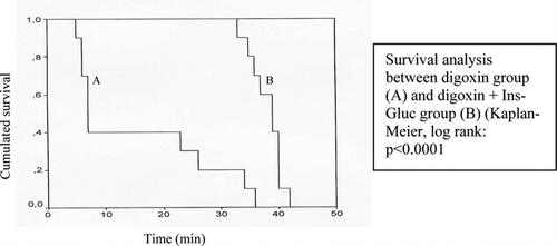
109 Pimozide Intoxication with QT Prolongation and Torsades de Pointes
Felgenhauer N (1), Jetzinger E (1), Pfab R (1), Eyer F (1), Roider G (2), Zilker T (1). 1. Department of Toxicology, Klinikum rechts der Isar, Munich, Germany; 2. Institute of Forensic Medicine, Munich, Germany.
Objective: It is well known that pimozide poisonings may cause prolongation of the QT interval and hypotension, but case reports with torsades de pointes (TdP) following an overdose are rather rare. Case report: A 38-year-old woman with paranoid schizophrenia took a total amount of 300 mg pimozide together with alcohol as a suicide attempt. On admission, 2 hours later, she was drowsy and partial agitated, her blood pressure was 110/50 mm Hg at a heart rate of 80 bpm. The ECG revealed a prolonged QTc of 0.65 sec. Toxicological analysis in serum showed a pimozide concentration of 63,7 ng/mL (therapeutic range 4–10 ng/mL), an ethanol concentration of 2.2 g/L and a lithium concentration of 0,43 mmol/L (within the therapeutic range). Minutes after admission the patient developed a self-limiting episode of TdP followed by two episodes which were terminated by electrical cardioversion with 150 J. Treatment with magnesium was started beginning with a bolus of 12 mval followed by an infusion with 1.33 mval/h. A transcutaneous external pacemaker was applied instituting overdrive pacing with a rate of 140 bpm. Hypotension necessitated infusion of epinephrine and norepinephrine with 10 μg/min each. In the following 36 hours the patient developed recurrent episodes of TdP requiring altogether 44 electrical cardioversions. In this stage magnesium infusion was increased to 1.92 mval/h and high doses of potassium (temporarily 30 mval/h) were substituted to keep the potassium concentration in serum at a level slightly above 5.0 mmol/L. Thereafter no further episodes of TdP occurred. Overdrive pacing could be stopped after 60 hours (pimozide concentration 15.3 ng/mL, QTc 0.58 sec). In the further course the patient developed pneumonia, pulmonary embolism and hemorrhagic infarction in the region of A. cerebri sinister with a homonymous hemianopia on the right-hand side. On day 17 extubation could be performed, on day 27 the patient could be transferred to the neurological department. The total halftime of pimozide in serum amounted 39.8 hours. Pimozide concentration in serum reached the therapeutic range on day 5. QTc returned to normal on day 6. Conclusion: Patients with pimozide overdose should be monitored carefully to detect and treat QT prolongation and torsades de pointes timely. Treatment includes high doses of magnesium, substitution of potassium to the upper limit of normal, catecholamines, overdrive pacing and electrical cardioversion. QTc corresponds well to pimozide concentration in serum.
110 Acute Hepatotoxicity Associated with Amiodarone Administration
Courtney J, Ganetsky M, Bird SB, Boyer EW. Department of Emergency Medicine, University of Massachusetts Medical School, Worcester, MA, USA.
Objective: Amiodarone, an iodinated benzofuran derivative, is occasionally associated with a mild transaminitis during long-term oral therapy. This hepatitis associated with chronic use is typically transient and has been described as “pseudoalcoholic liver disease,” with phospholipid accumulation, steatosis and portal fibrosis. In contrast, acute, severe hepatotoxicity from amiodarone loading is very rarely described. We report a case of acute hepatoxicity associated with amiodarone administration. Case report: A 78-year-old woman with a history of atrial fibrillation, cardiomyopathy and end-stage renal disease presented to the emergency department with generalized weakness and vomiting. The patient had been previously diagnosed with mild hyperbilirubinemia of unclear etiology; her baseline total bilirubin ranged from 2 to 3 mg/dL. Her physical examination was notable for an irregular heart rate of 150–160 bpm, respiratory rate of 26–30, blood pressure of 106/41, and bibasilar lung rales. Her electrocardiogram showed atrial fibrillation with a ventricular rate of 160 bpm and chest X-ray showed congestive heart failure. After no improvement with intravenous metoprolol, she received a loading dose of 150 mg of intravenous amiodarone, and was started on a 1 mg/min continuous infusion. Twelve hours after starting the amiodarone infusion the patient became febrile, icteric, moderately encephalopathic, and had abnormal liver function testing, with an ALT of 1199 IU/L and AST of 2065 IU/L. The amiodarone infusion was stopped, and she was given a three-day course of intravenous n-acetylcysteine despite the absence of detectable serum acetaminophen. Her liver transaminases peaked on hospital day #2 with an AST of 7716 IU/L, ALT of 4606 IU/L and total bilirubin mg/dL of 5.5, but returned to her baseline by hospital day #9. Conclusion: Our patient's pattern of liver function test abnormalities with markedly elevated transaminases is more consistent with acute drug-induced hepatitis than with peri-portal cholestatic disease seen with long-term amiodarone therapy (1). Possible mechanisms include heptatoxicity induced by the polysorbate-80 additive in intravenous amiodarone, an immunologic mechanism or a direct hepatotoxic effect (2,3). A previous case of acute amiodarone-induced hepatotoxicity described in the literature reported a liver biopsy demonstrating centrilobular necrosis, which is consistent with acute drug-induced hepatitis (4). References: 1. Lewis JH, Mullick F, Ishak KG, Ranard RC, et al. Histopathologic analysis of suspected amiodarone hepatotoxicity. Human Pathology 1990; 21:59–67. 2. Breuer HW, Bossek W, Haferland C, Schmidt M, et al. Amiodarone-induced severe hepatitis mediated by immunological mechanisms. Int J Clin Pharmacol Ther 1998; 36:350–352. 3. Alade SL, Brown RE, Paquet A. Polysorbate 80 and E-ferol toxicity. Pediatrics 1986; 77:593–597. 4. Kalantzis N, Gabriel J, Mouzas D, Tiniakos D, et al. Acute amiodarone-induced hepatitis. Hepato-gastroeneterol 1991; 38:71–74.
111 Morbidity from Paediatric Iron Poisoning
Donohoe E, Tracey JA. National Poisons Information Centre, Beaumont Hospital, Dublin, Ireland.
Objective: To examine the incidence and morbidity of paediatric iron poisoning in Ireland. Methods: A 5-year prospective study was carried out by a single observer to examine acute paediatric iron poisoning reported to the National Poisons Information Centre (NPIC) between January 2000 and December 2004. All enquiries involving ingestion of an iron preparation by children under 10 years of age were examined. The quantity of iron ingested was noted and cases were classed as either a) non-toxic if <20 mg/kg was taken, or b) potentially toxic if>20 mg/kg was taken. Cases involving unknown quantities of iron were classed as potentially toxic for the purposes of this study. Reported symptoms were noted in all cases. In potentially toxic cases, a follow-up call was made to the relevant hospital 24 hours post-ingestion to determine 1) symptoms, 2) plasma level, 3) use of antidote, and 4) outcome. Results: There were 253 cases of iron ingestion included in our study. 78% (n = 178) of patients had ingested <20 mg/kg. 14 of these had mild symptoms of gastrointestinal upset at the time of the call but no treatment was required. There were 75 potentially toxic iron ingestions during the study period. 22 cases were reported from members of the public or GP's but the patients did not attend an A/E Department. All of these patients were asymptomatic at the time of the call but no further information was available. Of the remaining 53 cases, all involved children under the age of 6 and the majority (71%) were under 3 years. Only 10 patients developed symptoms of toxicity. 1 developed severe symptoms including diarrhoea, haematemesis, hypotension and hyperglycaemia. He received IV desferrioxamine and was discharged clinically well 3 days post-ingestion. Plasma levels were available in 7 of these symptomatic cases and ranged from 49micromol/l to 82 micromol/l. 29 patients (57%) remained asymptomatic up to or beyond 6 hours post-ingestion. Plasma levels were available in 23 of these and ranged from 4.7micromol/l to 53micromol/l. No desferrioxamine was administered in any of these cases and the patients were discharged within 24 hours. Fourteen cases were lost to follow-up however 13 of these patients were asymptomatic at the time of the initial call. One patient had an episode of mild vomiting. Conclusions: The incidence of paediatric iron poisoning is low in Ireland and made up only 0.5% of total enquiries to the NPIC between January 2000 and December 2004. Desferrioxamine is rarely required and only one case of severe poisoning was reported during our study period.
112 Paracetamol Poisoning – Frequency, Severity & Treatment
Aakvik R, Jacobsen D. Department of Acute Medicine, Ullevaal University Hospital, Oslo, Norway.
Objective: Poisoning with paracetamol is common and may result in liver damage. We have assessed the frequency and severity of paracetamol poisoning and the use of serum paracetamol measurement as guidance for antidotal treatment in our tertiary care hospital. Methods: The hospital records of the Department of Acute Medicine were reviewed for the period from July 2001 until July 2004. All with ICD-10 diagnoses T4n and T50.9 were examined and all cases of suspected paracetamol poisoning were recorded. Liver damage was defined as ALT above 1000 U/l. Standard European treatment nomogram and IV antidotal regimen were used. Results: Of 869 admissions with acute poisoning, 158 (21%) were suspected paracetamol poisoning; 120 (76%) females and 38 (24%) males. Median time from ingestion to admission was 5 h. 107 (68%) of the patients were treated with N-acetylcysteine because of suspected ingestion of more than 10 grams (150 mg/kg) of paracetamol. Treatment was, however, discontinued in 84 (79%) because serum paracetamol was below the treatment line (). Only 17 patients (11%) had serum paracetamol above the treatment line 4–15 hours post ingestion. In two patients, an adverse reaction due to N-acetylcysteine treatment was suspected. Nine patients (6%) had liver damage, all of whom arrived later than 15 hours after ingestion. One patient admitted late died from acute liver failure. There were no other deaths from paracetamol poisoning recorded in this period. Conclusion: Poisoning with paracetamol is common, but rarely severe in our hospital. Liver damage is uncommon and associated with late hospital admission. Antidote administration based on history of ingested dose was associated with overtreatment.
FIG. 1
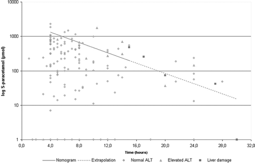
113 Delayed Presentation of Prolonged QTc Leading to the Diagnosis of Occult Citalopram Poisoning
Kirk MA (1), Eldridge DL (2), Holstege CP (1). 1. Division of Medical Toxicology, Department of Emergency Medicine, University of Virginia, Charlottesville, Virginia, USA; 2. Department of Pediatrics, East Carolina University, Greenville, North Carolina, USA.
Objective: Evaluating poisoned patients often requires careful search for objective findings that suggest poisoning because a given history maybe faulty or incomplete. We present a case of a patient with a clinical presentation suggesting alcohol withdrawal. However, after development of a prolonged QTc on his electrocardiogram, this patient was correctly diagnosed as a citalopram overdose. Case report: A 35-year-old male was brought to the emergency department following a new-onset seizure. Upon presentation, his blood pressure was 147/95 and his pulse was 120. He had hyperreflexia, clonus and a prominent resting tremor. The patient was sedated with intravenous (IV) midazolam. His wife related that the patient had a long history of alcoholism. She was unaware of any history of withdrawal symptoms. The patient and his wife vehemently denied any possibility of overdose. Between doses of midazolam, the patient had a grossly normal mental status. His presenting blood ethanol level was 83 mg/dL. His vital signs and tremor all improved with repeated doses of IV lorazepam. With this history, physical exam, and response to lorazepam, he was initially diagnosed with alcohol withdrawal. His initial electrocardiogram (ECG) revealed sinus tachycardia with a QTc of 381 msec. Approximately 16 hours after admission, his QTc was noted to have increased to 511 msec. Confronted with this new data, his wife returned home to find an empty bottle of her citalopram. The patient then admitted to the overdose. He recovered without sequelae and without the development of ethanol withdrawal. Conclusion: We present a case that substantiates the use of serial ECGs in the evaluation of potential drug overdose. In this case, an ECG taken later in this patient's hospital stay provided evidence leading to the diagnosis of citalopram overdose, a well-documented cause of prolonged QTc (1,2). References: 1. Catalano G, Catalano MC, Epstein MA, Tsambiras PE. QTc interval prolongation associated with citalopram overdose: a case report and literature review. Clin Neuropharmacol May-Jun 2001; 24:158–162. 2. Engebretsen KM, Harris CR, Wood JE. Cardiotoxicity and late onset seizures with citalopram overdose. J Emerg Med Aug 2003; 25:163–166.
114 Late Relapse in Tricyclic Antidepressant Poisoning
Rygnestad T. Norwegian Poisons Control Centre, Directorate for Health & Social Affairs, Oslo, Norway.
Objective: Tricyclic antidepressant poisoning can be serious due to CNS depression, cardiac arrhythmias and cardiovascular collapse. Three patients are presented who had a bi-phasic or prolonged course, which seemed to correlate with similar changes in serum concentration of the drug involved. Case series: In the three patients described the clinical course was recorded and several blood-samples were drawn for determination of drug levels. Patient 1 was a 29-year-old man admitted in deep coma 6 hours after taking 15 g of clomopramine. He had to be artificially ventilatied and needed vasopressive drugs. His condition gradually improved. The condition gradually deteriorated again after the first day and treatment had to be intensified. After the relapse he gradually improved. The deterioration correlated with a secondary rise in the blood level of the drug taken (). No other cause for the deterioration was found. Patient 2 was a 35-year-old female admitted in a deep coma several hours after taking an unknown quantity of clomopramine. She had to be treated with vasopressive drugs and was put on a respirator. Her condition gradually improved during the first day. After the initial improvement her condition deteriorated again and she needed cardiorespiratory support again. After the relapse her condition gradually improved and eventually she recovered. The deterioration correlated with a secondary rise in the blood level of the drug (). No other obvious cause of the deterioration was detected. Patient 3 was a 33-year-old man admitted in deep coma after taking an unknown quantity of amitriptyline. He had to be artificially ventilated and needed vasopressive drugs. His condition showed some improved during the first day. The condition did not improve further and he needed continued cardiovascular support. The deterioration correlated with a toxic level of the drug during the next 5 days (), after which he expired due to complications. Conclusion: Previous studies have reported that patients deteriorate shortly after admission in serious tricyclic antidepressant poisoning (1). Late relapse or deterioration has rarely been reported. Our patients show that both relapse and a prolonged course might occur after initial improvement. Possible cases are delayed and continuous absorption, redistribution and enterohepatic circulation of the drug (2). Drug levels can be of significant help to rule out other causes for the deterioration. References: 1. Epidemiology of fatal tricyclic antidepressant ingestion: implications for management. Ann Emerg Med 1985; 14:29–37. 2. Prescorn S, Irwin HA. Toxicity of tricyclic antidepressants – kinetics, mechanism, intervention: A review. J Clin Psychiat 1982; 43:151–156.
FIG. 1
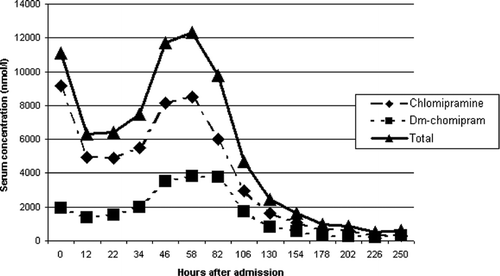
FIG. 1
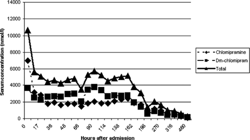
FIG. 1
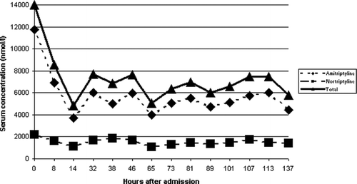
115 Suboxone® (Buprenorphine-Naloxone) Exposure in a Toddler
Schwarz KA (1,3,4), Vohra RB (1,2), Cantrell FL (1,3,4), Clark RF (1,2). 1. California Poison Control System, San Diego Division, San Diego, CA, USA; 2. Division of Medical Toxicology, University of California School of Medicine, San Diego, CA, USA; 3. University of California, San Francisco School of Pharmacy, San Francisco, CA, 4. University of California, San Diego Skaggs School of Pharmacy and Pharmaceutical Sciences, San Diego, CA, USA.
Objectives: We report the first case report of pediatric exposure to Suboxone®, a combination of buprenorphine and naloxone. Case report: A previously healthy 2-year-old, 13 kg male was found with 1 tablet of Suboxone® (8 mg buprenorphine/2 mg naloxone) in his mouth which was promptly removed by the parent. No other pills were reported missing. He had one episode of spontaneous emesis and became drowsy within 30 minutes. In the ED, the child was somnolent but arousable and miosis was noted upon examination. Vitals signs were: pulse 145 bpm, blood pressure 109/57 mmHg, respirations 20 per minute, temperature 37.0 degrees C, and room-air oxygenation saturation 97%. No interventions or antidotes were administered, the child was ambulatory at five hours post-exposure and was discharged 1 hour later. Conclusions: This case represents the first report of Suboxone® exposure in a pediatric patient. Suboxone® is a combination of buprenorphine and naloxone indicated for treating opioid addiction. Buprenorphine has partial mu agonist and kappa antagonist activity on opioid receptors; its potent respiratory and CNS depressant actions “plateau” with increasing doses (1). Deaths or severe toxicity attributed to oral buprenorphine alone and in combination with sedatives have been previously reported (1,2) in adult patients. Pharmakokinetic data suggest that buprenorphine has better bioavailability sublingually than orally due to extensive first-pass metabolism (3). Naloxone is added to Suboxone® to discourage parenteral misuse. Although IV/IM naloxone is a potent mu antagonist, its poor oral/sublingual bioavailability are expected to confer little protection against buprenorphine toxicity in oral Suboxone® exposures. Our patient developed mild signs of opioid intoxication from the buprenorphine portion of the tablet, most likely from sublingual absorption. The serum half-life of buprenorphine is 4 to 5 hours, but its high lipophilicity and the potency of its active demethylated metabolite (norbuprenorphine) may prolong duration of toxic effects (1). We thus recommend at least 6 hours of observation in symptomatic children exposed to Suboxone®. References: 1. Sporer KA. Buprenorphine: A primer for emergency physicians. Ann Emerg Med 2004; 43:580–584. 2. Tracqui A, Kintz P, Ludes B. Buprenorphine-related deaths among drug addicts in France: a report on 20 fatalities. J Anal Toxicol 1998; 22:430–434. 3. Mendelson J, Upton RA, Everhart ET, et al. Bioavailability of sublingual buprenorphine. J Clin Pharmacol 1997; 37:31–37.
116 Grapefruit Juice as a Potential Risk Factor of Intoxication by CYP2D6-Metabolized Drugs
Niewinski P (1), Jazwinska-Tarnawska E (1), Czarnik-Matusewicz H (1), Wiela-Hojenska A (1), Niewinska K (2). 1. Department of Clinical Pharmacology, Medical University, Wroclaw, Poland; 2. Chair of Emergency Medicine, Medical University, Wroclaw, Poland.
Introduction: Grapefruit juice interacts with a number of medications. As little as 250 ml of grapefruit juice can change the metabolism of drugs. Most drugs affected by grapefruit juice are known to be primarily metabolized by a form of cytochrome P450, CYP3A4. This drug-food interaction occurs because of a common pathway involving enzymes present in both the liver and the intestinal wall. Co-administration of grapefruit juice with CYP3A4 dependent drugs resulted in substantial increases in their bioavailability and higher risk of drug intoxication. Recently some studies show in-vitro inhibition of CYP2D6 metabolism by grapefruit juice. CYP2D6 enzyme is an important factor in the metabolism of 20–25% clinically important drugs, such as tricyclic antidepressants, neuroleptics, antiemetics, cardiologic drugs and opioids. Objectives: The aim of our study was to investigate the effects of grapefruit juice on human cytochrome CYP2D6 enzyme activity. Our goal was to assess the importance of grapefruit juice as a potential risk factor of increasing toxicity of drug which are metabolised by cytochrome CYP2D6. Methods: Study involved 24 unrelated individuals, healthy volunteers from the south-western region of Poland. Sixteen of them received 250 ml (one glass) of grapefruit juice daily for 5 days. Control group consist eight persons which received 250ml (one glass) of orange juice daily for 5 days. The CYP2D6 phenotype was analyzed before and after 5 days of juice/water ingestion, using sparteine as a model drug. Sparteine and its metabolites, 2- and 5-dehydrosparteine were determined in urine by gas chromatography method of Eichelbaum, using 17-ethylsparteine as internal standard. Metabolic ratio was calculated as the ratio of amount of parent drug to the total amount of metabolites. Results: Mean metabolic ratio (MR), measured after 5 days of drinking juice, increased by 34% in group receiving grapefruit juice, and by only 4% in control group receiving orange juice. Metabolic ratio increase by over 30% is an equivalent of similar decrease of detoxification potential of hepatic enzymes, involved in metabolism of CYP2D6 dependent drugs. Conclusion: Our results show relevant inhibition of CYP2D6 metabolism by grapefruit juice components, resulting in increasing risk of CYP2D6-metabolized drug toxicity.
117 Isoniazid-Induced Psychosis in an Adolescent Male
Gresham HW, Babu KM, Ali F, Bird SB, Boyer EW. Division of Toxicology, Department of Emergency Medicine, University of Massachusetts, USA.
Objective: Isoniazid (INH) induced psychosis is an extremely rare complication of tuberculosis therapy. We report the case of an adolescent male with INH-induced psychosis. Case report: A 15-year-old male presented to the emergency department with bizarre behaviors, including paranoia, shouting aloud and talking to inanimate objects. The patient was a recent immigrant to the United States, moving from Ethiopia three months prior to presentation. Approximately 7 weeks prior to presentation, the patient was started on isoniazid therapy after a positive PPD and subsequent negative chest X-ray. He had no other significant past medical or psychiatric history, and took no medications besides INH. He had no complaints of headache, fevers, chills, vomiting or diarrhea. On initial exam, the patient was afebrile, with a pulse of 165 beats per minute, and blood pressure of 164/71 mm Hg. The patient's neuropsychiatric exam was significant for agitation requiring restraints, however he could be redirected to answer questions appropriately. He was diaphoretic with mid-sized pupils, normal bowel sounds, and no evidence of clonus or hyperreflexia. Complete blood count, serum chemistries, and liver function tests were unremarkable. Non-toxicologic etiologies for the patient's psychosis were systematically excluded with MRI, lumbar puncture and EEG. A comprehensive urine toxicologic screen was negative for drugs of abuse. The patient was treated with pyridoxine, risperidone and benzodiazepines with gradual clearing of his sensorium. His mental status had returned to baseline by two weeks after cessation of the INH therapy. Conclusion: The mechanism for INH-induced psychosis is unknown. One theory suggests decreased GABAergic tone secondary to inhibition of glutamic acid decarboxylase. The second proposed mechanism stems from INH's monoamine oxidase inhibitor-like activity, creating increased sympathetic tone (1). Atypical antipsychotics have anecdotally been effective in ameliorating the symptoms of INH-induced psychosis, with prior case reports describing the use of risperidone (2). Prior cases of INH-induced psychosis have been reported in patients taking pyridoxine supplementation, leading to the theory that pyridoxine may not be effective in the treatment of this condition (3). While INH-induced psychosis remains a rare complication of treatment with this medication, its widespread worldwide use make awareness of this potential adverse effect essential. References: 1. Iannaccone R, Sue YJ, Avner JR. Suicidal psychosis secondary to isoniazid. Pediatr Emerg Care 2002; 18:25–27. 2. Duggal HS, Nizamie SH. Novel antipsychotic drugs and INH-related psychosis. Ann Pharmacother 1999; 33:1123–1124. 3. Chan TYK. Pyridoxine ineffective in isoniazid-induced psychosis. Ann Pharmacother 1998; 32:889–891.
118 Bradycardia and Hyperkalemia Associated with Octreotide Administration
Curtis JA, Greenberg MI. Drexel U. College of Medicine, Dept. of Emergency Medicine, Division of Toxicology, Philadelphia PA, USA.
Objective: Octreotide is a synthetic octapeptide which acts as somatostatin analogue. It is used to treat acromegaly, diarrhea, pancreatic fistulae, and has been recently used in toxicology for the treatment of hypoglycemia secondary to sulfonylurea overdoses. We report the case of a 16-year-old who developed hyperkalemia and bradycardia while being treated with octreotide. Case report: A 16-year-old man was hospitalized after an intentional ingestion of a unknown amount of nateglinide the previous evening. The patient was in no distress and had stable vital signs in the emergency department, but received intravenous octreotide for bedside glucose concentrations were determined to be 60 mg/dL. While in the intensive care unit, the patient became bradycardic, with a heart rate of 35 bpm. Serum potassium was measured at 6.27 meq/L. The patient was treated for hyperkalemia and octreotide was discontinued. The bradycardia improved, hyperkalemia did not recur and the patient was discharged in good condition. Conclusion: Octreotide is an effective agent for the treatment of hypoglycemia related to ingestion of oral hypoglycemic drugs. While generally considered safe, there have been 3 reports of possible octreotide-induced hyperkalemia in the medical literature. While an uncommon occurrence, toxicologists should be aware that hyperkalemia may be associated with octreotide therapy. References: 1. Brown RO, et al. Hyperkalemia secondary to concurrent pharmacotherapy in a patient receiving home parenteral nutrition. Journal of Parenteral & Enteral Nutrition 1996; 20:429–432. 2. Canada T. Octreotide- or trimethoprim-induced hyperkalemia?[comment] Pharmacotherapy 1995; 15:814–815. 3. Sargent AI, et al. Octreotide-induced hyperkalemia. Pharmacotherapy 1994; 14:497–501
119 Clobutinol Poisoning: A Case Series
Goossens E, Mostin M. National Poison Centre, Brussels, Belgium.
Objective: To describe a series of documented poisonings with the non-narcotic antitussive clobutinol. Case series: We describe 10 clobutinol overdoses in children (age 1 to 11 yrs, average 3 yrs) and 2 in adults (22 and 28 yrs). In children, poisoning occurred by accidental ingestion (n = 9) or medication error (n = 1). In adults, ingestion occurred in a suicidal attempt. The ingested amount in children was between 80 and 880 mg (average 450 mg) and 300 and 600 mg in adults. The very attractive presentation of the fluorescent pink sugar coated tablets probably explains the unusual high amount ingested by children. All the victims presented symptoms except a 14 months old child who ingested 80 mg (7,2 mg/kg). The following symptoms were reported: vomiting (6/12), seizures (6/12), tremor (3/12), myoclonia (2/12), coma (2/12), somnolence (2/12), ataxia (2/12), agitation (1/12), tachycardia (1/12), mydriasis (1/12), hallucinations (1/12), confusion (1/12), hypertonia (1/12), hypersudation (1/12) and nausea (1/12). In the majority of the cases, symptoms occurred within one hour after ingestion and persisted for about 12 hours. All victims recovered uneventfully. Conclusions: Main features of clobutinol poisoning are vomiting, stimulation of the central nervous system sometimes followed by central depression. References: 1. Ramirez MS, Rojas MD, Grand Mal Seizure and Clobutinol Overdose. Vet Human Toxicol 1993; 35:444. 2. Von Mühlendahl KE, et al. Vergiftungen im Kindesalter: 151. Thieme ed, 2003.
120 High Dose Tranylcypromine Withdrawal with Delirium and Thrombocytopenia, Effect of Physostigmine
Pfab R, Eyer F, Jetzinger E, Zilker Th. Toxikologische Abt., 2. Med. Klinik, Klinikum rechts der Isar, Muenchen, Germany.
Case report: A 37-year-old male with a history of polyvalent drug abuse (heroin, amphetamines, alcohol), but abstinent for six years, was prescribed 40 mg tranylcypromine daily because of migraine by his neurologist. Tranylcypromine relieved the migraine, but had an additional “peculiar effect” in that he could “concentrate better”, “think more clearly” and felt “less distracted by daily worries.” He increased the dose to 240 mg daily. After twelve weeks of this high dose consumption and several collapses he stopped the consumption and became delirious. He was admitted to psychiatry. Within two days the delirium worsened, he became combative and was transferred to our facility after sedation with diazepam. On arrival, he talked to imaginary policemen in English language (although a native German speaker) before he started to sing. Nevertheless, he followed simple commands. He had no other withdrawal-symptoms nor anticholinergic signs. His pupils were middle-wide. Laboratory tests were normal except: blood-glucose 50 mg/dl (Insuline < 2 μIU/ml C-peptide 0,6 ng/ml), thrombocytes 84000/μl, GPT 65, GOT 80, CK 2150 U/l. Screening the urine for drugs resulted negative except for benzodiazepines and tranylcypromine. Tranylcypromine was not detectable in plasma (<40 μg/l). Thromboctopenia had it's nadir with 52000/μl at day five, resolving spontaneously within four days. Global coagulation parameters (PTT, INR, D-Dimers) were monitored closely and remained within normal limits. He was treated with glucose iv, haloperidol iv, diazepam iv and midazolame as a continuous infusion. The delirium did not resolve within five days until 2 mg physostigmine iv on day six resulted in prompt recovery. The delirium returned after 30 min, the patient was treated with physostigmine by infusion 2 mg/h with interruptions every six hours in order to test his mental state until day eight. The patient left the hospital at day ten with no sequels. Discussion: Several cases of high-dose addiction to the irreversible MAO-Inhibitor tranylcypromine with delirium after discontinuation and thrombocytopenia are known from literature. Most of them had a history of substance abuse. The delirium typically starts about 20 hours after discontiuation of tranylcypromine and lasts three to twelve days. Treatment with haloperidol and benzodiazepines does not change the delirious state but may exhibit a helpful sedative effect. A novel feature in our case is the counteraction of the delirium by physostigmine. The effect of physostigmine was too prompt and too reproducible as to interprete it as just a coincidence with the natural wax and wane of a fading delirium. The effect of physostigmine is remarkable since our patient had no other anticholinergic signs.
121 Drug Mistakes at Home: Experience of the Marseilles Poison Centre During the First Three Months of 2005
Blanc-Brisset I, de Haro L, Hayek-Lanthois M, Arditti J. Centre Antipoison, Hôpital Salvator, Marseille, France.
Introduction: As drugs and medicines are sold in France in packaging instead of delivering the exact number of necessary pills, almost all French houses contain big medication stocks which are at the origin of numerous poisonings. Method: In order to illustrate this situation, the authors performed a retrospective study concerning all toxic problems directly induced by this French custom and observed during the first three months of 2005 (winter season which is a period of intense use of drugs at home). Statistical data were analysed with Microsoft Access® and Microsoft Excel®. Case series: 690 observations were studied, representing 12% of the Marseilles Poison Centre global activity during the chosen period (average number of 8 files a day, extreme 1 to 17 cases a day). Most of the poisonings took place during the night (56.5% between 6.30pm and 8.30am), representing an important part of the evening and night activity of our Poison Centre. Poisonings were the consequence of several circumstances (mistakes concerning the schedule, the route, the patient, errors of the chemist or of the physician, etc.), but the most frequent situations were dosage errors, drug misidentifications and automedications (about 60% of the whole case series for these three circumstances). The person responsible was mainly the patient himself (48% of all cases) or the patient's mother (30%). Situation, circumstances and evolution varied a lot depending of the victim age (children vs. adults vs. elderly). Conclusion: This study emphasises the toxicological consequences of the French habit locally named “home chemistry.” The poison centre role in such poisonings is really important as most of the patients can be managed at home and because cases usually happen during the night when other structures (chemistry, family doctors) are not available.
122 Ingestion of 6.3 Grams of Clopidogrel – A Case Report
Pedersen LM (1), Joensen H (2), Harreschou HE (1), Skanning PG (2). 1. Department of Internal Medicine, Glostrup Hospital, Copenhagen, Denmark; 2. Department of Anaesthesiology, H:S Bispebjerg Hospital, Copenhagen, Denmark.
Objective: Experience with high doses of clopidogrel is poor. To our knowledge the highest reported ingestion is 1050 mg (1). We present a case report in wich a patient ingests 6.3 grams of clopidogrel in suicidal purpose and survives without sequelae. Case report: A 56-year-old woman presented in the emergency department the day after ingestion of 84 tablets of clopidogrel 75 mg (a total of 6300 mg) to commit suicide. The tablets were taken the night before, more than 12 hours earlier. The patient showed up with an empty pill bottle (75 mg, 84 tablets) prescribed the day before, so the dose was certain. No other drugs were ingested apart from usual medication consisting of diclofenac, misoprostol, venlafaxin, furosemide, potassium, risperidone, zolpidem, topiramate, budesonide, estradiol, norethisterone acetate and vitamin supplement. Initial symtoms were nausea, regurgitation and diarrhoea. Blood samples showed a high blood reticulocyt count (741 billions/L). Plasma acetaminophen level was normal (below 66 uM) and ECG was normal. ABG measured the day after admission revealed compensated metabolic acidosis (pH 7,375, pCO2 3,9 kPa, BE -7 mM). Activated charcoal was not given due to the time from intake. Vital signs were normal and stable and no bleeding episodes were recorded during the hospital stay. Gastrointestinal symtoms ceased and the patient was discharged from the hospital after 2 days in habitual condition. Conclusion: It seems that clopidogrel is relatively unharmful in doses as high as 20 times the recommended dose. One could expect bleeding episodes. However the complexity of the processes inhibitng platelet aggregation (2) might suggest that clopidogrel plays only a minor role with respect to unwanted effects such as bleeding. In conclusion, clopidogrel in itself seems relatively non-toxic. References: 1.Wynn RL, Bergman SA. Clopidogrel (Plavix): dental considerations of an antiplatelet drug. General Dentistry 2001; 49:564–568. 2. Ulrichts H, Vanhoorelbeke K, Van de Walle G, et al. New approaches for antithrombotic antiplatelet therapies. Curr Med Chem 2004; 11:2261–2263.
123 A Severe Case of Olanzapine Overdose with Whole Blood Concentrations
Ballesteros S (1), Martínez MA (2), Ballesteros MA (3), Sánchez de la Torre C (2), Rodríguez Borregán JC (3). 1. Spanish Poison Control Centre, INTCF, Madrid, Spain; 2. Chemistry Department, INTCF, Madrid, Spain; 3. Intensive Care Unit, Hospital Universitario Marqués de Valdecilla, Santander, Spain.
Objective: Olanzapine is a relatively new antipsychotic drug used in intentional drug overdoses. At hospitals, blood serum is a specimen usually submitted for toxicological analysis, whereas in forensic toxicology whole blood is probably the most important biological sample. Different type of specimens and lack of adequate clinical information contribute to the difficulties of toxicological interpretation in forensic cases. We describe the clinical course and whole blood concentrations in a case of severe overdose with olanzapine. Case report: A 58-year-old woman with a 10-year history of paranoid schizophrenia and bad therapeutic compliance was found unconscious at home with rest of vomiting and 2 empty Zyprexa 10 mg® prescription vials, each vial containing 28 tablets, nearby. She was attended at the emergency department of the hospital 40 minutes later. Her vital signs were as follows: blood pressure 110/70 mmHg, pulse rate, 82 beats/min, in sinus rhythm; 20 breaths/min. Glasgow Coma Scale score of 7 with normal pupils. The patient was sent to the Intensive Care Unit (ICU) where the pulse rate was 160 beats/min, in sinus rhythm, QTc 0.423 sec (normal <0.4 sec). Relevant analytical findings: metabolic acidosis, leukocytosis, CPK 1992 mg/dL. Glucose was 207 mg/dL, 9 hours later 350 mg/dL and became normal at 24 hours. Treatment consisted of intubation and mechanical ventilation. Gastric lavage was performed and revealed no rest of pills. The patient needed antibiotics and insulin. At 7 hours after admission in the ICU, the patient started to move and was hemodinamically stable. At 36 hours she was conscious. After psychiatric evaluation, she was discharged from the ICU at day 3 and from the hospital after 30 days. Three whole blood samples were sent for measurement of olanzapine concentrations to our laboratory. Blood was submitted to solid phase extraction using Bond-Elut Certify columns. The sample extract was analyzed by gas chromatography with nitrogen-phosphorus detector for screening analysis and quantitation, and by gas chromatography-mass spectrometry for confirmation of the obtained results using a validated analytical method. At approximately 4, 8 and 12 hours postingestion, whole blood concentrations of olanzapine were 0.41, 0.34 and 0.38 mg/L, respectively. No other drugs were detected. Conclusion: This study reports an acute olanzapine monointoxication with severe toxicity and high blood olanzapine concentrations. Clinical and analytical data obtained in non-fatal life-threatening cases can be very useful when interpreting postmortmen cases.
124 Delayed Toxicity Resembling Serotonin Syndrome Following Massive Ingestion of Adderal XR
Bouchard NC, Wellington V, Pacheco-Fowler V, Skomorowsky AL. Columbia University Medical Center, New York, NY, USA.
Objective: Dextroamphetamine (d and l-amphetamine isomers) or “Adderall” was approved for unrestricted use in ADD/ADHD by the FDA in 1996 and is one of the more widely prescribed ADD/ADHD medications. It has also gained immense popularity among recreational drug users for its stimulant and appetite suppressant effects. Surprisingly, few cases of toxicity are documented in the medical literature. We report a case of delayed toxicity resembling serontonin syndrome from a self-reported massive Adderal XR ingestion. Case report: A 20-year-old man presented to the ER following a suicidal ingestion of 100 Adderall tablets shortly before arrival. He complained only of mild headache, dizziness, jitteriness and nervousness. His initial vital signs were: BP, 180/120 mmHg; pulse, 102 beats/min; respirations, 20 breaths/min; temperature, 36 C. He was treated with activate charcoal, IV normal saline and lorazepam 2 mg IV. Four hours later his vital signs were somewhat improved and his mental status was unchanged. His initial laboratory analyses were remarkable for a CPK of 83 U/L and WBC's of 15.0 × 10e9/L. Approximately 16 hours later, he became markedly more agitated, disinhibited and was delirious. His vitals signs were: blood pressure 195/112 mmHg; pulse 140 beats/mi; temperature 38.3 C. He demonstrated muscular rigidity with 3+ hyperreflexia and 3-beat ankle clonus. He was also noted to have opsoclonus, myoclonic jerks, a “shaking chorea” and “continuous bouncing” head movements, with his head held off the stretcher tonically. Over the next four hours he required an additional 50 mg of IV lorazepam for sedation. His CPK peaked at 2715 U/L and WBC's at 20.4 × 10e9/L. He was ultimately intubated and rendered comatose with propofol and was admitted to the ICU. Roughly 12 hours later he was extubated and clinically stable. He was discharged from the ICU 2 days later with a mild pneumonitis, presumed to be secondary to aspiration. It was determined by further history that the patient had ingested the Adderal XR formulation. Urine toxicology for drugs of abuse was positive for amphetamines and negative for cocaine and PCP. Serum levels of d and l-amphetamine could not be obtained. Conclusion: The growing popularity and easy access to prescription stimulant drugs via the internet will inevitably lead increased presentations to ER's of overdose-both recreational and suicidal. This case illustrates marked delayed significant toxicity resembling serotonin syndrome following reported ingestion of a non-sustained release preparation that was later determined to be a sustained release formulation. Clinicians should be alert for significant or delayed cardiovascular and neurologic toxicity following Adderall XR ingestions.
125 Persistent Disulfiram-Induced Vitamin K Dependent Coagulation Factors Deficiency
Madureira PR (1), Bizzacchi JMA (2), Vieira RJ (1), De Capitani EM (1). 1. Poison Control Centre, Hospital de Clinicas, State University of Campinas, Brazil; 2. Haematology Division, Hospital de Clinicas, State University of Campinas, Brazil.
Objective: Disulfiram has been used since the 1940s as an adjuvant drug in the treatment of chronic alcoholism. Some cases of toxic hepatitis with hepatic failure have been reported (1,2). We present a case of persistent deficiency of coagulation factors II, VII, IX and X following disulfiram use. Case report: RCP, a 24-year-old, male, presented himself at hospital complaining of haematuria for 3 days, with Hb = 12.8 g/dL; Ht = 37.8%, platelet count 280,000/mL; normal clotting time; enlarged prothrombin time, and PTT of 2.9 fold of the reference value. He developed gastrointestinal and gum bleeding. He was treated with 900 ml of frozen-fresh plasma and vitamin K, resulting in cessation of bleeding. Four days later, he had a new episode of coagulation deficit event, showing IRN of 7.6 and PTT of 1.9. Investigations for clotting inhibitor factors and for hepatopathy were negative, and he was referred to a haematologist. Ten days later, he came to the ER with haematuria, edema and pain at left limb and a Doppler showed partial femoral DVT with recanalization. Laboratory results showed enlarged PT and PTT. The bleeding event was again treated with vitamin K and frozen-fresh plasma. On the next day, laboratory results showed IRN = 2.29; R = 2.63; BUN = 13 mg/dL; creatinine = 0.93 mg/dL; Na = 137 mEq/L; K = 4.1 mEq/L; platelet count 357,000/mL; Hb = 8.7 g/dL; Albumin = 4.3 g/dL. As the patient could have been intoxicated with warfarin, despite the fact he denied ingestion, warfarin plasma concentration was measured and was negative. Psychiatric evaluation was normal. The only drug the patient was taking regularly, trying to control ethanol addition, was disulfiram. He had been taking the drug for the 7 months prior to the beginning of the symptoms. Vitamin K and frozen-fresh plasma was again given at the 8th day of treatment and patient was dismissed with a 2 vitamin K vials per day. One month later his IRN was 8.11, when vitamin K dose was increased to 3 vials a day. Three months later IRN was 1.88 with the patient taking 1 to 2 vials each other day. Only seven months later the coagulation tests were all normal and vitamin K was withdrawn. Conclusion: To our knowledge, this is the first clinical case of persistent disulfiram induced vitamin K dependent clotting factors inhibition presented in the literature. References: 1. Meir M, Woywodt A, Hoepr MM, et al. Acute liver failure: a message found under the skin. Postgrad Med J 2005; 81:269–270. 2. Mohanty SR, LaBrecque DR, Mitros FA, Layden TJ. Liver transplantation for disulfiram-induced fulminant hepatic failure. J Clin Gastroenterol 2004; 38:292–295.
126 Inadvertent Overdose with N – Acetylcysteine: An Unusual Case
Cassidy N, Tracey JA. The National Poisons Information Centre, Beaumont Hospital, Beaumont Road, Dublin, Ireland.
Introduction: N-acetylcysteine (N-AC) is the principal antidote used to treat acute and late-phase paracetamol poisoning. N-AC is administered intravenously over 3 infusions; 1. 150 mg/kg over 15 minutes, 2. 50 mg/kg over 4 hours, and 3. 100 mg/kg over 16 hours. N-AC is relatively safe, however, dose-related anaphylactoid reactions have occurred. There is limited data regarding overdose with N-AC and the toxic dose is not established. We report a case of inadvertent overdose of intravenous N-AC administered as treatment for paracetamol poisoning. Case report: A 27 year old female (61 kg) presented to a hospital emergency department 16 hours after completing a deliberate overdose of 24 g paracetamol and 3 g aspirin, ingested over a 6-hour period. She presented with vomiting and epigastric pain. Liver function was deranged on admission; ALT 181 IU/L, AST 170 IU/L, INR 1.3, and treatment with intravenous N-AC was commenced. The patient received the loading dose of N-AC (9 g/15 minutes), 30 g/4 hours (instead of 3 g/4 hours, a tenfold error in dose) and the 6 g/16 hours infusion was started. N-AC was discontinued once the medication error was discovered. The patient subsequently developed hypotension (98/56 mmHg), and right-sided chest pain but did not develop symptoms of an anaphylactoid reaction. Sixteen hours after admission her liver function had deteriorated further; ALT 1233 IU/L, AST 1233 IU/L, INR 1.8. Further administration of intravenous N-AC (100 mg/kg/16 hours) was given to limit hepatotoxicity. Peak hepatotoxicity occurred 40 hours after presentation; ALT 7000 IU/L, AST 6579 IU/L, INR 1.9. N-AC therapy was continued for 6 days and a total dose of 63 g N-AC was administered. The patient made a full recovery. Discussion: It is unknown whether hepatic impairment developed secondary to paracetamol poisoning or as a result of inadvertent overdose with N-AC. Liver injury was previously reported following high dose oral and rectal N-AC administration for meconium ileus in a cystic fibrosis patient (1). Our patient had transient liver impairment which normalised after 6 days following additional treatment with N-AC. Conclusion: Our patient recovered following paracetamol poisoning and inadvertent overdose with 10 times the dose of N-AC. The decision to continue with N-AC as antidotal therapy for paracetamol hepatotoxicity despite inadvertent N-AC overdose must be made on an individual basis. Reference: 1. Bailey DJ, Andres JM. Liver injury after oral and rectal administration of N-Acetylcysteine for meconium ileus equivalent in a patient with cystic fibrosis. Paediatrics 1987; 79:281–282.
127 Selective Serotonin Reuptake Inhibitors (SSRI): Unintentional Paediatric Ingestions
Balit CR (1), Gilmore S (1), Isbister GK (1,2). 1. NSW Poisons Information Centre, The Children's Hospital, Westmead, Australia; 2. Tropical Toxinology Unit, Menzies School of Health Research, Darwin, Australia.
Background: Selective Serotonin reuptake inhibitors (SSRIs) are widely available antidepressants. Unintentional paediatric ingestions of SSRI are subsequently becoming more common. Methods: Calls to the NSW poison information centre from January 2002 to July 2004 regarding paediatric ingestion of SSRIs were recruited and prospectively followed up. Information collected included: demographics (age, gender, and weight), type of exposure (unintentional, therapeutic error), details of the exposure (time of ingestion, ingested dose) and clinical effects. Doses were reported as defined daily doses (DDD). Results: From 96 calls for unintentional paediatric SSRI ingestion, 77 cases were included and 19 excluded due to uncertainty about the ingestion or loss to follow up. The median age was 2.5 years (IQR 2–3.5 years) and 34 (44%) were girls. Drugs ingested included sertraline (31), citalopram (17), paroxetine (15), fluoxetine (10), fluvoxamine (3) and escitalopram (1). The median ingested dose was a DDD of 1 (IQR 0.5–1). Symptoms were reported in 27 children (35%) including hyperactivity (15), drowsiness (7), vomiting (4), diarrhoea (8) and diaphoresis (2). The frequency of symptoms ranged from 20 to 67% for different SSRIs, with the highest rates in sertraline (52%) and fluvoxamine (67%). The median duration of effects was 12 hours (IQR 12 to 24 hours) and maximum duration was 48 hours. All recovered without any complications and there were no cases with severe effects. Conclusion: Most unintentional paediatric ingestions of SSRIs involve only half to two times the normal adult dose and resulted in minor symptoms in about a third of cases. The commonest effects were hyperactivity and diarrhoea and the spectrum of clinical effects was consistent with adverse effects reported for SSRIs. The majority can be managed at home with reassurance provided by the poisons information centre.
128 Pediatric Buprenorphine Ingestion with Subsequent Respiratory Depression
Geib A (1), Babu KM (2), Lai MW (3), Burns-Ewald M (1), Boyer EW (2). 1. Children's Hospital Boston, MA, USA; 2. University of Massachusetts Medical Center, Worcester, MA, USA; 3. American Association of Poison Control Centers, Washington, DC, USA.
Objective: To report a series of pediatric patients with respiratory depression after buprenorphine exposure. Case series: Three cases of pediatric buprenorphine (BUP) exposure with ensuing respiratory depression were reported to the MA/RI Poison Center in a one-year period. Details are presented in . Patients were ages 16, 20, and 22 months; two had ingested a combination product containing BUP and naloxone, and the third had ingested BUP only. All exposures were witnessed. One child was cyanotic, and all were somnolent. Two children had good response to naloxone, one required a naloxone infusion, and one required intubation and ventilatory support. Time to onset of respiratory depression was suspected to be within 6 hours of ingestion. None suffered any long-term consequences. Urine toxicology screening was negative for opioids. Discussion: Buprenorphine, an opioid partial agonist-antagonist, was recently introduced to the US market for opioid dependence substitution therapy. Adult fatalities have been reported mainly in the setting of concurrent benzodiazepine use (1). A single case report, and retrospective data described no respiratory depression after BUP ingestion in children (2,3). We suspect that due to the children's developmental stage BUP likely was absorbed transmucosally rather than enterally. The potential for delayed toxicity, BUP's long half life at the mu receptor, and children's inherent sensitivity to CNS depressants should increase the clinician's concern for potentially lethal toxicity. Naloxone has met with variable success in reversing buprenorphine-related respiratory depression and in the above cases dosing had to be repeated 4). Children with suspected high-dose buprenorphine ingestion should be observed for at least 6 hours; those developing toxicity should receive extended monitoring and antidotal therapy. References: 1. Kintz P. Deaths involving buprenorphine: a compendium of French cases. Forensic Sci Int 2001; 121:65–69. 2. Gaulier JM, Charvier F, Monceaux F, Marquet P, Lachatre G. Ingestion of high-dose buprenorphine by a 4-year-old child. J Toxicol Clin Toxicol 2004; 42:993–995. 3. Doyon S, Klein-Schwartz W, Welsh C. Toxicity Folowing Buprenorphine Ingestions. Clin Toxicol 2005; 43:640. 4. Gal TJ. Naloxone reversal of buprenorphine-induced respiratory depression. Clin Pharmacol Ther 1989; 45:66–71.
TABLE 1 Patient characteristics
129 Lack of Correlation Between Clinical Parameters and Clenbuterol Levels
Kirrane BM (1), Chan GM (1), Ginsburg BY (1), Halcomb SE (1), Tarabar AF (2), McGee M (3), Stajic M (3), Hoffman RS (1). 1. New York City Poison Center, NY, USA; 2. Section of Emergency Medicine, Yale University, New Haven, CT USA; 3. Office of the Cheir Medical Examiner, Department of Health, New York, USA.
Objective: Clenbuterol has been present in tainted heroin in the eastern US since early 2005. Clinical symptoms are caused by excessive stimulation of the beta-2-adrenergic receptor, and include tachycardia, palpitations, tremor, anxiety, agitation, and vomiting. Additional beta-2-adrenergic effects include hyperglycemia and hypokalemia. Unfortunately drug levels are not readily available, and do not return in a clinically relevant time frame. Thus in order to better understand the toxiocodyamic-toxicokinetic effects of clenbuterol we sought to determine if a correlation exists between common clinical abnormalities and clenbuterol levels. Methods: As a part of a larger investigation of clenbuterol toxicity blood and/or urine was requested on cases of potential clenbuterol toxicity reported to the regional poison center. These patients were initially identified based on a provisional case definition as described in the MMWR(1). Clinical information and treatment decisions were left to the discretion of individual physicians. Confirmatory clenbuterol testing was later performed on samples by liquid chromatography/mass spectrometry (LC/MS). Potassium, glucose and pulse were compared with blood and urine clenbuterol levels using a Pearson's Correlation Coefficient, with a p < 0.05 considered statistically significant. Results: A total of 11 patients had at least a blood or urine specimen for analysis (blood in 6, urine in 11). Reported ranges for clinical parameters were as follows: pulse, 110–150/min; potassium, 1.9–3.3 mEq/L; glucose, 6.0–19.5 mmol/L. For blood values, only the glucose showed a good correlation (r = 0.8), but was not statistically significant. For urine values (even when an outlier was removed) neither good correlations nor statistical significance was achieved with any parameter. Possible reasons for the lack of statistical significance include: small numbers of blood levels; variability between time of use and sampling, between therapy and sampling, between reported electrolytes and time of sampling; problems with interpretation of quantitative analysis of urine, and a failure of a correlation to exist. Conclusions: Neither urine nor blood clenbuterol levels seem to correlate with the common clinical parameters of clenbuterol poisoning. Data collection is ongoing. Reference: 1. CDC. Atypical reactions associated with heroin use-Five states, January–April 2005. MMWR 2005; 54:793–796.
130 Evaluation the Value of SAPS – II in Prognosticating the Outcome of Acute and Severe Carbon Monoxide Poisoning in Loghman – Hakim Hospital Poison Center
Shadnia S, Rezaie AR, Zareh S. Loghman-Hakim Hospital Poison Center, Shaheed-Beheshti University of Medical Sciences, Tehran, Iran.
Objective: Carbon monoxide is one of the most common causes of death from poisoning in the world. Unfortunately, despite the progress in pathophysiology and management of carbon monoxide poisoning, no signs, symptoms or laboratory findings have been found to predict outcome for carbon monoxide poisoned patients. Our aim was to evaluate the value of simplified acute physiology score (SAPS), which is a simple ICU Index used to predict mortalities and morbidities using the vital signs and laboratory results of the first 24 hours (14 items), in determining the outcome of acute and severe CO poisoning. Methods: This was a retrospective, descriptive and case-control study which was conducted in Loghman-Hakim Hospital Poison Center's ICU during the 6-year period from 1/1997 to 1/2003. SAPS scores were calculated for all the 55 patients and were compared. Data were abstracted from the medical records. All data were analyzed with statistical software SPSS, version 11.5. P values of 0.05 or less were considered to be statistically significant. Results: Our database covered 49 severely CO-poisoned patients who survived as controls, and 6 severely CO-poisoned patients who expired as cases. The SAPS scores of the cases (14.17 ± 2.86) were significantly higher than controls (7.45 ± 2.68) (p < 0.001 & Cl = 99%). We saw no mortality with SAPS less than 10; however, all the patients with SAPS score more than 14 died. 18 (36.7%) out of 49 survived patients experienced neuropsychological deficits. The SAPS score of those who experienced morbidity was not statistically significant (P = 0.51). Conclusion: Our results showed that SAPS scoring index is directly correlated with CO poisoning mortality (P < 0.001), although it has no significance (P = 0.51) for predicting psycho-neurological deficits occurring in these patients. SAPS can be used to predict the probability that a severely CO poisoned patients will survive or not. However, this index is not valuable in prognosticating the psycho-neurological morbidities. Further studies on larger populations and milder toxicities are recommended to elaborate the true role of this index in CO poisoning prognosis. References: 1. Mehta SR, Niyogi M, Kasthuri AS, et al. Carbon monoxide poisoning. J Assoc Physicians India 2001; 49:622–625. 2. Al-Moamary MS, Al-Shammary AS, Al-Shimemeri AA, et al. Complications of carbon monoxide poisoning. Saudi Med J 2000; 21:361–363.
131 The Incidence of Cardiovascular Symptoms Associated with Propoxyphene Exposures
Mrvos R (1,2), Krenzelok E P (1,2). 1. Children's Hospital of Pittsburgh, Pittsburgh, Pennsylvania, USA; 2. University of Pittsburgh, Pittsburgh, Pennsylvania, USA.
Objective: Propoxyphene is an opioid analgesic related structurally to methadone. It is available alone or in combination with acetaminophen, aspirin and caffeine. The number of propoxyphene-containing product exposures reported to the American Association of Poison Control Centers (AAPCC) Toxic Exposure Surveillance System (TESS) database relative to the number of human cases reported has not changed significantly over a 10-year period. In overdose propoxyphene has been reported to cause a variety of cardiovascular symptoms. A review was performed to determine the incidence of cardiac symptoms in overdose patients reported to the AAPCC TESS database. Methods: All exposures involving propoxyphene either alone or in combination with acetaminophen for a 10-year period were reviewed. Inclusion criteria included: human exposures, single substance ingestions and cases admitted to a health care facility. Data reviewed included age, gender, reason, symptoms, therapy, outcome and route. Results: A total of 3,769 cases were reviewed. Ages ranged from 1 year of age to 95 years (M = 34 years). There were 2,477 females (65.7%), 1,288 males (34.2%) and 4 were unknown gender (0.1%). Of those who developed symptoms (2,988–79.3%) 419 patients (14%) had a total of 596 cardiovascular symptoms including tachycardia 181, hypotension 136, bradycardia 78, conduction disturbances 66 (including prolonged EKG intervals or any degree of heart block), cardiac arrest 51, hypertension 40, dysrhythmia other 21, ventricular tachycardia 9, EKG changes other 5, chest pain 5 and asystole 4. 412 patients (98.3%) had definitive outcomes. Those included minor effect 58 (14.1%), moderate effect 211 (51.2%), major effect 88 (21.3%), fatality 47 (11.4%) and an unrelated outcome in 8 cases (2.0%). Of those who developed cardiac symptoms ages ranged from 11 months to 93 years with a mean of 39.36 years and a median of 37 years. Conclusions: Exposures reported to the AAPCC TESS database over a ten year period illustrate that propoxyphene exposures are associated with significant cardiovascular symptoms.
132 Evaluation of the Quality of Care Offered to Patients with Acute Poisonings in the Emergency Department
Nogué S (1), Amigó M (1), Gómez E (1), Sánchez M (1), Sanjurjo E (1), Puiguriguer J (2). 1. Toxicology and Emergency Departments, Hospital Clínic, Barcelona, Spain; 2. Emergency Department, Hospital Son Dureta, Palma de Mallorca, Spain.
Objective: To measure the quality of care offered in the Emergency Department (ED) to patients admitted due to acute poisoning. Methods: Twenty five quality indicators were designed: 6 structural indicators (referring to the readiness of the ED to care for these patients, e.g. the quantitative or qualitative analysis of the toxic substance), 15 functional indicators (referring to the clinical evaluation and the treatment given to intoxicated patients, e.g. adequate use of activated charcoal) and 4 administrative indicators (e.g. notification of attempted suicides to the legal authorities). For each indicator, a mathematical equation was established that defined a minimum standard of quality. Subsequently, the care received by intoxicated patients attended in the ED during one month was evaluated using the indicators and analyzing the medical and nursing notes and the administrative notes. Results: A total of 139 patients were evaluated. The majority were poisonings due to medicines or substances of abuse. A therapeutic protocol for the poisoning, the necessary antidotes and adequate gastric tubes were available in all cases. The qualitative toxicological analysis for the toxic substance involved was available in 89% of cases and the quantitative analysis in 49%. The time between arrival in the ED and the patient being seen was less than 15 min in 78% of cases and the time between arrival in the ED and the beginning of gastric decontamination (when indicated) was less than 15 min in 57% of cases. Adequate application of the various algorithms of clinical assessment, diagnosis and treatment varied between 50 and 95% of cases. A record of blood pressure, cardiac frequency, respiratory frequency and temperature was made in between 35 and 81% of cases. The legal authorities were correctly informed in 31% of cases of attempted suicide or occupational accidents. Psychiatric consultations were made in all cases of attempted suicide. No patient died. Conclusions: The Emergency Department of our hospital offers a satisfactory quality of care to intoxicated patients with respect to structural aspects (protocols and analytical techniques). However, the functional aspects (application of gastric decontamination techniques and the wait to be seen) should be improved, and there were deficiencies in administrative aspects, above all in notifications to the legal authorities.
133 ECMO for the Treatment of Severe Pneumonitis Following Lamp Oil Ingestion
Flohr-Madsen S (1), Damgaard K (2), Espersen K (3), Pott F (1), Hertel S (4). 1. Department of Anaesthesiology, Poison Centre, Bispebjerg University Hospital, Copenhagen, Denmark; 2. Department of Radiology, Rigshospitalet, Copenhagen, Denmark; 3. Department of Intensive Medicine, Rigshospitalet, Copenhagen, Denmark; 4. Department of Neonatal Medicine, Rigshospitalet, Copenhagen, Denmark.
Objective: In Denmark, approximately 150 poisonings from hydrocarbons (paraffin lamp oil, petroleum, turpentine) are registered annually, 63% of which are in children between the ages of 0 and 4 years. We report a case of severe paraffin lamp oil aspiration complicated by respiratory failure treated by extra-corporal membrane oxygenation (ECMO). Case report: A 13-month-old healthy boy was admitted immediately after ingesting an unknown amount of lamp oil. Two hours later he was drowsy with increasing respiratory insufficiency, an inspiratory oxygen fraction (FiO2) of 1.0 and oxygen saturation (SAT) of 82%. Blood gas analysis showed acidosis (pH 7.22, arterial oxygen and carbon dioxide tensions (PaO2) 7.4, (PaCO2) 6.2 kPa, lactate 1.3 mmol/l, base excess -9). The patient was intubated, transferred to the intensive care unit and started on mechanical ventilation, antibiotics and methylprednisolone 4 mg/kg/day. Circulation and other major organ systems were stable. The patient was placed in the prone position, which stabilized his respiratory status for about 12 hours. Pulmonary function deteriorated gradually (on day 3: FiO2 1.0, SAT 80–88%, PaO2 7.0 kPa, PaCO2 15.0 kPa, pH 7.07, lactate 2.5 mmol/l). On day 3 chest x-ray demonstrated severe air leak (pneumothorax, pneumomediastinum and subcutaneous emphysema) and extensive infiltrations indicative of pneumonitis. Pleural drains were inserted bilaterally, and ECMO treatment was initiated with a relative uncomplicated course. Following the fifth day of ECMO chest x-ray improved and air leak was significantly reduced. After 9 days of ECMO chest x-ray showed restitution of the lung parenchyma. ECMO treatment was discontinued after a total of 10 days and two days later the patient could breathe without respiratory support. The boy was discharged from hospital after 16 days. Four month later he had fully recovered, including a normal chest X-ray, and without signs of impairment. Conclusion: Ingestion of hydrocarbons by infants occurs frequently and accounts for 25% of all fatal ingestions in children (1). Due to low viscosity and weak surface tension of lamp oil the risk of aspiration to the lung is high. Pulmonary distress dominates the clinical picture. This case illustrates that in severe cases early transfer to a hospital with ECMO facility should be considered (2). References: 1. Minerva SV, Bhim SN. Hydrocarbon poisoning: A review. Pediatric Emergency Care 1987; 3:184–6. 2. Möller JC, Vardag AM, Tegtmeter FK. Intoxikation mit flüchtigen Kohlenwasserstoffen. Monatsschr Kinderheilkd 1992; 140:113–116.
134 Topical Brimonidine Toxicity in the Pediatric Patient
Murphy CG, Burns-Ewald M. Emergency Medicine/Toxicology, Children's Hospital, Harvard Medical School, Boston, MA, USA.
Objective: We report the case of a 6-month-old female who presented to the Emergency Department (ED) with altered mental status and bradycardia after receiving brimonidine tartrate 0.2% ophthalmic drops. Case Report: A 6 month old female infant presented to the ED secondary to extreme somnolence. The patient was a previously healthy full term infant who presented to Ophthalmology clinic for evaluation of anisocoria. The patient had 1 drop of brimonidine instilled into each eye to assess for Horner's Syndrome. Twenty minutes later the child was noted to be unresponsive; she was brought to the ED. On ED arrival, the patient was arousable only to pain. Her vital signs were normal except for bradycardia with a rate of 80. After generating a comprehensive differential, the patient's altered mental status and bradycardia were attributed to brimonidine. The patient was observed in the ED for 5 hours; because her mental status improved and her heart rate normalized to the 120s, she was discharged home. Brimonidine is an alpha-2 adrenergic agonist similar to clonidine that is used topically to decrease intra-ocular pressure in glaucoma patients by reducing aqueous humor production. It is highly lipophilic and absorbed quickly through the eye into the central nervous system (CNS) via the blood brain barrier. It is metabolized hepatically with a peak serum concentration time of 0.5–2.5 hours and a half-life of 2 hours. The safety and efficacy in children < 2 years of age has not been established. Past studies have shown infants to be at greater risk for systemic side effects from ocular medications as they are not weight-adjusted. Furthermore, a still immature blood-brain barrier makes these younger patients more vulnerable to CNS effects. In one pediatric study investigating brimonidine's side effects, the child's weight and age were the most significant factors in predicting CNS side-effects. Excessive lethargy was reported in up to 45% of patients weighing < 20 kg (1). Vital sign instability (hypotension, bradycardia, hypothermia, apnea) were reported in infant patients receiving this medication (2). Conclusion: Systemic effects from the topical application of ocular medications can occur with a clinically significant frequency in the pediatric patient. The immature blood brain barrier and lack of weight based dosing of the medication account for the potential CNS and cardiovascular toxic effects. References: 1. Al-Shahwan S, Abdullah A et al. Side-Effect Profile of Brimonidine Tartrate in Children. Ophthalmology 2005; EPub Article. 2. Berlin R, Lee U et al. Ophthalmic Drops Causing Coma in an Infant. J Pediatr 2001; 138:441–443.
135 Spontaneous Decrease of Elevated Blood Lead Levels Despite Retained Intracranial Lead Shotgun Pellets
Holstege CP (1), Eldridge DL (2), Kirk MA (1). 1. Division of Medical Toxicology, Department of Emergency Medicine, University of Virginia, USA; 2. Department of Pediatrics, East Carolina University, USA.
Objective: Elevated blood lead levels (BLLs) can result from retained lead foreign bodies in contact with cerebral spinal fluid (CSF). Surgical removal from the brain may be associated with significant risk of complication; yet leaving such retained lead foreign bodies can be associated with prolonged lead toxicity. We present a case of elevated BLLs resulting from retained intracranial lead foreign bodies that spontaneously resolved over time. Case Report: A 14-year-old girl suffered a shotgun blast to the face at close range. She survived multiple penetrating injuries to her head, neck, and thorax. Computerized topography of her head and neck revealed over 100 retained lead shotgun pellets. Three pellets were intracranial and in direct contact with her CSF, with the remaining pellets retained within the subcutaneous tissue. Thirty days after her injury, a venous BLL was 47 μg/dL and confirmed with a second venous BLL one week later. No other potential source of lead toxicity was found. Surgical removal of the intracranial pellets was considered, but due to the pellets location, the potential surgery complication risk was determined to be too high. The patient was started oral succimer for a standard 19-day course. Her BLL 13 days following the completion of the initial course of chelation was 3 μg/dL. She was subsequently lost to follow-up for greater then 5 years. A venous BLL was drawn 5 years and 5 months after the last recorded BLL and was 10 μg/dL. She had no findings associated with lead toxicity on examination or by laboratory test. Conclusion: This case illustrates the potential toxicity and management dilemma associated with retained intracranial lead foreign bodies. We suspect that the 3 pellets in contact with her CSF predominantly contributed to this patient's elevated BLL level. This case demonstrates that initially elevated BLLs associated with retained intracranial lead foreign bodies spontaneously diminished over time. We suspect this was due to gradual fibrosis eventually encompassing the lead foreign bodies thereby decreasing the pellets direct contact with the CSF.
136 Treatment of a Poisoned Child in Intensive Therapy Unit
Niewinska K (1), Sokolowski J (1), Poradowska-Jeszke W (2). 1. Chair of Emergency Medicine, Medical University, Wroclaw, Poland; 2. Lower Silesia Paediatric Centre, Wroclaw, Poland.
Objectives: Hospital admission rate for paediatric poisoning varies from 2–5%. The aim of our study was to establish the need for intensive therapy procedures in acute poisoning cases and frequency of admissions to intensive therapy unit (ITU) in paediatric population. Methods: We performed a retrospective analysis of acute poisoning cases admitted to Lower Silesian Paediatric Centre in Wroclaw, Poland, in years 2000–2003. The Centre is the biggest Children Hospital in the region with an average of 4500 admissions/year and serves as a paediatric poisoning reference centre for the whole region. We analyzed all cases of acute poisoning admissions with respect to exposure, type of poison, severity of symptoms and poisoning management in intensive therapy setting. Results: We analyzed 799 poisoning admission cases. Toxic ingestions were the main route of exposure (79,6%), followed by inhalation of gases or chemical vapours (13,3%). The exposure route remained unknown in 7,1%. 47 patients (5,9%) required admission to intensive therapy unit. The main reasons for admission were loss of consciousness and need for mechanical ventilation. Toxic agents involved were drugs (57,4%), carbon monoxide (10,6%), toxic plants (6,4%), household chemicals (4,3%), alcohol (4,3%). Toxic agent remained unknown in 17% of ITU cases. Poisoning Severity Score on admission was PSS = 2 (27,6%) and PSS = 3 (70,2%). Frequency of intensive therapy procedures is presented in . Mean endotracheal intubation time was 7,9 h (±6,92 h). After 24 hours 25 patients (53%) did not present any signs of poisoning, 16 patients (34,1%) presented mild and 2 patients (4,3%) moderate symptoms. Severe symptoms of poisoning were observed in 2 (4,3%) patients. We reported 2 deaths (4,3%) in first 24 hours. 2 patients, who presented severe symptoms after 24 hours of stay in ITU, died eventually due to multiorgan failure. Intensive treatment for longer than 24 hours was necessary in 8 patients (1% of poisoning cases, 17% treated in ICU). Mean ICU length of stay was 1,95 day (±0,59), and hospitalization time was 4,40 days (±1,79). Conclusion: In most poisoning cases in children there is no need for long term intensive treatment. Less than 6% of our patients required specialized monitoring and management in intensive therapy setting. Only 1% was treated in ITU for longer than 24 hours.
TABLE 1
137 Use of Quality Indicators in Toxicology: Gastrointestinal Decontamination Analysis in an Emergency Department
Puiguriguer Ferrando J (1), Amengual Rotger A (1), Barceló Martin B (2), Castanyer Puig B (2). 1. Emergency Department, Hospital Universitari Son Dureta, Palma Mallorca, Spain; 2. Toxicology Commission, Hospital Universitari Son Dureta, Palma Mallorca, Spain.
Objective: At the VIII National Meeting of Clinical Toxicology in 2004, we submitted a proposal with 13 indicators to measure and assess the quality of care we provide for poisoned patients. We consider that the various gastrointestinal decontamination approaches are a key point when trying to assess the overall efficiency of the process and the safety for the poisoned patient. Methods: We retrospectively examined the application of gastrointestinal decontamination measures in our setting (indicators 3, 4, 5 and 7) by using the Amigo, Nogue et al. criteria, Barcelona 2004: 3 – Availability of Faücher tubes for gastrointestinal decontamination, 4 – Indication for a proper gastrointestinal decontamination, 5 – Door to gastrointestinal decontamination time < 2 h, 7 – Patients with bronchoaspiration after gastrointestinal decontamination. Results: In the first period of time (13 months), June 2003 to June 2004, the care given to 349 patients was assessed. In 22% of the cases some kind of gastrointestinal decontamination was made and the percentage of inappropriate decontamination was 58%. Based on these results which can be considered rather unsuccessful or completely unsuccessful, the decision was made to train physicians and nurses in the Emergency Department. During the following 11 months (from June 2004 to June 2005) we were able to assess the care given to 472 poisoned patients, 14% of which underwent some kind of gastrointestinal decontamination, with an inadequacy rate of 35%. Despite a significant improvement, we were a long way off the optimal standards, so we decided to classify the causes of inadequacy and to measure them later in order to improve the most relevant aspects in future training. The main causes of inadequacy we found in gastrointestinal decontamination included: poor technique in the application of gastric lavage (50%), undue delay in the application of gastric lavage (37%), and lack of proper airway protection or intake of non-toxic product in 15% of cases. We did not find any broncoaspiration in either period of time. Conclusions: 1. The indicators developed have proven to be a specific, sensitive, useful and valid tool for the analysis of gastrointestinal decontamination, allowing an ongoing assessment, and thus the monitoring of the care process. 2. We have been able to find specific training deficiencies, so we now have the capability of improving training and avoiding unnecessary treatment actions as well as the risk for the poisoned patient.
138 Trend of Memory Recovery After Benzodiazepine Overdose
Gheshlaghi F (1), Ezadi-Mood N (1), Sabzghabaei AM (2), Montazeri G (3). 1. Medical Toxicology Department, Isfahan University of Medical Sciences, Isfahan, Iran; 2. Dept. of Clinical Pharmacy, Pharmacy, Isfahan University of Medical Sciences, Isfahan, Iran, 3. Centre of Legal Medicine, Isfahan, Iran.
Objective: Memory impairment due to use of benzodiazepines in contemporary medical practice is well defined in previous studies. we were interested to study the trend of possible amnesia after high dose ingestion, in Iranian patients. Methods: This was a prospective descriptive/analytic clinical trial which was conducted in Noor general teaching hospital (affiliated with Isfahan University of Medical Sciences). Relationships between memory variables (using the Weschler Memory Test) and dosage of ingested drug were assessed using Pearson correlation coefficients. Results: In this study, 51 patients have met the inclusion criteria and were evaluated. In 7 cases, the patient was intoxicated with more than 1 benzodiazepine and 3 others had multi-drug poisoning whom were excluded as well. The average score of memory immediately after wake up (t0) was 44.6 ± 7.1. This score was 76 ± 3.5 and 93.3 ± 8.18 in 12 and 24 hours after wake up (t12, t24), respectively. Using repeated measure test, the difference between the average memory score of t0 and t12 was statistically significant (p = 0.001) and this was the same between t12 and t24 (p = 0.02). The absolute trend of mean value changes for all three occasions was definitely ascending (). The average newly formed memory and old memory in all three stages of assessment were also (statistically) significantly different (p = 0.003) (). Conclusion: This study was conducted to clarify the trend of memory improvement following poisoning with long acting benzodiazepines. Results show that the memory status following wake up in these patients is definitely different in the course of recovery and ascending in an approximately linear way. These results are in concordance with the previous similar study. These results are in agreement with the existing wide body of literature in which verbal recall test results demonstrate memory impairment after the intake of benzodiazepines in a laboratory setting. Secondly, impairment was found in a photo recognition task. This result is surprising because a number of factors make memory loss in this task less probable. Patients were in contact much longer with the psychiatric resident than with the words of the verbal recall test, given that psychiatric examination of these patients takes 45 minutes on the average. In addition, in laboratory studies visual tasks are much less sensitive than verbal tasks, and recognition tasks are less sensitive than recall tasks (3). It means that we psychiatric consultation after intentional attempts of suicide should be postponed to at least 24 hours after wake-up of patients. These patients ordinarily get the psychiatric consultation obviously, if the time duration of memory assessments were extended this proposed lag time may be approximated more precisely. References: 1. Barbee JG. Memory, benzodiazepines, and anxiety: integration of theoretical and clinical perspectives. J Clin Psychiatry 1993; 54:86–97. 2. Curran HV. Tranquillising memories: a review of the effects of benzodiazepines on human memory. Biol Psychol 1986; 23:179–213. 3. Verwey B, Eling P, Wientjes H, Zitman FG. Memory impairment in those who attempted suicide by benzodiazepine overdose. J Clin Psychiatry 2000; 61:456–449.
TABLE 1
FIG. 1
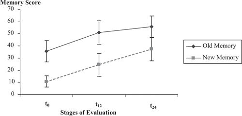
139 Rhabdomyolysis – Is it a Serious Problem?
Pap C (1), Karteszi M (1). 1. Department of Emergency Medicine and Clinical Toxicology, Péterfy S. Hospital, Budapest, Hungary; 2. Department of Emergency Medicine and Clinical Toxicology, Péterfy S. Hospital, Budapest, Hungary.
Background: Intoxications can cause rhabdomyolysis, which may lead to severe complications. Objective: to consider the range of rhabdomyolysis in poisoned patients. Patients and methods: Data of intoxicated patients treated at our department in the last 3 years were analyzed retrospectively. Patients after a suicide attempt, alcohol and/or drug abuse or accidental poisoning having a serum creatine kinase level more than 500 U/l with no acute coronary syndrome, cerebrovascular insult and acute abdominal crisis were enrolled in this study. The following parameters were recorded: age, gender, history of patient. The laboratory findings (serum potassium, CK, CKMB, LDH, ASAT, urea nitrogen, kreatinine, myoglobin, coagulogram and acid-base status) were determined at the admission to the hospital and at least 3 times while staying in hospital. Renal and soft tissue ultrasonography and an estimation of neurological status by a neurologist were performed within one day after admission and before emission. In case of compartment syndrome an examination by a traumatologist was performed. The therapy included vigorous hydration with diuretics, urinary alkalinization and if it was necessary, haemodiafiltration or haemodialysis treatment. Results: A total of 21,245 patients were admitted to our department because of intoxication over this 3 year-period and 138 (0.65%) of the patients were included in this study. The mortality rate of these patients was 12.3% (17/138). The further data can be seen in . Conclusions: Rhabdomyolysis is not frequent but not rare either in poisoned patients and can cause life-threatening complications. Early diagnosis and adequate therapy are of great importance for recovery.
TABLE 1 Causes, syndromes and complications of our patients treated because of rhabdomyolysis
140 Elevated Serum Ammonia as a Surrogate Marker for Ethylene Glycol Intoxication
Akhtar J, Johnston BD, Krenzelok EP. Pittsburgh Poison Center, Children's Hospital of Pittsburgh, Pittsburgh, USA.
Background: Drug and toxin induced causes of hyperammonemia include valproic acid, 5-fluorouracil, and ethylene glycol. Ethylene glycol ingestion results in the formation of organic acids which inhibit N-acetylglutamate synthesis. N-acetylglutamate is important in the incorporation of ammonia into the urea cycle. We report a case of ethylene glycol poisoning diagnosed based on the presence of an elevated ammonia level. Case Report: A 50-year-old man was found unresponsive, after he was noticed to have slurred speech and confusion. No other history was available. He was intubated for airway protection. Laboratory results revealed; Na158, K 5.9, Cl 107, CO2 8, BUN 16, Cr 3.3, Lactate 22. His ABG showed pH 6.92, pCO2 10.5, pO2 294, HCO3 2. An ammonia level done as part of his work up of encephalopathy was elevated at 157. The patient was started on a bicarbonate infusion and dialyzed for correction of the acidosis and hyperkalemia. The initial diagnosis was sepsis and multiple subspecialty consultations were obtained including medial toxicology. An ethylene glycol level was sent out at the request of the toxicologist, and the patient was empirically started on fomepizole. His ethylene glycol level was reported at 112 mg/dL. The patient recovered and admitted to ingesting fluid he believed to be ethanol that was in the trunk of his car. Conclusion: The diagnosis of ethylene glycol poisoning is based on indirect and direct laboratory measurement. Few institutions have timely access to direct measurement of ethylene glycol. As a result, diagnosis sometimes can be delayed and therapy initiated late. Hyperammonemia has been associated with ethylene glycol ingestion. An elevated ammonia levels in the presence of an anion gap acidosis may aid in making that diagnosis.
141 Early Poisoning Management in Paediatric Toxic Ingestion
Niewinska K (1), Niewinski P (2), Sokol-Ossowicz A (3), Poradowska-Jeszke W (3). 1. Chair of Emergency Medicine, Medical University, Wroclaw, Poland; 2. Department of Clinical Pharmacology, Medical University, Wroclaw, Poland; 3. Lower Silesia Paediatric Centre, Wroclaw, Poland.
Objectives: Toxic ingestions in paediatric population, either unintentional or intentional, account for 2–5% of paediatric hospital admissions. According to “Gastric decontamination: position statements” early management and decontamination procedures are supposed to shorten a length of stay in hospital and improve clinical outcome of the patient. The aim of our study was to assess the correlation between time of decontamination and clinical status of a poisoned child. Methods: We performed a retrospective analysis of acute poisoning cases admitted to Lower Silesian Paediatric Centre in Wroclaw, Poland, in years 2000–2003. The centre is the biggest children's hospital in the region with an average of 4,500 admissions per calendar year and serves as a paediatric poisoning reference centre for the whole region. We analyzed all cases of acute poisoning admissions with respect to exposure, type of poison, severity of symptoms and poisoning management in emergency department (ED). Results: We analyzed 799 paediatric poisoning admission cases. Toxic ingestions accounted for 636 cases (79.6%). Induced vomiting was only decontamination procedure performed in pre-hospital phase. Hospital phase decontamination included gastric lavage and/or activated charcoal administration. Important information noted were exposure time, time of decontamination procedures in pre-hospital settings, time of admission to ED and in-hospital decontamination, poisoning severity score (PSS) and hospital length of stay. The patients were included in statistical analysis provided they ingested a substance amenable for decontamination according to AACT and EAPCCT guidelines. Final statistical analysis was made for 496 cases of acute ingestion. We assessed risk factors of worse clinical grading of poisoning (PSS = 2 or 3) and longer time of hospital treatment (>2 days). Gastric decontamination procedures started after 1 hour post-ingestion resulted in 1,5 time bigger risk of higher PSS score and longer than 2 days hospitalisation time (odds ratio – OR = 1.65, confidence interval – CI = 1.06–2.67, chi2 = 5.01, p = 0.025). Conclusion: Better clinical outcome and shorter duration of treatment in paediatric toxic ingestions is directly dependent on early implementation of decontamination procedures.
142 Assessment and Management of Life-Threatening Conditions in Acute Poisonings
Vučinić S, Šegrt Z, Joksović D, Jovanović D. National Poison Control Centre, Military Medical Academy, Belgrade, Serbia and Montenegro.
Objective: Acute poisoning is a common medical emergency that requires accurate assessment and prompt therapy. Early identification of toxic substance can save time and decrease risk of toxicity and life-threatening, urgent conditions. Effective management of acutely poisoned patient includes initial stabilization, nonspecific, specific therapy and supportive care (1). Our objective was to evaluate urgent conditions in acute poisonings, and efficacy of therapeutic measures applied. Methods: A retrospective survey of acutely poisoned patients, treated in Clinic of Emergency and Clinical Toxicology and Pharmacology NPCC, during one year period. Results: Out of 1,318 hospitalized patients, 987 had acute poisoning. Beside being the most frequent, poisonings with drugs, pesticides and corrosives were the most severe, followed with urgent conditions such as toxic coma, convulsions, delirium, acute respiratory insufficiency, acute cardiocirculatory, renal and hepatic insufficiency (). Psychotropics, opioid drugs or pesticide poisoning were the most frequent cause of coma, often characterized by hypoxia, metabolic or respiratory acidosis. Toxicological analysis and blood glucose levels were available for majority of these patients. Most poisons that depressed consciousness had also impaired respiration. Stabilization of the patients was performed simultaneously with the initial physical assessment. Antidotal therapy was used only in 79 patients, and the treatment of most cases of poisoning was largely supportive. Mechanical ventilation was necessary in 58, and inotropic agents in 21 patients. Forced alkaline diuresis and hemodialysis were used for elimination of water soluble compounds and those with small volume of distribution. Fifty-one (5.2%) patients died: a majority of them with acute corrosive and pesticide intoxication. In some cases, prolonged hypoxia before the treatment had caused irreversible brain damage and lethal outcome. Conclusion: Continuous observation, re-evaluation, symptomatic and supportive treatment respresent the cornerstones of the management of the poisoned patient. Measurement of drug concentrations is clinically important for relatively few compounds. Decision about decontamination and specific antidotal therapy is based on toxin and patient's condition related factors. Inadequate ventilation, acute cardiocirculatory insufficiency and coma are the most common cause of serious morbidity or death in poisoning. Reference: 1. Krenzelok EP. New developments in therapy of intoxications. Toxicol Lett 2002; 127:299–305.
TABLE 1 Urgent conditions in poisonings by different toxic agents
143 Serotonin Syndrome-Associated Myocardial Infarction: A Case Report
Ganetsky M (1), Liang IE (2), Gresham C (1), Bird SB (1). 1. Department of Emergency Medicine, University of Massachusetts Medical School, Worcester, MA, USA; 2. Department of Emergency Medicine, St. Elizabeth's Medical Center, Boston, MA, USA.
Objective: We report the first case of serotonin syndrome (SS) associated with an acute myocardial infarction (AMI). Case report: A 31-year-old female presented with mental status changes. Three weeks before presentation her psychiatrist changed quetiapine for duloxtine as part of her treatment for depression and bipolar disorder. Two weeks after the medication change her mother described her as “twitchy.” On the day of admission she began vomiting, followed by somnolence and confusion. Her other medications included paroxetine, bupropion, and clonazepam. In the emergency department, her heart rate was 120 bpm and blood pressure was 160/104. On physical examination, she had confusion, nystagmus, a fine tremor, lower extremity hypreflexia, and inducible sustained ankle clonus. The patient had evidence of a non-ST segment elevation MI with peak troponin I of 3.83 ng/mL, which trended downward over the next three days. By hospital day #3, she developed inverted T-waves in leads I, aVL, II, aVF, V3-V6 of her EKG and an echocardiogram showed septal, anterior, and lateral hypokinesis with an ejection fraction of 30%. Her mental status improved and she was discharged home after four days, but returned one week later with pericarditis-type chest pain. During this second admission, she received a cardiac catheterization that showed patent coronary arteries and an ejection fraction of 74%. Conclusion: Cardiac ischemia is not a typical complication of SS or of SSRI therapy. On the contrary, SSRI use has been postulated to decrease the risk of AMI, possibly by inhibiting platelet activation (1). Our patient had cardiac biomarker leak and focal wall motion abnormalities that resolved after one week, which is consistent with myocardial stunning. The only case report of an SSRI associated AMI was a 69-year old with diabetes and CAD who developed an AMI five days after starting venlafaxine. This patient did not have clinical findings of SS and, unlike our patient, had evidence of coronary atherosclerosis on cardiac catheterization (2). As the number of approved serotonergic agents increases and as more patients are placed on combination serotonergic therapy, the incidence of SS will likely increase. We suggest that clinicians should consider the presence of myocardial ischemia in patients with serotonergic findings, especially those with known CAD or its risk factors. References: 1. Schlienger RG, Fischer LM, Jick H, et al. Current use of selective serotonin reuptake inhibitors and risk of acute myocardial infarction. Drug Safety 2004; 27:1157–1165. 2. Reznik I, Rosen Y, Rosen B. An acute ischaemic event associated with the use of venlafaxine: a case report and proposed pathophysiological mechanism. J Psychopharmacology 1999; 13:193–195.
144 Signs and Symptoms of Carbamazepine (CBZ) Overdose and CBZ Plasma Levels of 94 Patients Attended by UNICAMP Poison Center (UPC), Campinas City, S. Paulo State (Brazil), Jan. 1994–Oct. 2005
Vieira RJ, Shono EY, Bucaretchi F, Madureira PR, Toledo AS, De Capitani EM. State University Campinas (UNICAMP), Campinas, Brazil.
Objectives: As there is a recent increase in the use of Carbamazepine (CBZ) in Brazil, which has led to an increased frequency of overdoses, the objective here is to present a retrospective study considering the distribution of CBZ plasma level, CBZ ingested dose, symptoms, signals and evolution in 95 cases of CBZ poisoned patients attended by UPC from Jan 1994 to Oct 2005. Case series: Only 94 out of 903 cases attended by UPC had a determined CBZ plasma level, with the mean of 23.40 μg/ml. Out of 94 patients, 68 (72.34%) were attended at the University Hospital, while the other 31 (32.98%) at different medical centers. In terms of association with other drugs, 31 (32.98%) ingested only CBZ, and 63 (67.02%) had association with other drugs. The most frequent signs or symptoms of the 70 CBZ poisoned patients with CBZ plasma level less than 30 μg/ml were somnolence 30, tachycardia 13, coma 11, hypertension 9, conscience depression 9, dizzeness 8, agitation 8, mental confusion 7, mydriasis 7, vomiting 6, hypotension 6, ataxia 5 and 25 other signs or symptoms. 8 out of 70 patients needed tracheal entubation. The most frequently signs or symptoms of 30 CBZ poisoned patients with CBZ plasma level of 30 μg/ml or more, were somnolence 9, coma 7, agitation 6, myosis 6, tachicardya 6, conscience depression 5, hypotension 5, hypertension 4, convulsion 3, agitation 6, mental confusion 3 and 10 other signs or symptoms. From this group, 6 patients needed tracheal entubation and one died. Between the 31 patients poisoned only by CBZ, the most frequent signs or symptoms of the 21 CBZ poisoned patients with CBZ plasma level less than 30 μg/ml were somnolence 15, mental confusion 6, agitation 4, ataxia 4, vomiting 3, coma 2, dizzeness 2, diplophy 2, disartria 2, and 7 other signs or symptoms. The most frequent signs or symptoms of 12 CBZ poisoned patients with CBZ plasma level of 30 μg/ml or more, were coma 6, hypertension 4, myosis 3, convulsion 3, somnolence 2, tachicardya 2, hypotension 2, agitation 2, arrithmia 1, cyanose 1. From this group, 6 patients needed tracheal entubation and one died. Conclusion: The most frequent signs or symptoms of 12 CBZ poisoned patients with CBZ plasma level of 30 μg/ml or more, were coma 6, hypertension 4, myosis 3, convulsion 3. Reference: 1. Schmidt S, Schmitz-Buhl M. Signs and symptoms of carbamazepine overdose. J Neurol 1995; 242:169–173.
145 Poisonous Nightcap from Poppy Seeds
Bergmann I (1), Roether M (1), Werner KP (2), Roemhild W (3), Michalak H (4), Hentschel H (1). 1. Poisons Information Centre Erfurt, Erfurt, Germany; 2. Kreisklinik Aschersleben-Staßfurt, Aschersleben, Germany; 3. Institute of Legal Medicine, Otto-von-Guericke University, Germany; 4. Federal Institute of Risk Assessment, Germany.
Objective: Poppy seeds obtained from mature Papaver somniferum normally contain only small amounts of opium alkaloids. The alkaloid concentration of seeds is usually not under control by Food Administration. We report a poisoning developing disastrous consequences under a peculiar set of coincidences. Case report: A 6-week-old female baby (5 kg body weight) ingested about 75 mL of a milk nightcap prepared by her mother as an old household remedy for sedation. 200 grams of poppy seeds were cooked with 500 mL milk and honey. Three hours after ingestion the patient developed disturbances of respiration with CNS depression, miosis, tachycardia, and pale grey marbled skin. Intermittent respiratory arrest was noted ad time of hospital admission. Arterial oxygen saturation was 89% at hospitalization, and dropped further to 69%. The patient was intubated and ventilated. Administration of naloxon was successful to restore spontaneous breathing but repeated intravenous doses were needed for 10 hours (total dose 1 mg, 0,2 mg/kg, respectively). Fortunately, the patient awoke 15 hours after ingestion without hypoxic sequelae. Toxicological analysis (GC/MS) of urine confirmed morphine (18.2 micrograms/L on day 1; 627 micrograms/L on day 2) and codeine (317 micrograms/L on day 1; <5.0 micrograms/L on day 2) as cause of poisoning. Morphine was also found in serum (4.3 micrograms/L on day 2). The poppy seeds used for the preparation of the nightcap contained 0.1% morphine and 0.003% codeine. Subsequent investigations of 23 different poppy seed samples result variable morphine contents (range 0.0004% to 0.0228%) (1). Conclusion: Undoubtedly, the patient was poisoned by opium alkaloids from the extracted poppy seeds by the preparation of the nightcap. Apart from the overdose of morphine (estimated dose 6.0 mg/kg; usual oral single dose 0.125 to 0.225 mg/kg) the immaturity of blood-brain barrier, biotransformation and renal excretion resulted in the baby's life-threatening poisoning. As a consequence, the poison centre and Federal Institute of Risk Assessment and the Food Administration warned the public on this household remedy immediately. Furthermore, Food Administration takes measures to control the alkaloid content of poppy seeds to protect consumers health from opioid poisoning (2). References: 1. [Morphin in Speisemohn] http://www.lgl.bayern.de/de/left/fachinformationen/lebensmittel/morphin_speisemohn.htm. 2. [Risikoanalyse: Morphin und Codein in Mohnsamen für Back-bzw. Speisezwecke] http://www.lgl.bayern.de/de/left/fachinformationen/lebensmittel/morphin_speisemohn_risikoanalyse.htm.
146 Digoxin Effects After Snail (Helix Pomatia) Ingestion: A Case Report
Eleftheriou J (1), Bacis G (1), Mori D (2), Gechtman C (3), Zoppi F (3), Farina ML (1). 1. Bergamo Poison Control Center, Ospedali Riuniti, Bergamo, Italy; 2. Medicine Department, Ospedale di Treviglio, Italy; 3. Laboratory Unit, Ospedale Niguarda Ca’ Granda, Milan, Italy.
Introduction: Cardiovascular glycoside-like intoxication following ingestion of Nerium oleander or Digitalis purpurea is a common clinical problem and many authors reported cardiovascular symptoms similar to those observed after acute toxic digoxin administration. Modes of exposure to the plants include accidental ingestions as well as intentional administration in foods and drinks prepared from the leaves, but there are not published reports about poisoning after snails ingestion. Case report: The present case report involves two patients (female aged 43 and male aged 66) who showed gastrointestinal and cardiovascular symtoms following the ingestion of snails fostered with foxglove leaves. The initial hypothesis was an oleander intoxication as the patients found the snails near the N. oleander of their garden. At the time of admission, 12 hours after the ingestion, they presented with nausea, vomiting and diarrhea; the man presented ECG changes: ectopic beats and bradycardia (36 bpm) while woman's ECG was normal. The serum digoxin levels were respectively 0.89 ng/mL and 0.78 ng/mL (range 0.5–2.0 ng/mL) and the urine digoxin concentrations were respectively 0.29 ng/mL and 0.73 ng/mL. The potassium levels were normal in both of the patients (4.8 mEq/L). On the second day the serum digoxin concentration was 0.29 ng/mL in both of the patients and no therapy was performed. The digoxin concentration in the snail homogenized tissue, after freezing with liquid nitrogen, was 220 ng/g. The symptoms resolved on the second day and the patients recovered fully with a normal sinus rhythm on the third day. Conclusion: This is the first case report about an indirect digitalis intoxication, wich involves poisoned food. The snails may become poisonous by absorbing toxic compounds from their food and determine an acute human poisoning.
147 Poisoning by Scopolia Carniolica
Grenc D, Brvar M, Sarc L. Poison Control Center, University Medical Center, Ljubljana, Slovenia.
Objectives: Reports of poisoning by Scopolia carniolica are rare. The plant was named by Carl von Linné in honor of its discoverer, natural historian J. A. Scopoli (1723–1788) and the former province of Carniola, and is indigenous to some regions of Slovenia. Like several genera of the Solanaceae (Nightshade) family it contains tropane alkaloids which cause distinctive anticholinergic clinical syndrome in case of poisoning. Case report: We report a case of two female patients who consumed a meal containing what they thought were spinach leafs. As soon as 45 minutes after the meal they experienced the first symptoms of dry mouth and vertigo, one of them was unable to urinate. Other family members noted that both behaved erratically. Activated charcoal was administered in a primary care facility. Upon arrival in our institution, some 3 hours after the meal, they presented with delirium, agitation, tachycardia, mydriasis, warm, flushed skin and dry mouth. Both had diminished bowel sounds and one had urinary retention. They brought a plant from which they prepared the meal and which was later identified as Scopolia carniolica. Because of distinct anticholinergic signs and presumed decreased gastric motility we decided to decontaminate both of them. Upon gastric lavage we successfully removed a good deal of the meal. Initial treatment with intravenous diazepam was unsuccessful. Both patients continued to be agitated, delirious and had visual hallucinations, so physostigmine 2 mg intravenously was administered. Within minutes hallucinations, agitation and delirium reversed. Further recovery was uneventful and both patients were discharged the next morning, 20 hours after the meal. Conclusion: Poisoning by Scopolia carniolica is a rare occurrence mostly because it is not widely distributed and only rarely gets mistaken for other plants. Due to tropane alkaloids it causes distinctive anticholinergic clinical syndrome.
148 Overview of Amanita Phalloides Poisoning Treatment in PCC Ljubljana in the Last 30 Years
Jamsek M, Sarc L, Grenc D. Poison Control Centre, Ljubljana, Slovenia.
Objective: A study was performed to evaluate different recommendations about Amanita phalloides (APh) poisoning treatment. Methods: In a retrospective study mortality between 3 groups of patients, who underwent different therapeutic protocols in the last 30 years, was compared. In the first decade (1976–1985), patients were treated with high doses of crystalline penicillin and glucocorticoids. During the second decade (1986–1995), silibinin was added to the treatment protocol. In the last ten years (1996–2005), both penicillin and glucocorticoids were withdrawn from therapeutic guidelines; treatment relied on early silibinin and adequate symptomatic treatment. Concurrently we looked into possible prognostic value of prothrombin time and/or INR in predicting the severity and outcome of Aph poisoning. Patients were classified in 3 categories according to maximal values of prothrombin time (PT) and/or INR (>20%, 10–20%, <10% for PT and <4, 4–5,>5 for INR). Diagnosis relied on mushroom identification the characteristic course of clinical symptoms and signs; in a few cases RIA for amanitine or Aph spore identification were also used. Results: 323 patients were hospitalised due to poisoning by various mushrooms in our PCC from 1976 to Nov. 2005; 77 patients were poisoned by Aph. In view of three different therapeutic protocols used in past three decades, we found the following mortality rates: 14,1% (5 out of 34) for the first, 3,4% (1 out of 29) for the second and 7,1% (1 out of 14) for the third decade group. Regarding the prognostic value of PT and/or INR (for the first decade we have data for PT only) our findings were as follows: all patients with PT>20% survived, in the group with PT 10–20% 1 died and 5 survived and with PT <10% 4 died and 2 patients survived. In the last two decades all patients with INR < 4 survived. For patients with INR between 4 and 5, one died and 2 survived, and with INR> 5 one died and 2 survived. Since 1997 we recorded no new deaths due to APh poisoning. In 1998 a successful liver transplantation was performed in one patient due to fulminant hepatic failure caused by Aph poisoning. Conclusion: Although we reviewed a large number of patients poisoned by Aph, we cannot give reliable prognostic criteria about severity or outcome of poisoning because of the low number of deaths. This low number on the other hand is an indicator that current treatment is effective. Introduction of silibinin further decreased the mortality rate. However, due to the low numbers of letal outcomes we cannot objectively evaluate the advantage of silibinin monotherapy.
149 Prolonged Paranoid Psychosis after Amanita Muscaria Ingestion
Brvar M (1), Grenc D (1), Mozina M (1), Bunc M (2). 1. Poison Control Centre, University Medical Centre, Ljubljana, Slovenia; 2. Department for Cardiology, University Medical Centre, Ljubljana, Slovenia.
Introduction: Amanita muscaria contains ibotenic acid, muscimol, muscazone and other toxins such as muscarine. Ibotenic acid acts on glutamic acid receptors and has an excitatory action. On the other hand, muscimol acts on GABA receptors and has a depressant action. The concentration of muscimol increases with time due to ibotenic acid decarboxylation into muscimol. Muscazone and muscarine exhibit minor pharmacological activity. The duration of clinical manifestations after Amanita muscaria ingestion does not exceed 24 hours. We report on 5-day-long paranoid psychosis after Amanita muscaria ingestion due to a bet on mushroom species. Case report: A 48-year-old man, with no previous medical history, gathered and ate mushrooms presumed to be Amanita caesarea for dinner after making a bet with a friend about mushroom species. Half an hour later he started to vomit and fell asleep. He was found comatose having a seizure-like activity and soiled after having urinated and defecated three hours after ingestion. On arrival four hours after ingestion he was comatose, but the remaining physical and neurological examinations were unremarkable. Creatine kinase was 8.33microkat/l. Other laboratory results and brain CT scan were normal. A toxicology analysis revealed no drugs in his blood and urine. The mycologist identified Amanita muscaria between the remaining mushrooms. The patient was given activated charcoal. 10 hours after ingestion he awoke and was completely orientated. Eighteen hours after ingestion his condition deteriorated again and he became agitated and unco-operative. Afterwards visual and auditory hallucinations appeared and he became paranoid. During the second and third day paranoid psychosis with hallucinations persisted, and he became dehydrated due to refusing food and therapy, and exhausted after sleepless nights. At the end of the third day he was transferred to a psychiatric ward where he was physically restrained and treated with olanzapin. On the sixth day all symptoms of psychosis gradually disappeared and he was discharged. He was amnestic about hospitalization in PCC. Alcohol abuse was excluded. One year later he is not undergoing any therapy and had no symptoms of psychiatric disease. Conclusion: Paranoid psychosis with visual and auditory hallucinations can appear 18 hours after ingestion of Amanita muscaria and can last for up to 5 days. The reason for the delayed manifestation of psychosis could be muscazone that induce CNS effects with a longer latency period or delayed decarboxylation of ibotenic acid into muscimol, which is a real psychotropic substance in Amanita muscaria. The massive ingestion due to the bet coupled with late and insufficient gastric decontamination could also be the reasons for prolonged psychosis.
150 Poisoning with Tropical Euphorbiaceae: Two Case Reports
Blanc-Brisset I, Pommier P, Hayek-Lanthois M, Arditti J, de Haro L. Centre Antipoison, Hôpital Salvator, Marseille, France.
Introduction: Plants of the euphorbiaceae family can be toxic for humans for two reasons: the sap can induce skin irritation and digestive or ocular caustic lesions, and seeds contain toxalbumins which can be at the origin of systemic poisonings. Case Report 1: A 57-year-old man was snorkelling in Martinique Island (French West Indies) when he saw two fruits floating and looking like yellow apples. As he was thirsty, he ate the fruits and thought their taste was really good. He continued to swim for 15 minutes and then began to have throat and ocular burning sensations. He decided to go back to the beach and when he arrived on the sand, he had a malaise and intense abdominal pain. He realised then than the ingested fruits came from a big mancheneel tree (Hippomane mancinella) which was close to the sea. While he was preparing to go to the hospital, he had aqueous and painful diarrhoea which were corrosive and burned his buttocks and thighs. He finally arrived at the hospital one hour after the ingestion with face swelling, complete eyelid occlusion and multiple oral and pharyngeal ulcerations. He recovered after 5 days of symptomatic treatments in hospital. Case Report 2: A 22-year-old woman and a 21-year-old man, just arriving from a travel in Madagascar, decided to eat “aphrodisiac seeds” they bought in Antananarivo. The local plant name was “Valavelona” and the seller recommended to crunch one seed before love. During their first night in France, the decided to try and each of them crunched 6 seeds. One hour after the ingestion, they both had severe vomiting and aqueous diarrhea. They consulted their family doctor who observed the presence of blood in the diarrhea. With the indigenous name, the Marseilles Poison Centre discovered that the seeds came from a tropical plants of the euphorbiaceae family named Purging nut tree (Jatropha curcas). As the patients were dehydrated and suffered of violent abdominal pain, they were managed in the hospital where they recovered after 24 hours of symptomatic treatments. Conclusion: Plants of the euphorbiaceae family can represent a real danger for humans. In the case of the mancheneel tree, the pleasent taste can induce ingestion of big and toxic quantities at the origin of severe poisoning.
151 Herbal Medicines and Hepatotoxicity: Spanish Poison Control Centre Experience
Ramón MF, Ballesteros S, Martinez-Arrieta R. Spanish Poison Control Centre, INTCF, Madrid, Spain.
Background: The use of herbal medicines and remedies are becoming more common among Occidental population. Some of these herbs have been associated with hepatic disturbances in chronic cases essentially, over the years. The hepatic effects related to medicinal herbs are connected mainly to hepatitis (direct effects on hepatocytes or idiosyncratic reactions), and veno-occlusive disease. Our objective was to investigate the profile of herbal medicine exposures associated with hepatotoxicity recorded in our PCC between January 1991 and April 2005. Case series: Of 520 herb toxic exposures detected in our service during the study period, 65% have occured after 1999, and 252 of cases were due to herbs containing potential hepatic toxins described in the literature: antraquinones: Cassia acutifolia (23), Rhamnus sp. (20), and Aloe vera (4); aesculin or coumarin: Aesculus hippocastanum (31); pyrrolizidine alkaloids: Senecio sp. (4), and Symphytum officinale (1); ephedrine: Ephedra sp. (4); kava-lactones: Piper methyisticum (2); pulegones: Mentha pulegium (2); isoquinolines: Chelidonium majus (2); not identified toxins: Valeriana officinalis (154), Peumus boldus (3), and Echinacea sp. (2). A number of 9 toxic exposures have been related to hepatic disturbances. All of them were oral and chronic, except in one homeopatic preparation based on Symphytum sp., after the first intramuscular injection. These calls came from hospitals. Five cases corresponded to herbal medicine combinations including two homeopatic preparations. Only one case had cardiovascular antecedents. The outcome was favorable in 5 exposures. The principal data of these cases are shown in . Conclusions: Approximately, a half of herbal exposures detected in our service has a potential hepatotoxicity although only 3.6% of them has been associated with liver diseases. Recognition of herb induced hepatotoxicity depends on physician awareness of the problem. Prevention include patient's education in natural remedies.
TABLE 1
152 Epidemiology of Envenomations in Spain
Ballesteros S, Ramón MF, Martínez-Arrieta R. Spanish Poison Control Centre, INTCF, Madrid, Spain.
Objective: To determine the epidemiology of envenomation in Spain. Methodology: A retrospective review of documented cases of evenomation from 1991 to 2003. Data including patient age and gender, geographic location, timetable, place, and etiology of the exposures were recorded. Results: A total of 2,153 cases of animal envenomation were registered. Cases of envenomation appeared constantly all through the 10 years (mean 168 ± 40 cases per year). Saturday was the day with the highest number of calls (16.2%). They started increasing in May, and peaked in July and August (21% each month). January was the month with less accidents (1.6%). Most cases occurred along the Mediterranean coasts and the inner dry part of the country (45% each) and less frequent in the north and Baleares and Canary Islands. Adults were 75.4% of the victims (range 15–92 years, mean age 39), children between 3 and 14 years old were 19.1% (mean age 8 years), 3.7% were less than 2 years old (mean age 19 months), 1% were animals, rest unknown. The incidence of bites was higher in males (70% of human victims). Bites on the upper extremity were most common (61.5%) followed by the lower extremity (25.1%), head (7.3%), trunk (3.4%), and neck (2.7%). Arachnidae was the most frequent class of animals 37.3% (86% of them scorpions, and 12.4% spiders -16 of them black widow spider), followed by snakes 22.4%, insects 10.5% (83.3% of them Hymenoptera), marine animals 12.4% (84% of them fish, mainly Trachinus araneus and the rest invertebrates), centipedes 10.2%, amphibians 1.2%, other reptiles 0.5%, and unknown or not-venomous animals. All animals were indigenous to Spain except 33 exotic: 16 snakes, 8 fishes (catfish, globefish, turkeyfish, stonefish, lionfish), 4 scorpions, from Africa and Peru, 3 spiders, from Chile and Africa, 2 coral. Ten of them were children (3–13 years old), biten or stung by animals kept as pets. Number of animals involved in the envenomation ranged between 1 and 120. Higher number of animals involved were wasps, bees and scorpions. The place of envenomation was: nature 71.7% of occasions, home 14.8%, work 1.7% (including aquariums, ships and fish-shop), at the street, circus, cars, schools, restaurants, hospitals, etc. A percentage of 3% of victims were bitten or stung during the course of their job. At the time of the consult minor local symptoms were presented in 52.7% of cases, moderate systemic symptoms in 29.1% and life-threatening symptoms in 2.2% of occasions. Conclusion: This study helps to assess the magnitude of the problem since envenomations are not systematically reported and there are no recent statistical data available in our country.
153 Severe Bothrops Snakebite on the Head: Case Report
Bucaretchi F, Hyslop S, Mello SM, Vieira RJ. Poison Control Centre, Faculty of Medicine, State University of Campinas, Brazil.
Objective: Lanceheads (Bothrops species) account for 87.2% of the bites by venomous snakes for which the genus is officially notified in Brazil (lethality rate = 0.37%). Most of the bites occur on the extremities, with <0.5% involving the head and neck. Case Report: A previously healthy, 21-year-old female was admitted 5 hours after being bitten in the occipital area by a pit viper presumed to be Bothrops jararaca, the most important species in south-eastern Brazil. The patient was bitten while picking up a pen from the ground; the snake being curled around the branch of an avocado tree, approximately 1.7 m above the ground. Physical examination revealed marked cranial and facial oedema extending to the neck and dorsum, bilateral eyelid ecchymosis, and local and gingival bleeding. The patient was alert and complained of local, mild pain and nausea. The main laboratory findings on admission included incoagulable blood (based on the 30 min whole blood clotting time, PT, INR, APTT and R), a platelet count of 4,000/mm3, and a serum venom level (ELISA) on admission (T0) of 62.6 ng/mL (cut-off, 2.3 ng/mL). Sequential serum creatinine, BUN, sodium, potassium and calcium levels were normal. The case was classified as severe and 12 vials of undiluted bothropic antivenom [AV, F(ab')2, 10 mL/vial, equine origin, Instituto Butantan; 10 mL of AV neutralizes 50 mg of reference B. jararaca venom in mice] was infused IV (5–6 ml/min), preceded by the IV administration of ranitidine, chlorpheniramine and hydrocortisone. AV was discontinued after the infusion of 10 vials since the patient developed a severe early reaction successfully treated with adrenaline SC and hydrocortisone IV. Platelets replacement (7 units) was done after admission. Normal blood coagulation tests and an increase in platelet count (to 100,000/mm3) were observed 24 hours after AV administration. No circulating venom was detected at T6, T12, T24 and T48. The patient was discharged on the 4th day, with good clinical improvement and no signs of local infection. Conclusion: Crotalinae snakebites to the head and neck are uncommon and may be associated with airway compromise, high morbidity and, occasionally, deaths. Although this patient did not develop respiratory distress, she was at risk of severe systemic bleeding (very low platelet count, incoagulable blood and internal hemorrhaging), including brain hemorrhage. Treatment with AV restored normal coagulation and depleted circulating venom (confirmed by ELISA), with clinical improvement, despite the accompanying severe early reaction. Although B. jararaca is not generally arboreal, this case highlights the need for caution when moving close to low-lying branches and in dense undergrowth.
154 Accidental Oral Exposure to a Plant of Dieffenbachia Spp
Danescu I, Macovei R, Tudosie M. ICU II Toxicology, Emergency Clinical Hospital, Bucharest, Romania.
Objective: Exposure to the toxic plants is usually accidental. The most common species found in the home have limited toxicity. We report a case of oral exposure to the stalk of the plant belonging to Dieffenbachia spp. All parts of the plant contain idioblasts which are specialized pressure-sensitive organelles containing raphides, small needle-like crystals of calcium oxalate. When stimulated by a mechanical force such as biting or chewing, the idioblasts discharge the raphides as projectiles. The raphides, that contain proteolytic enzymes, penetrate the tissue and induce a local irritation due to local histamine release and proteolytic enzymes. The symptoms develop rapidly and include redness, swelling and local pain at the level of mouth and pharynx, potentially compromising the airway. The treatment is symptomatic, with copious irrigation, topical cooling, topical antihistamines and anesthetic; when the airway edema is present, systemic antihistamines or steroids may be necessary (1). Case Report: A 67-year-old female is admitted after accidental oral contact with a part of a plant stalk belongs of Dieffenbachia spp. She mistook the part of the plant with a cucumber, because the cucumbers and the plant were stored together by her daughter. After she bit the part of the plant, the female rapidly presented a burning sensation and intense pain at the level of oral cavity; shortly she developed edema. When she presented to the hospital, the clinical examination revealed a conscious agitated patient, with dysphonia and dysphagia, accusing burning pain and swelling of the oropharynx. The BP was 170/100 mmHg, pulse rate was 90/min. sinusal rhythm. Laboratory data showed no abnormality of arterial blood gases or other biochemical parameters. The examination of the oropharynx revealed giant edema of the lips, buccal mucous membrane, tongue, uvula and soft palate; redness and mild edema of the epiglottis and superglottic larynx at the laryngopharynx examination. After 6 hours from admittance the edema diminished, but vesiculous lesions appeared at the oropharynx level. The treatment was systemic with antiedematous, antiinflammatory, analgetic and antihistamines drugs and topical with local cooling and mixtures containing anesthetic, antihistamines and antiseptic substances. The lesions improved under treatment and the patient was discharged after 10 days. Conclusion: Dieffenbachia spp. cause local toxicity by way of calcium oxalate crystals that penetrate the oropharynx tissue, producing pain and swelling (1). The severity is due to potentially compromise of the airway by laryngeal edema. The treatment is symptomatic, topical in most cases. Reference: 1. Greller HA. Patient 35: A 2-year-old boy with a swollen mouth. In: Osterhoudt KC, Perrone J, DeRoos F, Henretig FM, Toxicology Pearls, USA: Elsevier Inc., 2004:110–112.
155 Stinging Catfish – Threat of Aquarists
Satora L, Targosz D, Szkolnicka B, Morawska J, Biskup-Bednarczyk A, Gawlikowski T. Poison Information Centre, Jagiellonian University Medical College, Kraków, Poland.
Objective: Heteropneustes fossilis (Bloch, 1794), commonly known as the stinging catfish are facultative air-breathers that inhabit the swamps of southeast Asia and are quite common. H. fossilis is dangerous due to the connection between the spines in the pectoral fin and the venom glands. When the spine penetrates the body of its prey, it presses its base against the cells, squashes them and squeezes the venomous contents into the wound. The venom appears to contain a few mouse-lethal fractions and two dermonecrotic fractions. The severity of the poisoning depends on the fish species, the number of stings and the amount of venom released. The potency of the venom is varied. Venom causes various inflammatory conditions with erythema, oedema, local haemorrhage and tissue necrosis. Other possible signs and symptoms are tachycardia, weakness, hypotension, nausea and vomiting, paraesthesias, dizziness and respiratory distress (1). More serious effects include radial artery injury, debilitating hand problem, chronic tenosynovitis and gangrene of the fingers requiring amputation. Case Series: A 17-year-old man, 33-year-old man, 15-year-old man, and 16-year-old man; were admitted the hospital (independently) after the hands stinging by H. fossilis. All cases happened during cleaning of the aquarium. First aid treatment for catfish stings was to remove all foreign material and irrigate with fresh water. Immersion of the injured part, as tolerated, in 450 C water for at least thirty to sixty minutes was beneficial to relieve muscle spasm, and inactivate some venom in the wound. A plain radiograph of the hands showed any foreign body in the wounds. The monitor cardiac function was included. The patients received an analgesics and a prophylactic short course of oral antibiotic therapy. As injuries inflicted by fish may result in delayed presentation of infection it was suggested that the patients be admitted for observation. After 24 hours of observation, the patients was discharged from the hospital. Over the next month, the wounds healed slowly by second intention. The hands had healed completely without any deficits in motor and sensory function. Conclusion: The stinging catfish has become a popular aquarium fish in Poland and is available in almost every pet shop. It is also bred in the aquarium on a large scale. There is a need for initiating educational activity among aquarists. Reference: 1. Satora L, et al. Stinging Catfish Poisoning. Clin Toxicol 2005; 43:903–904
156 Chelonitoxism: One Case of Collective Poisoning in French Polynesia
Fussy A (1), Pommier P (2), Bazire A (3), de Haro L (2). 1. Réanimation médicale, hôpital Mamao, Papeete, French Polynesia; 2. Centre Antipoison, Hôpital Salvator, Marseilles, France; 3. Centre Antipoison, Hôpital Fernand Widal, Paris, France.
Introduction: Chelonitoxism is a poisoning due to turtle flesh ingestion. As turtles are endangered and protected species for several decades, cases of chelonitoxism are extremely rare, and only observed in few areas where marine reptiles are eaten. The green turtle (Chelonia mydas) is considered as edible by indigenous populations of several Indo-Pacific Ocean coasts. Poisonings are usually described after misidentifications with ingestion of an other species, the hawksbill turtle (Eretmochelys imbricata) which is considered as poisonous. Case Series: In the Rangiroa Island (French Polynesia, Pacific Ocean), 19 members of the same Polynesian family ate the flesh of one turtle captured near the coast. It was identified as a little specimen of green turtle. All 19 patients quickly suffered digestive troubles (nausea, vomiting, diarrhoea), but 3 of them decided to have a second meal with this “delicious meat.” After a few hours, they felt bad enough to be evacuated to the hospital in Papeete on the main island of Tahiti. In the emergency unit, the clinical features of the 3 patients were the following: a 40 year-old-man had diffuse arthralgias, severe digestive disturbances and asthenia; a 64-year-old man had mild digestive troubles, renal failure, tetraparesis and coma; and a 26-year-old pregnant woman had multiple organ failure. The two men improved quickly with symptomatic treatments in intensive care unit, but the young woman died in a few hours. The police investigation proved that the turtle species was misidentified: it was an adult hawksbill turtle instead of an edible green turtle. Discussion: Chelonitoxism is a life-threatening marine poisoning which is mainly described in the literature with the carnivorous Eretmochelys imbricata. The toxins implicated in this species are not known. However, several chelonitoxism after ingestion of the herbivorous green turtle meat are clearly reported. Recent analytical studies in Japan showed that Chelonia mydas flesh can be contaminated with micro-algae toxins (lyngbyatoxins).
157 Is Gyromitra Esculenta Really a Conditionally Edible Mushroom? – Case Report
Sarc L, Jamsek M. Poison Control Centre, University Medical Centre, Ljubljana, Slovenia.
Objective: Gyromitrin, acetaldehyde N-methyl-N-formylhydrazone (ca 90%) and its 8 hydrazone homologues are toxic compounds of Gyromitra esculenta (GE). They hydrolyse in vitro and in vivo (a gastric phase) to N-methyl-N-formylhydrazine (MFH) and then to monomethylhydrazine (MH). Acute toxicity of MH and MFH is connected with blocking of the cofactors, containing carbonyl function group. Oxidation and alkylation of MH (and perhaps MFH) in hepatic phase, are responsible for hepatotoxicity and most probably for production of carcinogenic derivatives (1). Micheloth assumed that toxic substances of GE are heat-sensitive, volatile, water-soluble and they accumulate in the cooking juice or in the preservation liquid. The majority of toxicological textbooks cite, that toxins of GE are water-soluble and volatile; unclear and misleading are data about heat-sensitivity. Case Report: The three members of the family ate inadvertently for their supper roasted Gyromitras esculenta instead of morels. 9 hours after ingestion the 40-years old mother began with intense vomiting. During the next 24 hours her symptoms subsided. On the third day she became jaundiced and her urine became dark. On admission, she had icterus, body temperature of 37.8°C and pain under the right rib cage. Biochemical analysis of serum and urine showed signs of severe haemolysis and hepatotoxicity. Ultrasound showed diffuse increased density of the liver. Virus-markers for Hepatitis were negative. Biochemical markers of hepatotoxicity and of haemolysis reached a maximum level on the fourth day. The treatment was symptomatic only. Her 42-year-old husband and their 12-year-old daughter didn’t have any symptoms, but results of biochemical investigation showed signs of slight haemolysis. Conclusion: The comparison of the degree of poisoning by the three family members point to some physicochemical properties of the toxins. The daughter and her father developed slight biochemical signs of poisoning only, although they have eaten majority of prepared GE. Mother ate only a few mushrooms and a lot of gravy, which contained a high amount of toxins, which did not evaporate and were not inactivated by heat. Looking at the way the meal was prepared and the time of the onset of the symptoms, the inhalation exposure was unlikely. Poisoning of three members of the family indicated that toxins were accumulated in the gravy, but they were not inactivated by cooking. Due to a high acute toxicity and most probably also a carcinogenic effect, toxicological textbooks must provide more exact data on removing, destroying or inactivation of GE toxins. Manuals on Mushrooms need to stress that GE is a toxic mushroom. Reference: 1 Michelot D, Toth B. Poisoning by Gyromitra esculenta – a review. J Appl Toxicol 1991; 11:235–243.
158 Hyponatremia with Cerebral Edema – A Complication of Mushroom-Induced Gastrointestinal Distress
Hermanns-Clausen M, Stedtler U. Poisons Information Centre (PIC), Centre for Pediatrics and Adolescent Medicine, Freiburg, Germany.
Objectives: A large variety of mushrooms cause gastrointestinal distress within 1–2 h after ingestion. Symptoms are occasionally severe enough to cause significant dehydration. Most patients can be managed at home after receiving information regarding fluid replacement. Case Report: A 60-year-old man started vomiting 1 h after eating self-collected mushrooms. 4.5 h later he reported frequent vomiting. Seven hours after ingestion the emergency physician administered 5 mg midazolam iv assuming hyperventilation because of paresthesia. At admission the local hospital, the patient was comatose and showed focal convulsion which deteriorated to general tonic-clonic seizures. CT-scan showed cerebral edema. On arrival at the university hospital laboratory, results showed hyponatremic hyperhydration (serum sodium concentration 114 mmol/l, potassium 3.1 mmol/l, hemoglobin 11.2 g/dl, hematocrit 32). An infusion of isotonic saline was started. Sodium correction rate was 0.6 mmol/l/h. 16.5 h later serum sodium was 124 mmol/l. Two days after ingestion, neurological examination and CT-scan showed no pathological findings. The patient now reported that he felt sick 1 h after eating the mushrooms and drank at least 3 liters of tap water in 5–15 min. The mushrooms were classified as Tylopilus felleus. Conclusion: Expected complication in patients developing vomiting with/without diarrhoea is loss of water and sodium. But dilutional hyponatremia as in the presented case is another possible severe complication. Nausea stimulates ADH-secretion and elicits an immediate -50-fold increase in serum ADH even if it's not associated with vomiting (1,2). Under these circumstances drinking of a greater amount of tap water can cause dilutional hyponatremia. Recommendation of oral fluids in case of nausea with or without vomiting should therefore include the advice not to drink large amounts of tap water alone. In case of symptoms like vomiting, seizures and coma a possible severe acute hyponatremia should be considered. Poison centers as well as physicians in emergency departments must be aware that early recognition and treatment of acute severe hyponatremia are important in preventing bad outcome (3). References: 1. Robertson, G: Disorders of the neurohypophysis in Braunwald E, Fauci AS, Kasper DL, Hauser SL, Longo DL, Jameson JL. 15th ed. New York, 2001. 2052–2060. 2. Edwards G. Arginine vasopressin – a mediator of chemotherapy-induced emesis? Br J Caner 1989; 59:467–470 3. Sjöblom E, Höjer J, Ludwigs U, Pirskanen R. Fatal hyponatremic brain oedema due to common gastroenteritis with accidental water intoxication. Intensive Care Med 1997; 23:348–353.
159 Transference from a Lithraea Molleoides (Family Anacardiaceae) Dermal Lesion to Normal Skin by Contact: Stamp – Like Lesion
Vieira RJ, Rocha LM, Vieira Neto RJ. State University of Campinas (UNICAMP), Campinas, Brazil.
Objective: As Lithraea molleoides, (from the family Anacardiaceae) a native tree in the south, southeastern and west of Brazil, causes dermal lesion in sensitized people with erythema, infiltration, edema, papules, visiculation and pluritus on the exposed areas of the skin and there is doubt about how the lesion spreads (1–3). The objective was to test, in a voluntary sensitized man, if the dermal lesion from Lithraea molleoides passes to normal skin by contact. Case Report: A 68-year-old man, sensitized to Lithraea molleoides was intentionally exposed to a Lithraea molleoides leaf, in the internal face of his left arm, on July 30th at noon. At 23:35 a small erythema began to appear in the involved area. On July 31st, the skin lesion presented erytema, edema and vesicals not only in the directly exposed area, but in the neighborhood. After the first exposition he wore a long-sleeved shirt during the whole time, and on July 31st he took a bath. During the night from July 31st to August 1st, he slept without a shirt, with his left arm close to thorax. On August 1st at 8 a.m., an erytema appeared on the thorax and one day later, erythema, edema and vesicals in the area which had been in contact with the skin lesion of the left arm. Both lesions had similar slow evolution with erythema, vesicals and pluritus, but healed about three weeks. On August 28th the same experience, protection with long-sleeved shirt, was done in the internal face of his right arm. He developed erytema, edema and vesicals two days later. He slept without a shirt during the 2nd night, with the appearance of lesion in his contacted area of the thorax one day later. All the evolution was registered by means of photographs. Conclusion: Dermal lesion from Lithraea molleoides passes to normal skin by contact. The second lesion can be denominated as stamp-like skin lesion. References: 1. Alé SI, Ferreira F, González G, et al. Allergic contact dermatitis caused by Lithraea molleoides and Lithraea brasiliensis: identification and characterization of the responsible allergens. US J Contact Dermatitis 1997; 8:144–149. 2. Johnson RA, Baer H, et al. Comparison of the contact allergenicity of the tour pentadecylcatechols derived from poison ivy urushiol in human subjects. J Allergy Clin Immunol 1972; 49:27–35. 3. Lepoittevin JJ; Bezerra C, et al. Allergic contact dermatitis to Ginkgo biloba L: Relationship with urushiol. Arch Dermatol 1989; 281:227–230. 4. Roberts DW, Benezra C. Quantitative structure-activity relationships for skin sensitization potential of urushiol analogues. Contact Dermatitis 1993; 29:79–83.
160 Variability in Poison Center Recommendations for Lionfish Envenomations
Wiener SW (1), Halcomb SE (2), Hoffman RS (2). 1. SUNY Downstate Medical Center, USA; 2. New York City Poison Center, New York, NY, USA.
Objective: Although lionfish envenomations are common even in urban environments, there is little evidence to support any specific approach to therapy. A traditional approach to management includes immersion of the affected area in warm water. However, several case reports suggest that this may be insufficient to control the pain associated with these injuries. We reviewed all lionfish envenomations reported to one urban Poison Center from January 2000 to October 2005, the recommendations made, and the actual treatments offered. Methods: Data were retrieved from the Toxic Exposure Surveillance System (TESS) database using variations on the keyword “lionfish” as well as using the specific generic exposure code. All of the envenomations were either by pets or in a pet store, and therefore the fish was positively identified. Cases not involving human exposure were excluded. Recommended management strategies and actual treatment rendered were recorded for each case. Results: Forty-three human cases of lionfish envenomation were reported over the study period. Hot water immersion of the affected area was the most common recommendation, and was suggested in 38 cases (88.4%). Prophylaxis with tetanus toxoid (or evaluation of tetanus immunization status) was recommended in 27 cases (62.8%). Analgesia was recommended in 19 cases (44.2%). Radiographic evaluation to exclude a foreign body was recommended in 5 cases (11.6%). Treatments or evaluations actually performed included: hot water immersion (33/43, 76.7%), tetanus toxoid or evaluation of immunization status (19/43, 44.2%), analgesia (14/43, 32.6%), antibiotic prophylaxis (9/43, 20.9%), and radiography (6/43, 14.0%). Other interventions recommended or performed included: elevation of the affected area (2/43, 4.7%), irrigation with isopropanol (2/43, 4.7%), steroids (2/43, 4.7%), diphenhydramine (2/43, 4.7%), topical viscous lidocaine (1/43, 2.3%), warm compress (1/43, 2.3%). Follow-up data on treatments performed were unavailable for only five patients (11.6%). Conclusion: Although this represents one of the largest case series yet reported, poison center recommendations and current treatment practices for lionfish envenomation are inconsistent. Since data supporting a specific approach is currently limited, prospective studies are needed to develop appropriate evidence-based guidelines. It appears that in many cases, hot water immersion was insufficient to adequately treat the pain associated with these envenomations, and additional analgesia was required.
161 IN VITRO Comparison of Oral Glucomannan Dietary Supplement Formulations
Wu L, Haller C. California Poison Control System, Dept of Clinical Pharmacy, University of California, San Francisco, CA, USA.
Background: Glucomannan is a water-soluble dietary fiber derived from konjac root that is used in Asian cooking as a thickening agent. In recent years, glucomannan has been sold in pill form and marketed as an appetite suppressant for weight loss. It is thought to work by expanding in the stomach and causing early satiety. Reports of esophageal obstruction from glucomannan tablets led to it being banned in Australia, but it is readily available in the United States. We compared the expansion amount and rate of two glucomannan-containing products – one in tablet form, the other in capsule form. Methods: One glucomannan tablet was placed in 8 ounces of cold water (Fat Free, Starlight International, 500 mg glucomannan). Its dimensions were measured every 30 seconds for 30 minutes. This was repeated using a glucomannan capsule (Nature's Way 665 mg glucomannan root). Results: There was a slow and insignificant expansion of the capsule of glucomannan and a greater expansion of the tablet. The tablet increased in size from 3/4 × 3/4 ×1/4 in to 1 3/4 × 2 × 11/8 in. representing a 50-fold increase in volume from 0.07 in 3 to 3.9 in 3. There was also a notable change in consistency, as the pill became more gelatinous and amorphous with time. The capsule increased only slightly from 3/4 × 1/4 × 1/4 in. to 1 3/4 × 1/4 × 1/4 in. in 6.5 minutes and remained that size until the end of the 30-minute period. Conclusions: Glucomannan capsules do not expand significantly in water, but tablets increase in size and change in consistency over 30 minutes. Consumption of the tablets as directed (2–3 tablets with 8 ounces of water) could potentially lead to obstruction in some individuals with susceptible esophageal lumens.
162 Lilium Species Poisoning in Cats
Sturgeon K, Campbell A. Veterinary Poisons Information Service, Guy's and St.Thomas’ NHS Foundation Trust, London, UK.
Objective: A retrospective review of the cases of lily poisoning in cats reported to the Veterinary Poisons Information Service (VPIS). Method: Case details for all exposures to Lilium species in cats from 1994 to date were extracted from the VPIS database. Basic case details are recorded at the time of enquiry, with follow-up questionnaires providing more specific information and case outcome where possible. Identification of species was made by the enquirer and recorded as such. A literature review was also undertaken. Results: A total of 261 cases of lily exposure in cats were reported, 62 (23.7%) with follow-up data. In 25 (40.3%) of those 62 cases, the cat involved remained asymptomatic, with 3 receiving no treatment. Common early clinical effects observed were vomiting, inappetance, lethargy and anorexia. From 24 hours post ingestion some cats later developed elevated creatinine and urea, proteinuria, azotaemia, haematuria, anuria and renal failure. Five cats died, of which 3 had received treatment, 1 had not and in the other case this was unreported. Seven cats were euthanased with all but 1 receiving treatment. 2 cases were still ongoing at the time of follow-up. Conclusion: Our findings indicate that ingestion of any part of toxic lilies by cats can cause clinical effects and lead to nephrotoxicity. Literature reports indicate cats are peculiarly susceptible to poisoning by plants of the genus Lilium, with ingestion of 1 leaf enough to cause toxicosis (1). We established similar clinical time-courses to previous reports where gastrointestinal signs occurred 2–6 hours post ingestion and deterioration in renal function from 24 hours onwards. Successful management appears to centre on early decontamination (induction of emesis and/or activated charcoal) and aggressive IV fluid therapy (at least 48 hours at twice usual maintenance rate), to ensure adequate urine flow. Presentations more than 18 hours post ingestion and or development of anuria are associated with a poor outcome (2). Therefore the authors advocate early intervention for all potential Lilium spp. exposures in cats. As the exact compound(s) responsible for the nephrotoxic effects of lilies remain undetermined, treatment should aim to limit absorption and focus on the management of acute renal failure (3). References: 1. Rumbeiha WK, Francis JA, Fitzgerald SD et al. A comprehensive study of Easter Lily poisoning in cats. J Vet Diagn Invest 2004; 16:527–541. 2. Volmer PA. Easter lily toxicosis in cats. Vet Med 1999; 94:31. 3. Brady MA, Janovitz EB. Nephrotoxicosis in a cat following ingestion of Asiatic hybrid lily (Lilium spp.). J Vet Diagn Invest 2000; 12:566–568.
163 The International Health Regulations – A New Opportunity for Poisons Centres?
Tempowski J. International Programme on Chemical Safety, World Health Organization, Geneva, Switzerland.
Objective: To describe the newly-revised International Health Regulations and the possible role that poisons centres can play in their implementation. Method: The International Health Regulations (IHR) are a legally binding set of regulations for WHO Member States that aim to protect against the international spread of disease, while avoiding unnecessary interference with international traffic and trade. Until very recently the IHR applied only to three infectious diseases: cholera, plague and yellow fever. The revised IHR adopted by the World Health Assembly in May 2005 (1) have undergone a fundamental change from the obligation for disease-specific notification by Member States to one for notification of “public health emergencies of international concern.” While communicable diseases still remain the main focus, the IHR (2005) may now also apply to outbreaks of disease of chemical origin. In deciding whether an event falls under the IHR (2005) a decision instrument has been developed that asks four questions: 1) is the public health impact of the event serious; 2) is the event unexpected or unusual; 3) is there a significant risk of international spread; and 4) is there a significant risk of international travel or trade restrictions? An affirmative answer indicates the possibility of a public health emergency of international concern that should be notified to WHO by the Member State concerned, and that may require action by both parties to limit the threat to human health. Results: The implementation date for the IHR (2005) is 2007, by which time Member States should have in place the necessary capacity to detect, assess, notify and respond to public health emergencies of international concern. While it is likely that very few chemical events will meet the criteria of the IHR (2005), nevertheless the means for their detection needs to be in place, and poisons centres can fulfil this role. Many Member States have public health systems in place for surveillance, detection, verification and response to communicable disease outbreaks. Some Member States have recognized that such systems are also required for outbreaks of chemical origin, largely because of concerns about terrorism, and have started to integrate poisons centres into the wider public health network. The requirements for the implementation of the IHR (2005) should stimulate Member States to look at ways of strengthening existing systems to meet their obligations. Conclusions: The IHR (2005) provide a new opportunity for poisons centres to be recognized as an important component of an integrated public health system. References: Resolution WHA58.3. Revision of the International Health Regulations. In: Fifty-eighth World Health Assembly. Geneva: World Health Organization, 2005.
164 Methadone Overdoses Presenting to an Inner-City UK Hospital 2004–2005
Langford NJ (1,2), Bradberry SM (1), Vale JA (1), Ferner RE (2). 1. West Midlands Poisons Unit, City Hospital, Birmingham, UK; 2. West Midlands Centre for Adverse Drug Reaction Reporting, City Hospital, Birmingham, UK.
Introduction: Methadone is commonly used in the treatment of opiate addiction as a substitution therapy. However, there is a significant mortality and morbidity rate associated with its use. Objective: To review all methadone poisonings presenting to our hospital over the course of one year. Methods: A prospective audit of all admissions secondary to methadone poisoning requiring hospital admission was conducted between July 2004 and August 2005. The audit considered patient's demographics, amount ingested, co-ingestants, how obtained, whether patient was on a registered programme, treatment given within the hospital and length of stay. Results: 40 patients (26M:14F) were admitted to the West Midlands Poisons Unit (41 admissions) out of approximately 15,000 acute medical admissions per year. The patients were predominantly of a Caucasian background (75%) and lived locally (4 were of no fixed abode). The mean amount of methadone ingested was 122 mg (range 10–600 mg). Thirty patients consented to having a urine drugs of abuse screen. No patients who underwent a urine screen had taken a pure methadone overdose. Common co-ingestants are shown below (see ). Twenty-six patients were on regular methadone maintenance treatment with a mean daily dose of 60 mg, (mean volume dispensed 133 mg). Twenty-three patients were prescribed other medication, (17 benzodiazepines, 4 non-benzodiazepine hypnotics (zolpidem and zopiclone), 4 other opiates, and 6 tricyclic antidepressants). Eleven of those on regular programmes had been receiving methadone for less than 4 months. Only two patients were receiving methadone under supervised supply. Illegal acquisition of methadone was common with 43% of admissions having an illicit component. Two patients died as a consequence of a mixed overdose of methadone and heroin. Four patients were admitted to the critical care unit. Ten patients required a naloxone infusion. Eight patients had taken it deliberately as a suicide attempt, fourteen had taken it for a high, three patients had got their (illegal) dose wrong, one claimed for analgesia, and one secondary to a psychosis. Twenty-six patients took their own discharge against medical advice, with a mean length of stay of 13 hours, of the remaining 14 patients the mean length of stay was 21 hours. Conclusions: Methadone poisoning is a common problem. The lack of supervised supply and increasing availability of methadone on the black market suggests that it is widely available. The prescribing and dispensing of methadone needs better supervision.
TABLE 1 Frequency of drugs found in urine of methadone poisoned patients
165 What do Scottish Emergency Departments want from an Online Poisons Database?
Good AM, Bateman DN. NPIS Edinburgh, Scottish Poisons Information Bureau, Royal Infirmary of Edinburgh, UK.
Objective: To investigate what emergency department staff require from an online poisons database, and audit satisfaction with TOXBASE, the UK Internet poisons database. Method: A paper questionnaire, provided by the New Zealand Poisons Centre as part of a multinational survey of poisons database use, was sent to all hospitals in Scotland with an Emergency Department or Minor Injuries Unit. The number of questionnaires sent varied from 2 to 5 depending on the size of the unit and replies were requested from both doctors and nurses. The returned questionnaires were analysed in-house for Scottish data and sent to New Zealand for analysis and comparison with other participating countries. Results: 203 questionnaires were sent out to 60 hospitals in Scotland. 80 (39.4%) were returned from 32 hospitals (53.3%) [range 1–5, median 2]. Replies were received from 12 Consultants, 7 Specialist Registrars, 5 Staff Grades, 12 Senior House Officers, 5 other doctors, 3 Clinical Nurse Specialists, 15 Charge Nurses/Sisters, 16 Staff Nurses, 2 other nurses and 3 unidentified staff members. The number of poisoned patients seen/week in each department ranged from 1–75 (average 7.7, median 4 [using the maximum given if a range quoted]). TOXBASE was used at least once a week to obtain poisons information by 78.0% of respondents; an in-house protocol by 39.7%; a colleague 39.5%; British National Formulary (BNF contains a short poisons section) 38.7%; NPIS telephone advice 2.6%. These resources were rated excellent or good as follows: TOXBASE 96% (68.0% excellent); in-house protocol 57.7%; colleague 53.4%; BNF 49.2%; NPIS telephone advice 79.6%. Respondents were asked how TOXBASE could be improved and 41 replied. 19 (46.3%) said it was good as it was. Most common suggestions were expansion of the product database, inclusion of identification of tablets, plants and animals with pictures and inclusion of more toxic/fatal/observation doses for both adults and children. Some suggestions were for items already available on TOXBASE, e.g. generic names and trade-names, “sounds like” search and colloquial names for plants. Respondents were asked about the ideal poisons database. Most common replies suggested that it should be easy to access and be quick and easy to use for all grades of staff. It should be clear, concise, comprehensive and include clear toxic doses for all age groups. Conclusions: Emergency departments use TOXBASE frequently for poisons information and most consider it excellent. NPIS telephone advice was infrequently required but generally reported to be good or excellent when used. Expansion of the product database and inclusion of more toxic doses would improve TOXBASE. Some users are not aware of all the facilities available and education is required.
166 Impact of Electronic Poisons Information on Poison Centre Call Numbers
Fountain JS. National Poisons Centre, University of Otago, Dunedin, New Zealand.
Objective: Since their inception in the 1950s Poisons Information Centres (PICs) have provided telephone advice to both the lay public and health professionals. Over the ensuing 55 years, great advances in information technology have occurred allowing dissemination of information by means other than telephone. Such advances have not however been widely adopted by PICs for the delivery of core information service, despite both CD-ROM and Internet poisons information resources being highly regarded by health professionals (1,2). It is possible a barrier to wider implementation of advanced information delivery systems is concern over funding implications should an individual PIC's call numbers decline as a result. From September 1998, a CD-ROM mounted poisons information database was made available to New Zealand EDs by subscription; this was subsequently replaced by the TOXINZ Internet accessible database in September 2002 (1). As these electronic databases were very poorly adopted by general practitioners during this period, the impact on telephone calls from EDs and GPs can be usefully compared. This comparison will indicate the impact of information technologies on PIC telephone call volume. Methods: Annual reports from the New Zealand National Poisons Centre (NZNPC) from July 1995 to June 2005 were reviewed and telephone enquiry numbers extracted for: hospitals/emergency departments (EDs), medical centres/general practices (GPs), and total calls. Results: Call numbers to the NZNPC from EDs and GPs were both increasing prior to the advent of the CD-ROM and subsequent Internet accessible databases. Following their introduction, calls from EDs declined by 73% and GPs by 27%. Total call volume rose (). Conclusion: The availability of an electronic source of poisons information has led to a large reduction in use of the NZNPC telephone enquiry service by those health professionals (i.e. ED personnel) with access to the product. Interestingly, there was also a (smaller) decline in calls from other health professionals (GPs) not generally subscribing to the electronic source. Overall call numbers to the Centre increased during the period: likely due to the introduction of a free phone-line in 2002 and increased public awareness of the service. It is apparent that in New Zealand provision of electronic access to poisons information has reduced PIC telephone calls from health professionals. This has implications for Centres in other countries if similar information sources are introduced, or where uptake of existing electronic resources increases. References: 1. Fountain JS, Reith DM, Watts M. Comparison of CD-ROM and Internet access to clinical information. Int J Med Inform 2005; 74:769–77. 2. Bateman DN, Good AM, Kelly CA, et al. Web based information on clinical toxicology for the UK: uptake and utilization of TOXBASE in 2000. Br J Clin Pharmacol 2002; 54:3–9.
TABLE 1 Call numbers received by New Zealand National Poisons Centre: 1995–2005
167 Fatal Diltiazem Ingestion in an Infant: Parental Failure to Comply with Poison Center Advice and Poison Centers Responsibility and Role in Legal Proceedings
Bouchard NC (1,2), Vardakis D (2), Mercurio-Zappala M (2), Hoffman RS (2,3). 1. Columbia University Medical Center, New York, USA; 2. New York City Poison Control Center, New York, USA; 3. New York University Medical Center, New York, USA.
Objective: Incidents of significant legal precedent are important to establish appropriate levels of documentation, responsibilities and liabilities of poison centers. When callers fail to comply with poison center advice in potentially dangerous cases, the role of the poison center is unclear and varies by jurisdiction. We describe a fatal diltiazem overdose where computerized time-stamped poison center records, timely physician case notes, and voice recording were used to support the testimony of a poison information specialist in legal hearings to determine parental negligence following an overdose. Case Report: A call was received at the poison control call center at approximately 17h40 by a concerned mother regarding her 10-month male child with (sustained release diltiazem, capsule/microbead® possible Tiazac 300 mg formulation) ingestion. The infant had been found playing with the grandmother's pills (some pills in mouth). The poison center information specialist advised prompt referral to the nearest hospital. Despite numerous telephone calls to the home, the mother failed bring the infant for evaluation. He was ultimately brought to the ER approximately 5 hours later when the father returned from work and found the child lying face down, shaking and vomiting. On arrival to the ER he was crying and warm with a palpable pulse and a heart rate of 80 bpm. His initial blood glucose was>16 mmol/L. The infant rapidly deteriorated (bradycardia and hypotension) despite prompt and maximal medical therapy that included; intravenous saline boluses, calcium, glucagon, dopamine, high dose insulin and glucose, endotracheal intubation, epinephrine, atropine and activated charcoal with whole bowel irrigation. After several hours of resuscitation and multiple episodes of bradycardia requiring chest compressions the infant expired (asystolic at 06 h 20). Postmortem heart blood levels were 37200 ng/mL (therapeutic is 40–200 ng/mL). The small bowel showed 5 or more partially dissolved capsules. It was suspected that the older siblings were “pill feeding” the infant. The other children were evaluated for drug toxicity that night and had none. In lengthy legal proceedings the specialist's testimony was unconvincing without support by the extensive written and recorded records. The mother was found guilty of criminally negligent homicide and lost custody of her other two children. Conclusion: Rigorous real time documentation in an unmodifiable database and digital telephone recordings were critical in establishing the true sequence of events in this case. Poison centers should strive to attain the highest degree of rigor and real-time data and advice documentation for calls.
168 Survey of Antidote Holdings in Accident and Emergency Departments in UK
Dev S (1), Dines AM (2), Collignon U (1), Jones AL (2), Dargan PI (2). 1. Emergency Department, Guy's and St Thomas’ NHS Foundation Trust, London, UK; 2. Guy's Poisons Unit, Guy's and St Thomas’ NHS Foundation Trust, London, UK.
Objective: The timely and appropriate supply of antidotes is crucial in the care of poisoned patients. However there is currently no authoritative UK guideline on the stocking of antidotes in the Accident & Emergency (A&E) department. The aim of this study was to determine the holding and availability of antidotes in A&E departments in hospitals in UK with a view to developing recommendations on antidote stock. Methods: The British Association of Emergency Medicine (BAEM) directory was used to identify 197 hospitals with an A&E department in UK. A questionnaire, based on a list of 38 drugs/agents used in the treatment of poisoning, was sent to the Chief Pharmacist of each of the hospitals in January 2005, with a reminder sent out a month later. Results: 128 (65%) hospitals returned questionnaires. Of these 14 (11%) had a resident pharmacist, while for 15 (12%) out of hours the pharmacist was available within 30 minutes, for 73 (57%) the pharmacist was available within one hour and for 13 (10%) the pharmacist was available within two others. For 13 (10%) of the hospitals, the information was not given. The following antidotes were held by 95% or more of the responding hospitals:- absolute alcohol; activated charcoal; atropine; calcium chloride; calcium gluconate (injection); desferrioxamine; flumazenil; glucagon; N-acetylcysteine; naloxone; phytomenadione (vitamin K, injection); procyclidine (injection); sodium bicarbonate (injection). 120 (94%) hospitals held antidotes for cyanide. Usually it was dicobalt edetate (114, 89%) but many (106, 83%) held this in combination with sodium thiosulphate and sodium nitrite. 28 hospitals (22%) also held Cyanokit® (hydroxocobalamin 2.5 g/250 ml). Data on minimum stock levels was inconsistent and difficult to interpret, as was information on the speed with which drugs could be obtained if they were not held in the hospital. It was therefore not possible evaluate how easily clinicians would be able to obtain the less commonly used drugs in many hospitals. Conclusion: The response rate to the comprehensive questionnaire was high and indicated that the availability of antidotes to Accident and Emergency departments in UK is variable. However, as anticipated, most hospitals held those drugs that are commonly used. The information collected on the stocking levels by hospitals was inconclusive but indicated that guidance would be beneficial. Concise, practical guidelines on what antidotes should be readily available to A&E departments should be developed.
169 Identification of Mushrooms in Poison Centres and the Use of Mycologists and Digital Imaging
Borgeraas J (1), Thrane V (1), Mohn Jenssen K (2). 1. Poisons Information, Directorate for Health and Social Affairs, Oslo, Noway; 2. Mycoteam, Oslo, Norway.
Objective: Identification of mushrooms in poisonings is often a difficult challenge firstly because the caller and in part poison centre workers lack knowledge in mushroom identification. Secondly, to communicate morphological and structural features of the mushrooms over the phone without visual sighting is very hard. This often leads to unnecessary treatment and uncertainty for the patient. Methods: The Poisons Information in Norway has solved this problem by collaboration with specially-trained mycologists and by use of digital imaging. When the Poisons Information is contacted about a possible mushroom intoxication, the workers first try to identify the mushroom with focus on excluding the most toxic species. If the worker assesses that a more reliable identification is necessary in order to give a good advise, the caller is asked to dial a special number to reach the mycologists. The operators of this phone are not only mycologists, but are specially trained to identify mushrooms on phone and communicating with people with minimal mushroom knowledge. The caller is often encouraged to take photographs or video clips of the mushroom in question. The commonly use of digital cameras and mobile phones with camera, makes this approach an easy and powerful tool in mushroom identification. If the mycologist identifies the mushroom to be toxic, the caller calls back to the Poisons Information in order to receive adequate advice and information on symptoms and treatment. Results: In the period from November 2004 to October 2005, the Poisons Information in Norway had 616 calls concerning human acute mushroom exposures. Of different reasons 385 (62%) of these were impossible to assess. Due to uncertain identification, the mycologists were contacted in 280 of the cases. In the same period, the mycologists received digital pictures of mushrooms in 103 cases. However, only 15 poisonings by toxic mushrooms were observed. Conclusion: The use of specially trained mycologists and digital imaging is a powerful tool in evaluation of possible mushroom poisonings. The procedure outlined here helps to more correct treatment advice, and save patients from unnecessary being sent to health care units for decontamination and intensive care.
170 Grape Poisoning in Dogs – A Case Series from the Veterinary Poisons Information Service, London
Sutton NM, Campbell A. Veterinary Poisons Information Service, Guy's & St Thomas’ NHS Foundation Trust, London, UK.
Objective: the study examined incidents of grape poisoning in dogs reported to the Veterinary Poisons Information Service (VPIS). Method: a retrospective study of enquiries received by VPIS from 1994–2005 regarding the ingestion of Vitis vinifera fruits (grapes, raisins, sultanas) by dogs. Case data were gathered at the time of the original enquiry, and further information was subsequently collected by follow-up questionnaire to determine the circumstances and outcome of the cases, clinical effects that developed, results of laboratory investigations, and the medical management employed by the treating veterinarians. Results: During the study period 141 cases in total were reported; 59 were received in 2005 alone, and a general trend of increased reporting was noted. Outcome data revealed that 3 dogs died, whilst a further 7 had deteriorated severely enough to merit euthanasia. Twenty-two remained asymptomatic and 16 developed signs but made a full recovery. Initial clinical effects frequently reported were vomiting diarrhoea, anorexia, lethargy and abdominal tenderness. Approximately 24–72 hours post ingestion evidence of renal failure developed with associated oliguria and anuria in some cases. In four of the fatal cases laboratory data were available. These showed increased creatinine, urea, phosphate, WBC, ALKP and glucose. Post-mortem revealed liver damage and multifocal proximal tubule necrosis and renal calcification. Analysis of quantities ingested in all the fatal cases show deaths occurring following ingestion of 10–60 g/kg body weight of raisins. Eighty percent (8 out of 10) of those animals that remained asymptomatic throughout were given an emetic and/or adsorbents. Notably, none of the fatal cases (natural/euthanased) received any form of gastric decontamination. Conclusions: The discovery that grapes (Vitis vinifera) are toxic to the canine species is a relatively recent discovery. The fatal doses reported in this case series are similar to those reported elsewhere in the literature (1,2). It is evident that Vitis vinifera is nephrotoxic and therefore early and aggressive IV fluid therapy needs to be instigated to support renal function. The authors stress the apparent importance of gastric decontamination and early diagnosis in improving chances of a positive outcome. Raised awareness of grape toxicity should improve the prognosis of cases if they are caught early. References: 1. Gwaltney-Brant S, Holding JK, Donaldson DW et al. Renal failure associated with ingestion of grapes or raisins in dogs. J Am Vet Med Assoc 2001; 218:1555–1556. 2. Penny D, Henderson SM, Brown PJ. Raisin poisoning in a dog. Vet Rec 2003; 152:308.
171 Animal Owners in Finland Calling the Veterinarian are Often Only Referred to Call the Poison Information Centre
Mustonen H, Nyman T, Hoppu K. Finnish Poison Information Centre, Hospital District of Helsinki and Uusimaa, Helsinki, Finland.
Objective: No Poison Information Centre (PIC) dedicated to animal poisonings exists in Finland. In large cities private veterinarians provide service for small animals. Veterinary services in rural areas are fully occupied treating large animals. The increasing number and proportion of calls related to animal poisonings (1) is a concern to the Finnish PIC, funded by the human health care. We performed a study to assess the role of our centre in the care of animal poisonings. Methods: A prospective survey of all persons making an inquiry to the Finnish PIC during 1.12.2004–31.1.2005 concerning actual or suspected exposure of an animal. A structured interview was performed after the advice was given. Results: Of the 522 calls concerning animals, 463 concerned exposures. Of these, 364 were included in the study, 99 were not interviewed. Most of the calls (463) concerned dogs (380; 82,1%) and cats (68; 14,7%). A veterinarian called in 46 (12.6%) and the owner in 318 (87.4%) inquiries. 134 (42.1%) of the owners had called a veterinarian first. In 96 (71.6%) cases the veterinarian instructed to call the PIC but only in 22 (16.4%) gave some treatment instructions. In 27 (20.1%), the veterinarian gave treatment instructions without advising to contact the PIC, but the owner did it anyway. In 11 (8.2%), cases, the owner could not reach the veterinarian or did not get any advice. The geographic distribution of all calls received by the PIC, animal calls and calls made after a contact with a veterinarian did not differ. If the PIC would not answer calls concerning animals 154 (48,4%) of the owners would have called a veterinarian. The 46 veterinarians would have consulted colleagues or various information sources. Of the veterinarians 4 (8.7%) did not know what they would have done, if the PIC had not been available. Conclusions: The Finnish PIC is a human health service resource without veterinary expertise. By answering calls related to animal poisonings PIC has become a reference centre for treatment to such an extent that veterinarians often advise the animal owner to call the PIC without giving any treatment advice. Providing good service has for the PIC been counterproductive. Due to the increasing number of calls measures have to be found to shift the responsibility for animal poisonings to the animal health care system. References: 1. Nyman T, Hoppu K, Kuisma P. Inquires to the Finnish Poison Information Centre con-cerning acute poisonings in animals during 1973–2002 (Abstract). J Toxicol Clin Toxicol 2004; 42:533–534.
172 Effects of Changing Prescribing Patterns on Hospital Admissions and Poisons Centre Enquiries Following Co‐Proxamol Poisoning
Lawler JM (1), Masters G (2), Thomas SHL (1). 1. National Poisons Information Service (Newcastle), Wolfson Unit, Newcastle upon Tyne, UK; 2. Regional Drug and Therapeutics Centre, Newcastle upon Tyne, UK.
Objective: A phased withdrawal of the prescription medication co-proxamol (paracetamol 325 mg/dextropropoxyphene 32.5 mg) was announced in the UK in January 2005 following a period of consultation (1). This study was performed to ascertain the pattern of co-proxamol prescribing and of poisons enquiries and hospital admissions related to co-proxamol overdose before and after these changes. Method: Data for co-proxamol prescriptions in the North of UK were obtained for the period January 2001 to March 2005 and are expressed as average daily quantities (ADQs). Data collected by the Clinical Toxicology Service in Newcastle upon Tyne were used to review hospital admissions of adult patients (over 16 years) following deliberate self-poisoning during the study period in order to identify all cases where co-proxamol had been taken in overdose, on its own or with other agents. Details of all enquiries to the National Poisons Information Service (Newcastle) involving co-proxamol, either on its own or with other agents, were retrieved. Data were also collected for co-codamol (paracetamol/codeine phosphate) in order to identify whether changes might be part of a general trend present with other compound analgesics. Results: A slight decrease in prescriptions for co-proxamol is apparent between January 2001 and October 2004. Following this there has been a substantial reduction of about 50%. A slight increase in prescriptions for co-codamol appears over the study period particularly in the last 3 months (). The proportions of poisons information enquiries and hospital admissions involving co-proxamol have decreased since 2001 without there being identifiable trends for co-codamol ().
FIG. 1 North of England ADQ usage
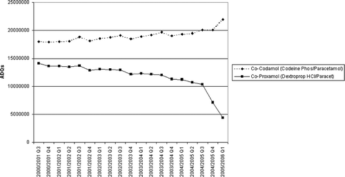
FIG. 1 Hospital admissions and poisons centre enquiries
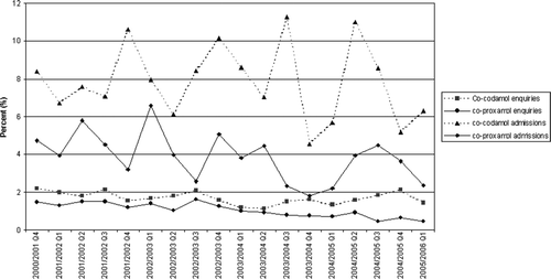
Conclusions: A gradual decline in co-proxamol prescribing in the North of UK has occurred since January 2001 and this appears to be associated with reductions in poisons enquiries and hospital admissions. Since October 2004 there has been a marked decrease in co-proxamol prescribing but it is too early to demonstrate the effect of changes in prescribing practice on the use of co-proxamol in overdose. Reference: 1. Committee on Safety of Medicines (2005). Withdrawal of Co-proxamol products and interim prescribing information CEM/CMO/2005/2.
173 Comparison of the Circumstances of Pesticide Exposures and the Perceived Seriousness of the Incident, Results from Toxbase Online Surveillance Project
Adams RA, Good AM, Bateman DN. NPIS Edinburgh, Scottish Poisons Information Bureau, Royal Infirmary of Edinburgh, UK.
Objectives: To evaluate the relationship between perceived seriousness of pesticide exposures with actual symptoms displayed, type of exposure and class of pesticide. Methods: All patient related accesses to pesticides of interest (174 pesticides and products) on TOXBASE (UK clinical toxicology database) between 1 April 2004 and 30 September 2005 were automatically notified to NPIS, Edinburgh in real time as part of a pilot project. Users accessing these pesticides were requested to complete an online form or a postal questionnaire. All pesticide enquiries to the NPIS, Edinburgh telephone enquiry service were followed up by a postal questionnaire to obtain further details about the incident. Enquiries from outside the UK, those involving animals and those involving deliberate self-harm were excluded. Data was analysed for perceived severity by respondent, class of product and type of exposure for adults and children. Results: Thirty-three telephone enquiry follow-ups, 355 electronic follow-ups and 658 postal follow-ups were available for analysis (1,046). There were 467 adults and 521 children (<13 yrs), 58 age not known. 295 (28.2%) of patients were exposed while using the product themselves. 143 (13.7%) were exposed while another person was using the product, 211 (20.2%, of which 64.9% children) involved later exposure eg. slug pullets, ant killer, rat poison. 144 (13.8%, of which 82.6% children) involved unsatisfactory storage (Note not = 100% as respondent could select no or multiple options). Overall severity ratings – 322 (72.5% of those who answered) were considered minor (51.9% no symptoms), 61 (13.7%) were considered moderate (34.4% no symptoms), 5 (1.1%) were considered major (all symptomatic), 56 (12.6%) were considered uncertain (41% no symptoms), no severity recorded 602, (55.6% no symptoms). For adults exposed to professional products (72) – 4.2% were considered major (all symptomatic), 18.1% moderate (92.3% symptomatic) 58.3% minor (88.1% symptomatic) and 19.4% uncertain (85.7% symptomatic). For adults exposed to home/garden products (123) – 0 were considered major, 13.8% moderate (all symptomatic), 76.4% minor (73.4% symptomatic), 9.8% uncertain (83.3% symptomatic). For children exposed to professional products (20) – 0 were considered major, 5.0% moderate (all symptomatic), 80.0% minor (43.7% symptomatic) and 15.0% uncertain (66.7% symptomatic). For children exposed to home/garden products (179) – 0.6% were considered major (all symptomatic), 13.4% moderate (20.8% symptomatic), 74.8% minor (17.2% symptomatic), 11.2% uncertain (20.0% symptomatic). For occupational exposures (60), -1.7% were considered major (all symptomatic), 15% moderate (all symptomatic), 66.7% minor (82.5% symptomatic) and uncertain 16.7% (80% symptomatic). Non-occupational exposure (384), 1% major (all symptomatic), moderate 13.5% (59.6% symptomatic). minor 73.4% (43.3% symptomatic), uncertain 12.0% (54.3% symptomatic) Conclusions: Exposures in children were more likely to be considered serious despite the lack of symptoms whereas in adults severity grading more closely reflected presence of symptoms. Patients involved in occupational exposures were more likely to be symptomatic than those involved in non-occupational exposures.
174 TOXBASE Enquiries about Exposures to OTC Head Lice Preparations
Good AM, Adams RA, Bateman DN. NPIS Edinburgh, Scottish Poisons Information Bureau, Royal Infirmary of Edinburgh, UK.
Objectives: To investigate the nature of enquiries concerning over-the-counter (OTC) head lice preparations in the UK. Background: In April 2004 NPIS, Edinburgh instituted an alert system on TOXBASE, the NPIS Internet database, as part of a research project to improve the reporting of pesticide related incidents. As a result a number of reports of exposure to head lice preparations containing pesticides were collected. In the UK, head lice (Paediculus humanus capitis) infestations are usually treated with OTC preparations containing organophosphorus (malathion) or pyrethroid (permethrin or phenothrin) insecticides in the form or aqueous liquids, creams, alcoholic lotions or shampoos. Carbamate (carbaryl) preparations are prescription only, infrequently used and not included in this study. Methods: Accesses on TOXBASE to specified pesticide products are automatically notified to NPIS, Edinburgh in real time. Users accessing these pesticides on TOXBASE for a patient related enquiry are invited to complete an on-line form or, if no form is received, are sent a postal questionnaire. Results: For the period 1 April 2004 to 31 October 2005 103 completed questionnaires about exposures to head lice preparations were received. The products involved contained malathion (92), phenothrin (8) and permethrin (3). 84 patients (83%) were aged less than 10 years, 7 aged 10–19 years, 8 aged 20–69 years and 2 70+ (2 not known). 66 (64%) were female, 37 (36%) male. Most exposures were accidental. Two were due to deliberate self-harm in adult females, both taking a malathion containing preparation (12 mL; 25 mL). Neither had symptoms. In 90 replies it was possible to determine cause: 25 were due to poor supervision and/or unsatisfactory storage; 23 accidental exposure (mouth, skin, eye) during normal use; 18 unspecified accidental; in 15 cases the product was given or taken orally in mistake for other medication. Three enquiries were considered unrelated to use, 3 related to adverse effects, 2 mistakes in application and one to chronic use. Most exposures were considered by the respondents to be of low toxicity, with only one moderate, involving red painful eyes. In 70 incidents (68%) there were no symptoms. Features in others included eye irritation (11), gastrointestinal symptoms (7), skin rash/burns/blisters (5), burning mouth/throat (3). 30 of 35 who replied to the question on treatment said that no specific treatment was required. The only treatments mentioned were for eye and skin contact. Conclusions: Exposures to head lice preparations do not usually result in severe symptoms. Care while using the products, in storage, in supervision of children and care in ensuring that the correct medication is being given or taken would reduce the number of exposures considerably. Data collection using this approach seems feasible and potentially useful in monitoring such products.
175 Ethylene Glycol Poisoning: A 5-Year European Toxicological Information Centres Survey
Krenová M, Pelclová D. Toxicological Information Centre, Charles University, General University Hospital, Prague, Czech Republic.
Objective: In 2003, the American Association of Poison Control Centres Toxic Exposure Surveillance System received 5,081 reports of ethylene glycol (EG) ingestion, 16 of which related to fatalities (1). Nevertheless, there is not a similar surveillance system in Europe. The objective of our survey was to obtain data concerning EG poisoning during a 5-year period from European Toxicological Information Centres (TICs). Methods: In the summer 2005 a questionnaire has been sent by e-mail to 67 European TICs in 32 countries. Following questions were asked: 1. How many human cases of EG poisoning have you been dealing with during the last 5 years (2000–2004) in total (fatal and surviving)? 2. What percentages of cases were due to EG antifreeze fluids? 3. Is there any law or other restriction of using and selling EG antifreeze products in your country? 4. Is fomepizole available in your country for treatment of EG poisonings? Results: During the four-month period 14 (20.9%) TICs from 10 countries responded (). They reported 2292 cases of EG poisoning; 95% cases were due to ingestion of EG antifreeze. Twenty-nine fatalities were recorded. There are no restrictions concerning EG products in any country. In all but two countries fomepizole is available, but ethanol is still the most commonly used antidote. Conclusion: According to the replies EG poisonings belong to frequent causes of poisoning in the European countries. However, TICs do not receive all discharge summaries from hospitals. The true number of severe poisoning is probably higher. Limitations of the study are: an e-mail study, retrospective data, and no detailed clinical data. Reference: 1. Watson WA, Litovitz TL, Klein-Schwartz W et al. 2003 Annual report of the American Association of Poison Control Centers Toxic Exposure Surveillance System. Am J Emerg Med 2004; 22:335–404.
TABLE 1 Overview of EG poisonings in the European states
176 Changing Pattern of Poisoning in Ireland Over 40 Years
Tracey JA. National Poisons Information Centre, Beaumont Hospital, Dublin, Ireland.
Objective: To analyse changing pattern of poisoning in Ireland. Case Series: A review was carried out of 40 years data accumulated since the foundation of the (NPIC) to identify changes in agents taken and in fatalities from poisoning. The NPIC was opened in Dublin in 1966 and produced its first report the following year for its initial 6 months activity, with a total of 209 enquiries. The following year (1967) there were 869 calls to the Centre and 11 fatal cases. 2/3 of the calls concerned children and 1/3 concerned adults with 60% being defined a deliberate. The annual reports continued to recorded more detail with the 1969 report (total 1649 calls) giving a breakdown on agents into Drugs (892), Household (505), Plants and Agriculture products (112). The drugs were further divided into subgroups of barbiturates (39), aspirin (118), hypnotics (47), psychotropics (243) and Iron (27). There were a large numbers of fatalities reported in these early years with paraquat featuring prominently, in a number of cases secondary to decanting. In 1974, a coding system was introduced and details were collected on 28 different medications. By 1977 the Centre was generating computerised reports with detailed age breakdown. Numbers of calls to the Centre continued to rise over the next 30 years finally levelling off in 2001 and decreasing after that, probably secondary to the introduction of TOXBASE to Emergency Departments throughout the country. In the 39 years (1966–2004) there were a total of 295,477 calls to the Centre with 747 fatalities with an average of 19 fatal cases per annum. Of these fatalities 315 were due to Paraquat ingestion. Looking at the data on fatalities highlights the changing patterns in poisoning. In 1967 out of a total of 869 calls there were 11 fatalities. The agents involved were drugs, (2) both barbiturates, household (2), town gas and bromadiolone rat poison, Industrial (1) occupational Argon gas exposure and Agriculture products (6), three of paraquat ingestion, (including one accidental poisoning in a child) and the other three took metaldehyde, mortweed (2,4D) and sodium chlorate respectively. In 2004, out of total of 13,000 calls, there were 14 fatalities, eight involved drugs (including co-proxamol, TCA's, venlafaxine and Paracetamol) three involved paraquat (again one accidental poisoning in a 3-year-old), one household product (Fairy Liquid) and two from drugs of abuse (Ecstasy, GHB). Conclusion: Over 40 year the number of calls to the NPIC have increased dramatically but, the number of fatal cases each year remain approximately the same. Reference: Woodcock JA, The Poisons Information Centre, Journal Irish Medical Association 1968; 61:439–441.
TABLE 1
177 Use of the Internet Poisons Information Database TOXBASE by Pharmacists in the UK
Good AM, Gordon LD, Bateman DN. NPIS Edinburgh, Scottish Poisons Information Bureau, Royal Infirmary of Edinburgh, UK.
Objective: To investigate use of an Internet clinical toxicology database (TOXBASE) by pharmacists. Background: The UK Medicines Information (MI) guidelines (1) state that poisons enquiries should be referred directly to the National Poisons Information Service (NPIS). Nevertheless, 7.5% of registrations for use of TOXBASE, the UK NPIS Internet poisons database are from pharmacy staffed departments. Method: Postal questionnaires were sent to 377 registered pharmacist users of TOXBASE asking about their service, poisons information resources available, and use of and satisfaction with TOXBASE. For some questions a scale of 1–6 was used with 6 being greatest satisfaction. Means and interquartile ranges were calculated. TOXBASE usage statistics for the period 1/1/4–31/8/5 were analysed to confirm what information these users most frequently access. Results: In the time period studied pharmacy users accounted for 5.1% of total (652,215) database log-ons, individual departments logging onto the database 1–777 times (median 39, average 88). 207 completed responses were received (54.9%) from hospital departments incorporating both pharmacy and MI (85), hospital pharmacy only (32), MI only (58), Medicines Management / Pharmaceutical Advisor teams (23), others (9). Users reported they accessed TOXBASE “most days” (27–13.1%), “once a week” (54–26.1%), “once a month” (43–20.7%), “less frequently” (75–36.3%). Units also contacted the NPIS telephone service (40.6%), British National Formulary (68.2%), other databases and on-line resources (19.3%), books (10.6%) and other sources (8.7%). Other sources included local emergency departments, regional MI units, and manufacturers. 133 (64.3%) of users reported referring NHS poisons enquiries to the NPIS telephone line; 43.9% used TOXBASE themselves for answering poisons enquiries; 17.8% would telephone NPIS for advice; 24.2% referred NHS callers to TOXBASE. Units accessed TOXBASE most frequently for teratology information (56.1%), poisons information (15.9%) and antidote information (7.3%). TOXBASE was generally considered “easy to access” (median 6, interquartile range 5–6); with sufficient information (median 5, range 5–5); overall satisfaction scored median 5 (range 5–6). Most useful items were teratology (50.3%); poisons monographs (14.5%); both (24.5%); and antidotes (13.5%). Of the 76 suggestions for additions to TOXBASE 90% concerned teratology information; 51 additional monographs; 31 more frequently updated information; and 17 more detailed information on the management of common conditions in pregnancy. Top 5 accesses on TOXBASE were use in pregnancy of antibiotics, antiemetics, fluoxetine, amitriptyline and “antidepressants – neonatal withdrawal syndrome.” Conclusions: Most pharmacist TOXBASE users do not routinely receive acute poisons enquiries. When poisons enquiries are received pharmacists will often use TOXBASE to answer simple queries but refer more enquirers about more severe poisonings to TOXBASE or directly to the 24 hour NPIS telephone service. Pharmacists use TOXBASE primarily as a source of teratology information, and would like to see the range of teratology information on TOXBASE increased. Reference: 1. UK MI training workbook. http://www.ukmi.nhs.uk.
178 Herbal Products: Opinions, Perceptions and Behaviours of Callers to the New Zealand National Poisons Centre
Lee J (1), Temple WA (2), Smith NA (3). 1. Department of Pharmacology and Toxicology, University of Otago, Dunedin, New Zealand; 2. National Poisons Centre, University of Otago, Dunedin, New Zealand; 3. School of Pharmacy, Griffith University, Gold Coast, Australia.
Objective: Consumers often view herbal products as natural, safe and non-toxic. This study followed-up general public callers to the National Poisons Centre (NPC) regarding their herbal product (HP) exposure. It investigated their opinions, perceptions and behaviours concerning the use and storage of HPs; how well the NPC handled HP enquiries; and why HP use and adverse reactions were underreported. Methods: Follow-up telephone calls using a standardized questionnaire were made to the 98 general public callers who contacted the NPC between July 2002 and November 2003 regarding exposure to a HP. Results: The NPC receives approximately 10 herbal inquiries each month; most involve acute poisonings following child home exploration, with 20% referred for medical treatment. Of the 60 survey respondents, 95% were female, 73% were aged 21–40 years, one-third were University educated and most (93%) were and New Zealand European. Two-thirds of respondents recalled receiving no advice when purchasing HPs, consistent with almost half obtaining HPs from outlets such as supermarkets. HPs were used primarily in disease prevention (66.7%). Most respondents (58.3%) did not believe that HPs were more efficacious than conventional medicine (CM), but favoured HPs for their perceived safety. More that half (55%) combined HPs with CM to increase efficacy, compared to using either independently. Nearly all (86%) HPs were in bottles but few (15%) used child safety packaging. Whilst 43% of HPs were stored with CM few (13%) of HPs were actually locked away. Most respondents were willing to tell health professionals about their HP use and adverse reactions, if asked. Respondents considered NPC advice to be very useful (70%), sufficient in quantity (63%) and very clear (62%). Almost all (98%) were satisfied with the NPC service and would recommend it to others. Conclusion: Health professionals should discuss safe use and storage of HPs with patients, and caution them against over-estimating HP safety. Child safety packaging for HPs needs wider promotion. The increasing popularity of HP use is an indication for the NPC database to enhance its information on HPs.
179 Drug Exposures in Children – A Follow-Up of Drug Exposures in Small Children in Cases Where the Poisons Information Centre was Contacted
Torbergsen TE (1), Muan B (2), von Krogh A (2), Andrew E (2). 1. Department of Pharmacy, University of Tromsø, Norway; 2. Department for Poisons Information, Directorate for Health and Social Affairs, Oslo, Norway.
Objective: Little is known about the course and outcome of unintentional drug exposures in children in Norway. The national Poisons Information Centre (PIC) receives about 4000 of these calls per year. The present study was undertaken to 1) survey what happened with the exposed children after the PIC had been contacted, 2) assess whether the recommendation given by the PIC was followed, and 3) assess the callers’ degree of satisfaction with the service given by the PIC. Methods: Callers who contacted the PIC regarding children under 5 years of age, who had been unintentionally exposed for drugs May–June 2004, were asked to participate in the study. A student without affiliations to the PIC conducted the interviews 2–5 days after the call to the PIC; posing standardized questions about type of drug, development of symptoms and the caller's degree of satisfaction. Data recorded routinely during the consultation regarding the circumstances of the exposure, risk classification and recommended treatment, were also included in the study. Results: 200 callers were included in the study, comprising 211 children with drug exposures. The exposure was due to an accident in 201 cases and to erroneous administration in 10 cases. Fluoride-tablets, paracetamol, antihistamines, local anaesthetics, NSAIDs and cough preparations were the most frequent drug-exposures. The PIC assessed the risk as follows: poisoning unlikely in 163 cases, minor poisoning possible in 30 cases, moderate poisoning possible in 5 cases and severe poisoning possible in 3 cases. 44 of the children developed symptoms, mostly caused by fluoride-tablets. None of the symptoms observed were surprising given the drug-exposure. Paracetamol was the most frequent cause of potentially serious poisonings. The recommendations given by the PIC were: no treatment in 133 cases, home treatment in 69 cases, treatment by a general practitioner in 4 cases and hospital treatment in 5 cases. These recommendations were followed in 206 of the cases. The callers’ satisfaction with the PIC was very good with respect to communication, friendliness and inspiration of confidence, moderate with respect to response time. Conclusion: The drug most frequently associated with unintentional exposure and also causing symptoms most often was fluoride-tablets. The symptoms were mild and probably caused by the sorbitol in the tablets. Paracetamol caused the potentially most serious poisonings. The callers had a high compliance with the recommendations given by the PIC, and their satisfaction was good.
180 Availability of Antidotes in Acute Poisoning: System Developed by the Belgian Poisons Centre
Smet H, Daeleman W, Mostin M. National Poisons Centre, Brussels, Belgium.
Objective: To describe the development of our system to make antidotes available throughout the country. Methods: Since it's set-up in 1963 the Belgian Poisons Centre (PC) has taken on the responsibility to supply antidotes (AD). According to the law an antidote as a medicine must be registered by the Department of Health or the EMEA prior to marketing. Unfortunately a number of AD are not registered and are not commercially available. In order to make them available to any patient the following strategy has been set-up, 1) A supply of selected AD at the PC. In the case of acute poisoning we deliver the AD on prescription in a sufficient amount to start the treatment. The hospital pharmacist receives by telephone all necessary information on dose, administration and how to purchase that particular AD. 2) We have established a network of 22 collaborating hospitals all over the country. The PC keeps an overview of their available AD in a databank. It contains information on the quantity and the place in the hospital where it is stored. Ideally any change in their stock should be reported to the PC. 3) In order to make antidotes available to any pharmacist, the Belgian Association of Pharmacists (APB) keeps AD available in a central and several regional depots. This information is also entered in the database. Regular contacts between PC and APB provide them with information on AD, e.g. if new antidotes become advisable. 4) Recently we published online a list of 29 currently recommended AD. We inform healthcare professionals about indications, dose, administration and addresses of suppliers. Conclusion: The antidote system currently used in the Poisons Centre provides instant information on the location and the stock levels of antidotes in the country.
181 Pregnancy Outcome of Women Using Selective Serotonin Re-Uptake Inhibitors
Eleftheriou J, Carrara M, Bacis G. Bergamo Poison Control Center, Ospedali Riuniti, Bergamo, Italy.
Introduction: Selective Serotonin Re-Uptake Inhibitors (SSRIs) are widely used antidepressant drugs. Many studies of antidepressant use during pregnancy reported that those drugs are not associated with an increased risk of major malformations above the baseline of 1–3% in the population. Results: In 2004, Bergamo Poison Control Center, received 3050 phone calls about requests for potential effects of drugs and chemicals in pregnancy. 724 (24%) of the questions were related to drugs of the Central Nervous System and 303 patients of these (41.9%) were classified as the group exposed to antidepressant drugs. The SSRIs group was the most commonly represented: 219 patients (72%) used SSRIs during any time of the pregnancy as a monotherapy or in association with benzodiazepines and other psychotropic drugs. We completed follow-up on 106 women, with a median age of 33 (19–41) years old, exposed to SSRIs during the first trimester of pregnancy and two groups were evaluated: 1) women exposed to SSRIs (79 patients) vs. a non teratogen group and 2) those taking SSRIs and other psychotropic drugs except benzodiazepines (27 patients) vs a non teratogen group. Paroxetine and citalopram were the commonly used drugs (40 and 25 patients respectively), followed by sertraline (19), fluoxetine (19) and fluvoxamine (3). Among the outcomes of these pregnancies there were 81 live births, one ectopic pregnancy, nine pregnancies electively terminated and 15 spontaneous abortions: 12 spontaneous abortions were observed in the first group (15%) and 3 in the second one (11%). No major or minor malformations were recorded. The outcome was compared to that of 49 pregnancies with exposures known to be not teratogenic. 45 women of this group had normal deliveries and 4 (8%) spontaneous abortions were identified. Conclusion: Our data suggest these drugs do not increase the malformation rate, although are not sufficient to state that there is no risk at all, and the magnitude of spontaneous abortions risk increases in offspring exposed to polytherapy. Because of the increase in the number of spontaneous abortions observed in both antidepressant groups, additional studies are needed to separate the effects of the psychiatric condition from that of the drug therapy. Reference: Hemels ME, Einarson A, Koren G, Lanctot KL, Einarson TR: Antidepressant use during pregnancy and the rates of spontaneous abortions: a meta-analysis. Ann Pharmacother 2005; 39:803–809.
182 A New Interactive Website Improved Response to Nuclear Accidents
Leenders MEC (1,2), Riel AJHP Van (1), Groot R de (1), Kok-Palma YS (1). 1. Poisons Information Centre, National Institute for Public Health and the Environment, Bilthoven, NL; 2. Department of Anaesthesia, University Medical Hospital, Utrecht, The Netherlands.
Introduction: During the last decade safety measures for nuclear power plants have been improved. On the other hand threats in the world increased. The latter was the reason to renovate the response network for incidents with ionising radiation. A Planning and Advice network (EPAn) for immediate response in case of nuclear incidents was erected. This network is imbedded in the normal infrastructure for disaster management. The network represents several disciplines and knowledge institutes. Depending on the kind of nuclear accident the EPAn is confronted with, different disciplines or knowledge institutes are contacted. Information and knowledge within the network is shared via a newly developed, secured interactive website, “Calweb.” There is a hierarchy in the way information is concentrated in order to advice the government. Support centres gather their information and concentrate this to a manageable amount of data. These data are transferred to back offices. The poisons centre is one of the back-offices. Via the interactive website exchange of information takes place on, e.g. number and kind of casualties, water and food contamination, real time weather conditions, the dissemination of radionuclides etc. Besides text, maps of the actual weather condition, maps of possible spread of radionuclides, and video information can be transferred. The back offices concentrate the data further to condense information for the front office and the governmental policy teams. These teams decide about the measurements to be taken. Their decisions are also disseminated via the interactive website to the local authorities, responsible for the execution of the measurements. In May 2005, this interactive website was tested in a national nuclear accident exercise. Over 1,100 administrators, officials and relief workers from municipalities, provinces, ministries and emergency services were involved to deal with an accident at a nuclear power plant. All day a realistic scenario was unrolled, with the use of the interactive website. The exercise gave a good impression of the feasibility of the diverse measures and the time needed to implement them. Conclusion: The EPAn structure, the interactive website and the exercise gave an enormous impulse to all institutions involved to further improve their preparation for nuclear accidents. Calweb proved to be a very good tool to upgrade the responsiness to nuclear accidents.
183 No Hope? Intoxications with High Mortality Rate
Pap C (1), Karteszi M (2). 1. Department of Emergency Medicine and Clinical Toxicology, Péterfy S. Hospital, Budapest, Hungary; 2. Department of Emergency Medicine and Clinical Toxicology, Péterfy S. Hospital, Budapest, Hungary.
Objective: Nowadays the majority of intoxications have a good prognosis. However, there are poisonings with high mortality rate despite our therapeutic efforts, perhaps due to the lack of appropriate antidote therapy. Case Series: Based on data of patients who died at our department during the last 10 years we revealed the toxins which might have caused fatal outcome directly. Patients who died in consequence of direct effects of the toxic substances were included. The exclusion criteria were the following: death because of any complication (pneumonia, cerebral hypoxia etc.) not caused by direct toxicity of the poison, and death after 3 days of hospitalization (except for paraquat). Afterwards, based on data of all patients treated at our department during that period we determined the mortality rate for each type of intoxication. Results: A total of 1,209 patients died at our department over this period and 276 (22.8%) of patients were included in this study. Paraquat showed the highest mortality (21 deaths/22 cases, 95.4%) followed by hydrogen fluoride (5/10, 50%) and verapamil (21/51, 41.2%). Organophosphates (38/200, 19%), ethyleneglycol (43/323, 13.3%), digoxin (8/69, 11.6%) and antidepressants (28/1927, 1.4%) seemed to be less fatal. There were some intoxications with a mortality of 100% (barium polysulfide, 2 deaths/2 cases and calomel, 2 deaths/2 cases) but the number of these cases is very small for an appropriate analysis. Conclusion: Nowadays in Hungary the intoxications caused by paraquat (the occurrence rate of which is also decreasing), hydrogen fluoride and verapamil have the highest mortality rate of all poisonings. Early diagnosis, adequate risk-assessment and new therapeutic approaches are required to decrease the number of deaths caused by these toxins.
184 Outreach to the Spanish Speaking Constituency: Language Program Results After 4 Years
Gracia R (1,2), Ray M (2), Ramirez J (2), Yudizky M (2). 1. The University of Texas Southwestern Medical Center, Dallas, Texas, USA; 2. North Texas Poison Center, Dallas, Texas, USA.
Objective: The purpose of this longitudinal study was to develop and evaluate methods in a Spanish language Poison Prevention and Awareness program. An estimated 10% of our designated poison center service area is “Spanish-only” or cannot speak English very well. Previous reports address the need to target “Spanish-only” speaking individuals in Poison Center interventions. We present a comparison of techniques used for a language outreach program spanning a four year time period. Methods: Methodologies incorporated into the Spanish Language program progressed from Spanish printed material mail outs, stationary bill boards, and isolated television/radio news briefs to recruiting a Spanish speaking educator and incorporating Hispanic community partnerships. The program now solicits Latino health fairs and lectures, as well as utilizes more mainstream media including mass transit/mobile marketing to targeted residential areas and scheduled radio announcements on prominent entertainment platforms. Program success is measured by tracking “Spanish-only” callers to our regional poison center. This study was approved by the University's Institutional Review Board. Results: Data was complied as “Spanish-callers” in a month by call type report for all calls originating from our regional poison center area. Baseline data from the year 2000 shows minimal poison center utilization by Spanish speakers; there were no documented Spanish callers and less than 100 Spanish material mail-outs. The Spanish Language program was initiated in 2001 with 8,144 Spanish material mail outs and a subsequent increase in Spanish calls to 118 (0.2% call volume). Spanish radio and television news briefs resulted in limited peaks of increased utilization. Involvement of Hispanic community partnerships in 2004 and the addition of a Spanish speaking educator in 2005 were key to understanding how to best reach Spanish speaking communities. After progression to mass transit/mobile marketing, a greater than 50% rise in poison center utilization was noted. Data from 2005 demonstrates a now thriving Spanish Language program with 723 (0.7% call volume) calls documented. Conclusion: Results from our experience suggest that outreach to Spanish speaking communities is most effective when provided in more mainstream media forms. Distribution of Spanish printed materials without direct teaching does not appear to be a sufficient means of outreach. Since call volume is not the only way to evaluate outreach programs, we are currently in the process of developing community surveys as an additional measure of program performance. In effort to improve outreach to underutilized populations, we recommend meeting with community leaders and representatives to identify optimal program methodologies as well as incorporating evaluation techniques to indicate progress.
185 Acute Poisonings Treated in Serbia National Poison Control Centre
Todorovic V, Jovic-Stosic J, Joksovic D, Jovanovic D, Šegrt Z. National Poison Control Centre, Military Medical Academy, Belgrade, Serbia & Montenegro.
Objective: Clinic of Emergency and Clinical Toxicology is the part of National Poison Control Centre and it is the single institution in Serbia specialized for the treatment of acute poisonings. The aim of this study was the estimation of the incidence and severity of acute poisonings in our country. Methods: Retrospective analysis of patients treated during one year period. The severity of poisoning was estimated by Poisoning Severity Score (PSS). Results: The exact data were obtained for the area of about 2 millions inhabitants (Belgrade and surroundings). During follow up period 3,039 patients (15 per 10,000) were treated for acute poisoning. Most cases were managed in emergency room (69.6%) as the outpatients, while 30.4% were admitted to hospital for further treatment. Distribution of patients according to causative agent is shown in . In the group of admitted patients 2% were asymptomatic (PSS 0), 49% had mild intoxication (PSS 1), 23% reached PSS 2 and 26% were severely poisoned (PSS 3). All fatalities were among the PSS 3 group, and the total mortality was 5%. Conclusion: Incidence of acute poisonings in Serbia is not high, but it is probably underestimated due to the lack of proper recording. Drugs are the most common cause of acute poisoning among hospitalized patients as well as among the outpatients.
TABLE 1
186 Challenging an Expert – Driven Organization to Adopt a Consumer – Focused Communications Strategy
Simeonov IM (1,2), Heard SE (1,2). 1. California Poison Control System, San Francisco, CA, USA; 2. University of California San Francisco, School of Pharmacy, San Francisco, CA, USA.
Objective: Shift a traditional clinician-driven public health education program towards a consumer-focused model based on conventional product and service marketing. Create acceptance of this approach, which places a high emphasis on consumer beliefs, values and perceptions, within an organization of seasoned medical experts unfavorably disposed to traditional marketing principles. Methods: Listening to consumers, and being open to their criticism, helped shape a line of products and a new communications strategy for the California Poison Control System, which serves a highly diverse group of residents. Standard components of marketing, advertising and promotion (including positioning, market segmentation, creative strategy, and message design and testing) replaced a previously didactic approach, established a direct benefit for the consumer. Advance a new way of thinking about consumers among the organization's management group by using the same principles to achieve organizational goals. Establish a connection between meeting organizational goals and determining and satisfying the needs of consumers. Recognize that self-interest on the part of the consumer and the organization is critical to the process, and that a perceived benefit on either side can motivate behavioral change. Results: Consumers and health educators were receptive to new approach, tone of voice, and design of literature describing poison center services and this new model was cited as “best practices” in the Institute of Medicine report Forging a Poison Prevention and Control System. Internally, organizational conflict surfaced around consumer-driven changes considered overly simplistic and not properly reflective of the organization's professional status. A conflict resolution strategy encouraging increased participation by clinician-managers was adopted. Conclusion: Acknowledge the legitimate role of self-interest in forging an exchange between the organization and its constituency. Each party can advance its own interest while accommodating the other's and achieve its own goals. Shifting attention from an organization's needs to the audience's is best achieved by underscoring the benefits and understanding that what the public will or will not accept is critical in achieving our own goals of providing a health advice service dependent on voluntary use.
187 Epidemiology of Toxic Deaths in a Medical Intensive Care Unit in France
Mégarbane B (1,2), Belal-Rekkab D (1), Résière D (1), Karyo S (1), Deye N (1), Rettab S (1), Guerrier G (1), Baud F (1,2). 1. Réanimation Médicale et Toxicologique, Hôpital Lariboisière, Paris, France; 2. INSERM U705, Hôpital Fernand Widal, Paris, France.
Objectives: Acute poisonings are still responsible of a significant mortality rate (2,600 deaths per year in France), despite an optimal management in Intensive Care Unit (ICU). Our objectives were to prospectively evaluate the circumstances and etiologies of toxic deaths following admission to a toxicology-specialized ICU and to analyze the evolution in relation to time of etiologies and treatments. Methods: Prospective study over 8 years (1998–2005) of all toxic deaths in a toxicological ICU; clinical and toxicological data collection on admission and during ICU stay; result presentation as median [10%-90% percentiles]; patient comparisons using Chi-2 and Mann-Withney tests. Results: During an 8-year-period, 83 patients (49 F/34 M, age: 46 years [26–82], SAPS II: 73 [42–93]) died in our ICU in relation to a voluntary (87%) or an accidental (13%) acute drug ingestion. Among these patients, 57% had significant psychiatric past histories (depression: 52% or psychosis: 5%), 12% were drug-users, and 2% chronic alcoholic. The main ingested drugs were cardiotropic drugs [50%, including chloroquine (17%), antidysrhythmics (8%), beta-receptor blockers (10%), calcium-channel antagonists (7%), digitalis glycosides (5%), or colchicine (4%)] or psychotropic drugs [36%, including benzodiazepines (14%), cyclic antidepressants (12%), neuroleptic agents (6%), drugs of abuse (6%), or meprobamate (4%)]. A cardiac arrest was observed on the scene where the patient was found (29%), or secondarily in emergency room or ICU (47%). On admission, arterial pH was 7.29 [7.03–7.47], plasma lactate 6.8 mmol/l [1.9–19.9], PaCO2 42 mmHg [30–63] and PaO2/FiO2 ratio 237 mmHg [63–516]. Supportive treatments were associated with antidotes (43%) and since 2003, with extracorporeal life support (20%). Death occurred 2 days [1–3] after ICU admission, following multiorgan failure (42%), cerebral anoxia (20%), sepsis (13%), persistent asystole (7%), refractory ventricular fibrillation (5%), hemorrhage (5%), pulmonary embolism (5%), acute respiratory distress syndrome (2%), or coronary spasm (1%). Comparative analysis showed no significant modification of the number of deaths and the nature of intoxicants in relation to time. Conclusions: Acute poisonings are responsible in ICU of an important and incompressible mortality rate (around 3%), mainly related to the ingestion of cardiotropic drugs following a suicidal attempt. To improve management and prognosis, it is mandatory to develop not only in France but also in Europe a registry of toxic deaths in ICU.
188 Alcohol Intoxication in Children and Adolescents Requiring Hospital Admission in 13 Major Children's Hospitals in Germany Between 2000–2002
Meyer S (1,2), Steiner M (3), Wurm D (1,2), Nunold H (2), Gortner L (1,2). 1. Dept. of Pediatrics/Pediatric Intensive Care, University Children's Hospital of Saarland, Homburg/Sa; 2. Poison Control Center, University Children's Hospital of Saarland, Homburg/Saar, Germany; 3. Prognos AG, Basel Germany.
Objective: Drug and alcohol (C2H5OH) abuse is a common phenomena in children and adolescents throughout the world. However, precise epidemiological data on acute C2H5OH intoxication in children and adolescents requiring hospital admission are rare (1,2). Methods: An ex-post analysis was performed to assess the number of children and adolescents (age 10–17 years) with acute C2H5OH intoxications who required in-patient treatment (24 hours) in 13 major children hospitals in Germany between 2000 and 2002. The diagnosis of C2H5OH intoxications was made on the basis of the discharge summary of the participating hospitals, and was defined according to the International Classification of Diseases (ICD)-10. Results: The number of children and adolescents with acute C2H5OH intoxications requiring in-patient hospital treatment increased from 227 in 2000 to 313 in 2001 (+38%) and 350 in 2002 (+12%). The percentage of female patients increased from 34.1% in 2000 to 41.9% in 2001 and 49.8% in 2002. As far as age is concerned the most significant increase in intoxicated patients was seen in adolescents aged 15–17 years. Mean time spent in the hospital was 1.5 days (range: 24 h –>4 days). In conjunction with data from federal German health institutes, the overall annual incidence of acute C2H5OH intoxications requiring hospital admission in children and adolescents age 10–19 years in Germany can be estimated to be 1/1000. Conclusions: Our study provides epidemiological data on the incidence of alcohol abuse in children and adolescents. It demonstrates an increase in the incidence of acute C2H5OH intoxications requiring hospital admission in children and adolescents in Germany between 2000 and 2002. Apparently, gender differences seem to play a minor role in alcohol abuse. Our data may be indicative of an overall rise in alcohol consumption and abuse in this specific age cohort. Specific intervention programmes including short-term (eg, increasing awareness of potential harm of alcohol) and long-term measures (eg, legislation) are mandatory. References: 1. Woolfenden S, Dosseter D, Williams K. Children and adolescents with acute alcohol intoxication/self-poisoning presenting to the emergency department. Arch Pediatr Adolesc Med 2002; 156:345–348. 2. O'Farrell, Allwright S, Downey J, et al. The burden of alcohol misuse on emergency in-patient hospital admissions among residents from a health board region in Ireland. Addiction 2004; 99:1279–1285.
189 Paradoxical Reaction to Benzodiazepines Reversed by Flumazenil
Yadav K (1), Khodorkovsky B (2), Su M (1). 1. Emergency Medicine, State University of New York Downstate Medical Center, Brooklyn, NY, USA; 2. Emergency Medicine, Staten Island University Hospital, Staten Island, NY, USA.
Objective: Benzodiazepines are sedative-hypnotic agents commonly used in pediatric sedation. Effects include anxiolysis, sedation, and antegrade amnesia. Rarely, benzodiazepines produce a paradoxical reaction, characterized by emotional instability, aggressive behavior, and excessive movement (1). Without a clear cause or treatment for this phenomenon (2), we describe an approach to management of this condition in children. Case Report: A 20-kilogram 5-year-old male with history of attention deficit hyperactivity disorder and expressive language disorder presented to the emergency department (ED) for evaluation of a fall in school. His initial vital signs were within normal limits. The patient was unable to give a clear history because of his language disorder. In the presence of the school aide, the patient was awake, alert and in no acute distress, but during physical examination, he was uncooperative. There was a 1 cm linear laceration to his right upper lip violating the vermillion border. The rest of his exam, including spine, chest, heart, abdomen, extremities, and neurologic system, were unremarkable. To assess for traumatic brain injury, the patient was to undergo a computed tomography (CT) scan of the brain. To avoid patient distress, he was administered 1 mg of lorazepam intravenously (IV) 10 minutes prior to performing the CT scan (T0 min). Another 1 mg of lorazepam IV was given just before the CT scan (T10 min). The patient became very agitated, and was kicking and screaming despite the arrival of his father. An additional 2 mg of midazolam was administered IV (T30 min), however the patient remained aggressive and uncontrollable. Upon returning to the ED, the patient was placed in soft restraints (T55 min) and received another 2 mg of lorazepam (T65 min). His agitated state did not improve and his father was unable to control his behavior when left alone with the patient. He was then administered 0.01 mg/kg flumazenil, or 0.2 mg IV (T105 min). Within minutes of administration (T115 min), his positive symptoms ceased and he became sedated. Subsequently, the patient had successful completion of the CT scan, which demonstrated no evidence of intracranial bleed, mass, shift, edema or fractures. He returned to his mental status baseline and went on to receive ketamine IV as procedural sedation for primary repair of his laceration. He was observed in ED for 8 hours total and discharged home without complications. Conclusion: Optimal management of paradoxical reactions to benzodiazepines is not known. Our case report suggests that low-dose flumanzenil may be effective in treating the positive effects while allowing sedation to set in. Further study is needed. References: 1. Hall RW, Zisook S. Paradoxical reactions to benzodiazepines. Br J Clin Pharmacol 1981; 11:99–104S. 2. Mancuso CE, Tanzi MG, Gabay M. Paradoxical reactions to benzodiazepines: literature review and treatment options. Pharmacotherapy. 2004; 24:1177–1185.
190 Veterninary Needlestick Injuries Reported to the National Poisons Information Service (Cardiff Centre)
Brooke N, Davies JT, Cooper GA, Thompson JP. National Poisons Information Service (Cardiff Centre), Llandough Hospital, Penarth, UK.
Objective: To determine the number of telephone enquiries made to the NPIS (Cardiff Centre) concerning accidental needle stick injuries with veterinary medicines. Methods: Telephone enquiries to NPIS Cardiff are recorded on a standardised database (UKPID). This database was searched to identify enquiries made concerning accidental injection, and in particular accidental injection involving the use of veterinary medicines between 1998 and 2003. Results: A total of over 198,000 telephone enquiries were received during the six year period 1998 to 2003. 1,835 enquiries (0.9%) involved accidental needle stick injuries and 298 of these (16%) involved veterinary medicines. Calls were received from accident and emergency departments, general practitioners’ surgeries, veterinary practices and NHS Direct, a public access health advice service. Many calls involved products with low inherent toxicity and produced only minor ill-effects. However, 31 calls involving 27 patients concerned the use of timicosin, a veterinary antibiotic which has been reported in association with death following accidental injection of small amounts. Three telephone calls involved etorphine, an opioid sedative. Discussion Injecting animals with veterinary medicines carries the risk of accidental self injection, or injection of a person helping restrain the animal. This may result in ill-health either as a result of local damage at the site of injection, absorption of the medicine into the circulation, or infection transmitted to the operator from a previously used dirty needle. Injectable veterinary medicines may contain mineral oils which can cause severe local tissue damage, especially if injected into a digit. Some veterinary medicines are well recognised to be toxic systemically (e.g. etorphine) and should only be used with an assistant who is capable of administering a reversing agent present. Few calls were received about accidental injection with this agent. However, other medicines (e.g. tilmicosin) may be potentially fatal without their potential toxicity being widely appreciated and more calls were received about this medicine than for etorphine. Concern has been expressed about the need to be aware of the potential seriousness of accidental injection of veterinary medicines and the need to report such incidents to national pharmacovigilance schemes1. Incidents happen regularly: 28% of suspected human adverse reactions to veterinary antimicrobial products reported since 1985 to the Veterinary Medicines Directorate were due to accidental injection2 and underreporting occurs, weakening the strength of such schemes. Conclusion: Accidental injection with veterinary medicines occurs regularly and may be serious. Operators need to be aware of the risks involved and the need to report incidents. References: 1. Skilton D, Thompson JP. Needlestick injuries. Vet Rec 2005; 156:522. 2. Appraisal Panel for Suspected Adverse Reactions to Veterinary Medicines, Annual Report 2004, Veterinary Medicines Directorate. http://www.vpc.gov.uk/reports/vpcapar04.pdf.
191 Longitudinal Trends in the Incidence of Organophosphate Exposures in the United States
Sudakin DL, Power LE. Department of Environmental and Molecular Toxicology, Oregon State University, Corvallis OR, USA.
Objective: The implementation of the Food Quality Protection Act of 1996 resulted in a decision by the United States Environmental Protection Agency to phase out and eliminate the use of organophosphate insecticides in residential environments. The decision was based, in part, upon concerns with respect to reducing exposure to sensitive populations, including infants and children. The process of phasing out residential uses of organophosphates began in the year 2000, and concluded at the end of 2005. The purpose of this study was to utilize national Poison Control Center statistics to investigate whether the phase out of organophosphates from residential uses has affected the incidence of organophosphate exposures in the United States. Methods: Insecticide and organophosphate exposure data were extracted from Annual Reports of the American Association of Poison Control Centers Toxic Exposure Surveillance System (TESS) for the years 1995–2004. Organophosphates incidents were examined by total exposures for each year, and stratified by age range, and reason (unintentional and intentional). Organophosphate incidents were also examined as a proportion of all insecticide exposures for each year. Results: summarizes the organophosphate incident data from 1995–2004. There was a statistically significant reduction in the average number of exposure incidents involving organophosphates when comparing the time periods before (1995–1999) and after (2000–2004) their phase out from residential use (Mann-Whitney, p = 0.008). The percentage of organophosphate incidents among ages less than six, between six and nineteen, and greater than nineteen years decreased 70, 59, and 43 percent, respectively, when comparing incident statistics from 1995 and 2004. There was a substantial decrease in both intentional and unintentional incidents involving organophosphates from 1995–2004. The proportion of all insecticide exposure incidents involving organophosphates consistently decreased for each year from 1995 (33.0%) to 2004 (13.3%). Conclusion: TESS data demonstrated a clear and consistent decrease in incident cases involving organophosphates in association with their phase out from residential uses. This effect was observed across all age categories, but was most clearly demonstrated among children under 6.
TABLE 1 Organophosphate exposures in the United States (TESS Data, 1995–2004)
192 Prevalence, Pattern, and Outcome of Adverse Drug Events Leading to Emergency Department Visit
Yang CC (1,2), Yen HJ (2,3), Kao WF (2,3). 1. Department of Medicine, Taipei Veterans General Hospital, Taipei, Taiwan; 2. Faculty of Medicine, National Yang-Ming University, Taipei, Taiwan; 3. Department of Emergency Medicine, Taipei Veterans General Hospital, Taipei, Taiwan.
Objective: Adverse drug events (ADEs) due to medical error or drug interaction are common among both outpatients and inpatients, and relevant research works regarding medical error/potential drug interaction are markedly increasing in recent years. Although some studies had previously focused on potential drug-drug interactions at the emergency department (ED), there are few, if any, studies that have specifically looked at the pattern and outcome of ADEs among patients who visit EDs. Methods: We conducted a study using data from the Taipei Veterans General Hospital. The medical charts of all patients who visited the medical ED of the Taipei Veterans General Hospital from March 1, 2004 through August 15, 2004 were reviewed. Cases with ADEs were identified after the diagnosis was agreed by at least 2 researchers. Information on relevant variables was collected by reviewing the medical charts and by using a telephone interview. Descriptive data on the prevalence, pattern, and outcome of ADEs among patients who visited the medical ED was then calculated and reported. Results: During the study period, 534 cases (3.5%) were identified from a total of 15,438 ED patients. Among the 534 cases, most of the ADEs were related to the use of hypoglycemic agents, cardiovascular medications, and analgesics. Males (63.5%) outnumbered females during this period and the mean age of the 534 cases was 66.4 years. After the development of an ADE, 234 cases (43.8%) were hospitalized and remained inpatients for 1 to 131 days (median 8.5 days). Severe or fatal ADEs were noted in 102 patients (19.1%) and 13 cases (2.4%) with ADE eventually died. Four hundred and seventy-five ADES (89.0%) were classified to be definitely or possibly preventable events. Conclusion: In the study, we found that 3.5% of all medical ED visits were related to ADEs, and most ADEs were caused by the use of hypoglycemic agents and cardiovascular medications, both findings are consistent with previous literature reports on ADEs-related hospital admissions. Because most ADEs in the study were preventable and because many patients with ADEs were hospitalized, it is important for health care professional to prescribe drugs more appropriately so that the medical cost associated with ADEs could be minimized. More studies are also needed in the future to better understand the potential predictors of ADEs leading to ED visit and to adopt preventive measures.
193 Epidemiology of Drug – Associated Hypotension Reported to a Poison Control System
Wu L, Olson K, Kearney T. California Poison Control System – University of California, San Francisco, USA.
Background: Hypotension is a common presenting sign of patients whose cases are reported to the poison control center. We analyzed the causes and consequences of drug-associated hypotension over a one-year period. Methods: We conducted a retrospective review of calls to the San Francisco division of the California Poison Control Center during the year 2003 in which hypotension occurred in the setting of poisoning or drug intoxication. For each case, the poison center chart was reviewed to determine: the drug(s) involved; the degree of hypotension; treatment measures attempted and the response to treatment; and the medical outcome. Results: The total number of poison center calls associated with hypotension was 215. The leading individual drugs associated with hypotension were trazodone (20 cases, 9.3%), quetiapine (18 cases, 8.4%), ethanol (14 cases, 6.4%), acetaminophen/hydrocodone (11 cases, 5.1%), risperidone (10 cases, 4.7), and olanzapine (10 cases, 4.7%). The leading drug classes associated with hypotension were benzodiazepines (49 cases, 22.8%), atypical anti-psychotics (42 cases, 19.5%), SSRIs (29 cases, 13.5%), opioids (26 cases, 12.1%), beta-blockers (22 cases, 10.2%), calcium-channel-blockers (19%, 8.8%), and TCAs (17 cases, 7.9%). Multiple potentially hypotension-inducing drugs were involved in 53.5% of the cases. Vasopressors were used in 35 (16%) cases. Suicide attempts accounted for 133 (61.8%) of the cases. There were 17 deaths. Of these death cases, the leading drug class was opiates (5 cases, 29.4%), followed by methamphetamines (3 cases, 17.6%). Intubation (11 cases, 64.7%), dialysis (3 cases, 17.6%), and vasopressor therapy (10 cases, 58.8%) were often performed. Complications associated with hypotension and death included unstable dysrhythmias (7 cases, 41.2%) and renal failure (7 cases, 41.2%). Conclusion: Trazodone was the drug most commonly associated with hypotension. Newer anti-psychotics such as quetiapine, risperidone, and olanzapine have emerged as drugs often associated with hypotension. The drugs most commonly associated with death were opiates and methamphetamines.
194 Tricyclic Antidepressant Poisoning in Wales 1999–2003
Wood KL, Thompson JP. National Poisons Information Service (Cardiff Centre), Llandough Hospital, Penarth, UK.
Objective: To determine the pattern of tricyclic antidepressant poisoning in Wales between 1999 and 2003. Method: Telephone enquiries to NPIS (Cardiff Centre) concerning tricyclic antidepressant poisoning were collated as were the number of ‘hits’ on the TOXBASE internet information database. Health Solution Wales and the National Statistics Board provided statistics on accident and emergency department admissions and mortality from tricyclic antidepressant poisoning. These data were examined for the years 1999–2003. Results: Telephone enquiries to NPIS (Cardiff Centre) concerning tricyclics antidepressants declined form 740 in 1999 to 338 in 2003. However, internet enquiries increased substantially from only 23 in 1999 to 638 in 2003. Admissions from A and E departments declined substantially but this fall was not reflected in the number of deaths which showed little change. gives data for tricyclic antidepressant poisoning in Wales between 1999 and 2003. Discussion: The change in telephone and internet use to obtain information about poisoned patients reflects national trends and the promotion of TOXBASE as a first tier database for poisons information. The lack of a change in deaths is disappointing and is likely to reflect the early mortality which occurs with severe tricyclic poisoning, with the majority of deaths occurring outside hospital. Conclusion: These data demonstrate a fall in hospital attendances with tricyclic antidepressant poisoning without a corresponding fall in mortality. Intervention strategies to decrease mortality must address this issue. Altered prescribing practice offers an opportunity to decrease mortality. Provision of advice once a patient has presented to hospital is likely to have a more limited effect on mortality.
TABLE 1 Tricyclic antidepressant poisoning in Wales 1999–2003
195 High Risk Behaviors and Hospitalization Among Gamma Hydroxybutyrate (GHB) Users
Kim SY (1,2,5), Dyer JE (1,2,5), Anderson IB (1,2,5), Barker JC (3,5), Blanc PD (1,4,5). 1. California Poison Control System (CPCS)-SF Division; 2. Department of Clinical Pharmacy; 3. Department of Anthropology, History, Social Medicine, 4. Department of Medicine, 5. UCSF, San Francisco, USA.
Objective: As part of a multi-factorial investigation of GHB, we are conducting structured telephone interviews of GHB users to query specific beliefs and practices related to GHB. We hypothesize that users who had ever been hospitalized for GHB related events were more likely to exhibit risky behaviors characterized as: frequent lifetime GHB use (defined as>20 times vs. less), ever driving a motor vehicle while under the influence of GHB, having unsafe sex while under the influence of GHB, co-ingestion of GHB with alcohol, and co-ingestion of GHB with other drugs (from a specific checklist including cocaine, amphetamines, ecstasy, benzodiazepines, opiates, LSD, mushrooms, and carisoprodol). Methods: The participants are enrolled through one of two methods: 1) GHB cases identified through California Poison Control System surveillance of hospitalized patients, and 2) recruitment from the general public for adults who are using or have used GHB, via advertisement through flyers, internet postings, and others. Those recruited from the general public were asked screening questions to eliminate non-users. Trained interviewers conducted structured telephone interviews using computer-assisted telephone interview (CATI) software. Results: A total of 108 interview subjects were available for analysis. Of these, 80 were male (74%); ages ranged from 18 to 59 (mean 31 years). Twenty-two subjects (20%) had been admitted to hospital at least once for GHB-related adverse events. High risk behavior prevalence was: frequent GHB use 43(40%); driving on GHB 33(31%); unsafe sex on GHB 44(41%); ethanol co-ingestion 58(54%); and any drug co-ingestion 43(40%). Of these high-risk behaviors driving under the influence of GHB was associated with a three-fold increased risk of GHB-related hospitalization (Odds Ratio (OR) = 2.9; 95% Confidence Interval (CI) 1.1–7.6). The age and sex-adjusted driving-associated risk was even greater (OR = 3.6; 95% CI 1.3–10.1). Although frequent GHB was also associated with increased hospitalization risk, this was not statistically significant (age and sex adjusted OR 2.4; 95% CI 0.9–6.4; p = 0.08). Neither alcohol (OR 1.3; (95% CI 0.5–3.4) nor drug co-ingestion (OR 0.7; 95% CI 0.3–1.9) were significantly associated with increased hospitalization risk. The frequency of hospitalization was the same (20%) among those who reported or did not report unsafe sex while using GHB. Conclusion: Driving under the influence of GHB was a common risk (one in three subjects) and was strongly associated with reported GHB-related hospitalization. More frequent GHB use was marginally linked to hospitalization. Other high-risk behaviors that we analyzed were not strongly related to hospitalization.
196 Poisoning by Antidepressants Reported to the Swedish Poisons Information Centre – a Ten Year Perspective
Palmborg M, Sjöberg G, Persson H. Swedish Poisons Information Centre, Stockholm, Sweden.
Objective: To document the pattern of poisonings by antidepressants during the year 2000 and to compare with the situation ten years earlier. Methods: Case records sent to the Swedish Poisons Information Centre (SPIC) from Swedish hospitals concerning overdoses with antidepressants among adults and adolescents (age> 10 years) during 2000 were studied retrospectively. Epidemiological data was documented and the cases were graded according to the Poisoning Severity Score (PSS). The results were compared to data from a corresponding study 1990. Results: The number of case records received by the SPIC concerning poisoning by antidepressants were 158 in 2000. Selective serotonin re-uptake inhibitors (SSRI) were ingested in nearly 50% of the cases. The rest of the poisonings were in equal proportions due to tricyclic antidepressants (TCA) and “other antidepressants” (mirtazapine, mianserin, venlafaxine etc). Among the patients with TCA overdose 25% developed severe symptoms (PSS 3) with arrhythmias and deep unconsciousness. One death was reported. In the group of patients with SSRI overdose 3% had severe symptoms, mainly seizures. Minor ECG-changes, but no severe arrhythmias could be registered. No severe cases were reported in the group “other antidepressants” in 2000. The total number of poisonings by antidepressants, estimated by the number of case records sent to the SPIC, has been halved from 1990 until 2000 (n = 292, n = 158 respectively). The severe cases were also fewer, 11 cases compared to 72 ten years earlier. The total number of poisonings caused by antidepressants as well as severe cases decreased despite an increased use. Discussion: Poisonings by SSRI and other new antidepressants are more common and more benign than poisonings by TCA. Severe cardiovascular symptoms are rare in cases caused by SSRI and the most dramatic effect is seizures which however are easy to treat. Poisonings by TCA have become less frequent but the morbidity is persistently high. Current forensic statistics show that TCA still cause more deaths in Sweden than SSRI in spite of covering only a minor share of the market. Conclusion: Poisonings by antidepressants in Sweden have become both less frequent and less severe. This favourable development can be explained by the fact that new effective but less toxic medical products have been introduced and the use of more toxic drugs gradually has decreased. Reference: Jonsson A, Holmgren P, Ahlner J. Fatal intoxication in Swedish forensic autopsy material during 1992–2002. Forensic Sci Int 2004; 143:53–59.
197 Intentional Poisoning in Adolescents 1998–2004
Spears RA, Thompson JP. National Poisons Information Service (Cardiff Centre), Llandough Hospital, Penarth, UK.
Objective: The objective of this study was to investigate trends in self-harm through poisoning in adolescents over a seven year period using call data from NPIS (Cardiff Centre). Methods: Computerised records of all telephone calls made to NPIS (Cardiff Centre) between 1998 and 2004 were reviewed. Calls involving adolescents (aged 10 to 15 years) who had taken any agent intentionally were examined and data on age, gender and need for hospital treatment was noted. Results: shows a summary of results. The proportion of calls regarding intentional poisoning by adolescents increased from 1.6% to 2.9% over the seven year period studied. The ratio of male to female patients decreased with approximately one quarter of calls involving males in the early years, but only 15% of calls in 2004 involved males. Few calls were received regarding intentional poisoning in ten year-old individuals (total = 55). The number increases significantly with each year in age with the biggest rise in calls from age twelve (total = 415) to fourteen (total = 2009). Of all calls regarding intentional poisoning adolescents accounted for 7.4% of the total. An average 73% of all intentional poisonings by adolescents were considered ‘treatable’, the minimum requirement being a period of observation in a hospital environment. Conclusion: Proportionally, the number of adolescents intentionally self-harming through poisoning doubled between 1998 and 2000 and then remained stable to the present. The reason for this sudden rise is impossible to ascertain from our data. A large proportion of these adolescent patients receive attention in hospital due to their actions.
TABLE 1
198 Toxic Coma in Children-Clinical Study in a Pediatric Toxicology Department in a Period of 5 Years
Nitescu VG, Ulmeanu CE, Moldovan M, Dinu M. Toxicology Department, Children's Hospital Grigore Alexandrescu, Bucharest, Romania.
Objective: To analyse the main types of toxic coma in children in a Pediatric Toxicology Department for a period of 5 years. Methods: We have analyzed all cases of toxic coma admitted in our department between 2001–2005, taking into consideration the medical records and using the following criteria: poisoning etiology, age, sex, evolution and hospitalization duration. Results: 3,250 children with acute poisoning were hospitalized between 2001–2005 of wich 155 cases with toxic coma were registered, representing 4.46%. The etiology of poisoning causing coma was the following: ethanol 88 cases, carbamazepine 15 cases, medicine combinations 15 cases, tricyclic antidepressants 11 cases, barbiturates 7 patients, pesticides 6 cases, carbon monoxide 3 cases, benzodiazepines 3 cases, mushrooms 3 cases, neuroleptics 2 cases, ethanol + medicines 1 case, clonidine 1 case. Classification by age revealed the following distribution: 0–1 year 1 case,1–5 years 26 cases, 6–12 years 13 cases, and over 12 years 115 cases. Six deaths were registered (pesticides 3 cases, carbon monoxide 1 case and mushrooms 2 cases) and all the other children recovered. Three patients needed mechanical ventilation. The median hospitalization duration was 4 days. Conclusion: Although morbidity by acute poisoning in children is still high the number of cases with severe evolution – coma – is low(4.46%). The main etiology of toxic coma in children in our statistics is ethanol followed by carbamazepine and medicines combinations. The majority of patients were over 12 years old (115 cases) and almost all of our cases (149 cases) completely recovered.
199 Occupational Poisoning: A One-Year Prospective Study
Kristinsson J (1,2), Gudjonsdottir GA (2), Snook CP (3), Blondal M (4), Palsson R (5), Gudmundsson S (6). 1. Institute of Pharmacy, Pharmacology and Toxicology, University of Iceland; 2. Iceland Poison Information Center, Landspitali University Hospital, Reykjavik; 3. Division of Toxicology, Department of Emergency Medicine, University of Cincinnati Medical Center; 4. Department of Emergency Medicine, Landspitali University Hospital, Reykjavik; 5. Department of Medicine, Landspitali University Hospital, Reykjavik; 6. Directorate of Health, Reykjavik, Iceland.
Objective: To examine the incidence and nature of occupational poisonings presenting to hospitals and health care facilities in Iceland. Methods: The study was prospective and included all visits due to toxic exposures in the workplace during the twelve-month period from April 2001, until March 2002. Information collected included age and gender of each patient, previous poisoning history, time and location of the toxic episode, causes and circumstances of the episode, type and amount of poison, route of exposure, symptoms, treatment and outcome. Results: One hundred and twenty-eight occupational exposures were recorded representing an average rate of 0.81 per 1,000 employed persons per year and 11.2% of all toxic exposures overall in Iceland during that year. The exposure rate was highest among workers younger than 25 years of age; 35% of exposures occurred in this age group. Patient age ranged from 16 to 67 years (median 28 years) old. Males outnumbered females 98 to 30. One hundred and twenty three patients were discharged after minor treatment or consultation. Five were referred for further treatment by specialists (e.g. ophthalmologists). The most common routes of exposure were eye contact (52%), inhalation (27%) and skin contact (14%). The toxic agents included alkaline cleaning agents (23%), smoke and gases (23%), organic solvents and paints (17%) and strong acids and alkalis (14%). At least 40% of the agents were potentially caustic or strongly irritating. The Iceland Poison Information Center was consulted in only 12% of the cases. The highest rate of toxic exposure occurred in the fish processing industry and was in most cases associated with the use of cleaning agents. Conclusion: Data on occupational poisonings in Iceland reveal a particularly high incidence of toxic exposure in the fish processing industry. Young workers were more likely than others to suffer toxic exposure. This finding could reflect either inexperience or the roles which younger workers are asked to play in the workplace. The high proportion of caustic exposure, particularly involving the eye, suggests a need for further use of personal protective equipment such as goggles in the workplace and better education of workers. Further utilization of available resources such as the poison center could serve to reduce occupational toxic exposure in Iceland.
200 Trends in Corrosives Exposures in Czech Republic
Rakovcová H, Pelclová D. Toxicological Information Centre, Dep of Occupational Medicine, General University Hospital, Prague, Czech Republic.
Objective: To evaluate the trends and severity of exposures to corrosives in the calls to the Toxicological Information Centre for the Czech Republic. Methods: Data concerning corrosives exposures and hospitalizations were extracted from the database of the calls to the Czech Toxicological Information Centre (TIC) and from the Institute of the Health Care Statistics of the Ministry of Health. Results: From 1995 to 2004 the increase of the calls due to corrosives was about three-fold. During past 5 years the number of calls increased 1.5-fold (by 143 calls), hospitalizations 1.3-fold (by 31 hospitalizations). In the years 2000–2004 TIC received 3,059 calls concerning corrosives exposures in 1,452 adults and 1,607 children; total 645 hospitalizations were registered. During this time interval 24% exposures concerned acids (hydrochloric, sulphuric, phosphoric and nitric acids) The number of calls due to acids slightly decreased (163 in 2000 vs. 104 in 2004). 21% concerned alkalis (esp. sodium and potassium hydroxide). Their number increased slightly (115 in 2000 vs. 133 in 2004). 55% concerned other corrosives (sodium hypochlorite, hydrogen peroxide, cationic detergents, etc.). These calls increased substantially (204 in 2000 vs. 439 in 2004). In adults, 71% of subjects were exposed by ingestion, 20% by inhalation during mostly inappropriate use of products at work or in the household, including mixing bleaches with acids. About 1.5% concerned skin or eye exposures, the rest of calls more rare or combined exposures. Adult's exposures were in 64% unintentional, 10% involved households’ activities, 6% suicidal attempts, 6% occupational, and 14% unknown. Only 0.3% of the calls concerned fatalities. In children, ingestion occurred in 86%, combined and non-specified exposures in 11.5%. Skin or eye exposures were exceptional (1.3%), similarly to inhalation exposures (1.2%). Exposures in children were unintentional in 94.6%, 5.4% unknown. Conclusion: In the Czech TIC the number of calls substantially increased since the year 1995. The severity of exposures, as measured by the numbers of hospitalizations in the years 2000–2004, does not correspond to the increase of inquiries during the past five years. Higher number of calls may reflect a better awareness about the TIC, however better preventive measures should be taken to reduce exposures.
TABLE 1 Number of calls to TIC and hospitalizations due to corrosives in the years 2000 – 2004
201 Poisonings Due to Attempted Suicide in Iceland: A One-Year Prospective Study
Gudjonsdottir GA (1), Kristinsson J (2), Snook CP (3), Blondal M (4), Palsson R (5), Gudmundsson S (6). 1. Iceland Poison Information Center, Landspitali University Hospital, Reykjavik, Iceland; 2. Institute of Pharmacy, Pharmacology and Toxicology, University of Iceland; 3. Division of Toxicology, University of Cincinnati Medical Center, Cincinnati, USA; 4. Department of Emergency Medicine, Landspitali University Hospital, Reykjavik, Iceland; 5. Department of Medicine, Landspitali University Hospital, Reykjavik, Iceland, 6. Directorate of Health, Reykjavik, Iceland.
Objective: To examine the incidence and nature of poisonings due to attempted suicide in Iceland and to compare their prevalence to that of other means of attempting suicide. Methods: The study was prospective and included all visits due to suicide attempt poisonings to hospitals and health care facilities in Iceland during the twelve-month period from April, 2001, until March, 2002. Information collected included age and gender of each patient, previous poisoning history, time and location of the incident, type and amount of poison, route of exposure, symptoms, treatment and outcome. Results: Four hundred and fourteen suicide attempts were recorded providing an incidence of 1.4 per 1000 inhabitants per year. This accounts for 69% of the estimated total suicide attempts per year in the country. Females outnumbered males 304 to 110. 23% of females were under 20 years of age compared to 15% of males. Fifty-eight cases (14%) involved children and adolescents less than 18 years of age. The majority of the toxic ingestions (n = 408) involved either multiple drugs or drugs in combination with alcohol; other substances were used in 6 cases. The most frequently used drugs were antidepressants and non-opioid analgesics. Most incidents (80%) occurred in the patient's home and ingestion was the most common route of exposure. One hundred and eighty two patients (44%) had attempted suicide by self-poisoning before. Fifty-two patients (13%) were admitted to intensive care units while 236 (57%) were managed in acute care wards. The remaining 126 patients (30%) were discharged from the emergency department after treatment. Conclusions: Self-poisoning accounts for the majority of suicide attempts in Iceland. Suicide attempts by self-poisoning are more common among females than among males and the rate of recurrent attempts is high.
202 Acute Exposure to Cleaning Products in Slovakia
Plackova S, Caganova B, Klobusicka Z, Kresanek J, Batora I. Toxicological Information Centre, University Hospital, Bratislava, Slovakia.
Objective: The aim of this study was to describe the risks of the misuse of cleaning products in the household environment in order to propose preventive measures. To obtain more information we performed a retrospective analysis of all telephone calls in our Toxicological Information Centre (TIC). Methods: Review of cases reported to the TIC in the years 2000–2004. The following data were analysed: age, sex, route of exposure, aetiology, symptoms and signs and clinical severity. Results: During the 5-year period acute accidental exposure to household cleanings represents 695 cases (7% of the total cases recording by our TIC). In 83% of the occasions our TIC was consulted by doctors, 17% by private persons. The cleaning products most frequently implicated in decreasing order were: toilet bowel cleaners (50%, including drain, bathtub and sink), floor polish (13%), dishwashing agents (11%), laundry (10%), decalcifying agents (5%), windows (2%) and other disinfectants. 52% of the total number of cleaning products were due to agents containing sodium hypochlorite as a bleaching cleaner and disinfectant (concentration around 6%). Children made up the majority of cases (73%). 64% of them were less than 2 years old. 52% of the studied cases were female. Accidents were the cause in 99%. The route of exposure was oral (81%), inhalation (15%) and dermal (4%). 49% of patients were asymptomatic, 46% of patients developed minor symptoms, moderate symptoms occured in 4% and severe symptoms in 1% of all cases. The most relevant route of exposure to bleach for children was the ingestion of small amounts of product. Depending on amounts ingested, symptoms range from none at all to mild and transient discomfort (nausea, vomiting, abdominal pain). 33% of the inhalation exposure was mostly caused by the mixture of sodium hypochlorite plus acid. Clinical manifestations after exposure to mixtures were more significant and severe than those occurring with sodium hypochlorite alone. Symptoms involve dyspnea, coughing, eye, nose and throat irritation. Conclusion: The exposure of humans to bleaches results in minor adverse health effects or no effects at all. Medical attention is required in certain cases of ingestions of large volumes and high concentration of bleach. Inhalation exposure requires medical intervention, a follow-up is often desirable. The appropriate cautionary warnings should be located markedly on the labels of consumer products because of their necessity in educating consumers to handle household products safely and correctly.
203 Repeat Enquiries to the National Poisons Information Centre of Ireland
Cassidy N, Tracey JA. The National Poisons Information Centre, Beaumont Hospital, Beaumont Road, Dublin, Ireland.
Objective: To determine the incidence and reasons for multiple telephone enquiries, concerning a single poisoning case, to the poisons information centre. Methods: A 3-year prospective study of telephone enquiries to the National Poisons Information Centre was conducted from January 2001 to December 2003 inclusive. A single investigator reviewed telephone enquiry records to identify multiple enquiries relating to the same case. Information on enquiry source, patient age, clinical features, type and number of agent(s), and the reason for the first subsequent enquiry was collated. Results: During the study period, the poisons centre received 45,024 enquiries concerning 27,179 cases of poisoning. 2580 (15%) of these cases precipitated at least one further enquiry to the poisons centre. Multiple enquiries were received on 1,160 adults, 1,373 children aged under 18 years, and there were 47 additional requests for information only. The majority of cases which resulted in multiple enquiries to the poisons centre involved a second call (84.5%), however, 316 cases generated a total of 3 enquiries and 83 cases generated 4 or more enquiries. The principal drugs involved included paracetamol, benzodiazepines, antidepressants, and anti-inflammatory drugs. The main chemicals included alcohol, corrosive agents, essential oils, and rat poison. 54.8% of adults were poisoned with a single agent while multiple agents were involved in 45.2% of cases. The majority of children (90.3%) were poisoned with a single agent. Overall, 78.4% of adults were symptomatic compared to 42% of children. The majority of symptomatic patients developed minor features, however, 267 adults (23%) developed moderate to severe features compared to 50 children (3.6%). The principal reasons for multiple enquiries included; the arrival of the patient at a healthcare facility following a poisons centre referral (35.7%), requests for advice regarding further patient management (25.1%), requests for more information regarding the agent(s) involved (10.0%), a different healthcare professional contacted the poisons centre regarding the same patient (5.9%), another agent(s) or different agent(s) was confirmed (5.6%), requests for the initial information to be repeated (4.7%), the patient was transferred to another hospital (3.2%), repeat enquiries from a member of the public (2.6%). There were 226 subsequent enquiries requesting advice on the interpretation of laboratory results, 82 subsequent enquiries regarding the use of decontamination procedures, and 64 subsequent enquiries regarding antidotal therapy. Conclusion: A substantial number of poisoning cases resulted in multiple enquiries to the poisons centre. In most cases the subsequent enquiries were essential as they concerned patient management, however, a small number of cases generated additional unnecessary enquiries.
204 Pattern of Acute Opioid Poisonings in North East Iran, 1993–2000
Afshari R (1), Balali-Mood M (2). 1. Medical Toxicology Centre, Imam Reza (p) Hospital, Mashhad University of Medical Sciences, Iran; 2. Medical Toxicology Centre, Imam Reza (p) Hospital, Mashhad University of Medical Sciences, Iran.
Background: Despite heavy regulation, illicit drugs are almost easily available in eastern Iran as a neighbor country to Afghanistan, which produces the majority of opium worldwide. Therefore, acute opioid poisonings, either as intentional or accidental and also drug abuse/addiction are common in Iran (1–3). The frequency of illicit drugs cannot be easily quantified in Iran, since there is no national center for poison control and surveillance It was thus aimed to undertake a study on the pattern of opioid poisoning in Mashhad Poisons Centre, which covers North East Iran. Methods: The files of a systematic randomly selected ten percent of all hospital-referred poisoned patients from 21 March 1993 to 20 March 2000 in Imam Reza (p) University Hospital of Mashhad (71589 cases) were screened retrospectively. Results: Opioid-related cases (503) accounted for 7.0% of all acute poisonings. Young adults (29.9%) were the most vulnerable group. Mean age was 28.8 (19.5) (mean (S.D.). e) years with a minimum of less than one and a maximum of 80-years-old. A male predominance (66.5%) was found. Intentional poisonings were more common (59.9%) than accidental exposures. Four cases of criminal poisoning were recorded. Ingestion (95.7%) was the most common route of exposures, followed by inhalation (3.1%) and injection (1,2). The majority (81.9%) of patients were from urban areas. Most patients (53.4%) were treated in the Emergency Toxicology Clinic and discharged, 20.2% were temporarily hospitalized, and 23.3% were hospitalized for at least 24 hr; 1.95% died and 0.8% left with personal consent. The overall number of poisoned patients was higher in the spring and summer (54.1%). Conclusions: Acute opioid poisonings, particularly self-poisonings, were common in North East Iran. Since medical documentation is not routinely provided in this country the results of this retrospective study can be used for surveillance. A fluent data gathering and analysis within the local health system should be established. References: 1. Afshari R, Majdzadeh R, Balali-Mood M. Pattern of acute poisoning in Mashhad Iran 1993–2000. J Toxicol Clin Toxicol 2004; 42:965–975. 2. Afshari, R. Descriptive Epidemiology of intoxication in Mashhad, Iran. 56–111. 2001. Health Faculty, Tehran University of Medical Sciences. Ref Type: Thesis/Dissertation. 3. Ahmadi J, Maharlooy N, Alishahi M. Substance abuse: prevalence in a sample of nursing students. J Clin Nurs 2004; 13:60–64.
205 Acute Toxic Exposures to Chloralose Rodenticides
Esteban M, Ballesteros S, Martínez-Arrieta R, Ramón MF. Spanish Poison Control Centre, INTCF, Madrid, Spain.
Background: Alpha chloralose (anhydro glucochloral) is a chemical derived from chloral. It is used in the control of bird pests and to kill mice, rats and moles. The toxicity profile of this agent resembles that of chloral hydrate and strychnine. Chloralose paradoxically possesses both central depressant effects producing sedation and anesthesia as well as a stimulant action. It is sold at concentrations of 4% and toxic dose is about 1 g in adults and 20 mg/kg in infants and suicidal fatalities have been reported. It is metabolized firstly to chloral and after to trichloroethanol which is a central nervous system depressant. Methods: All chloralose exposures registered in the Spanish Poison Control Center were evaluated retrospectively from 1991 to 2003. Age, gender, etiology, exposure routes, clinical manifestations, and severity of symptoms were recorded. Results: During the study period, there were 2,596 exposures to rodenticides. A number of 61 of them (2.4%) were chloralose exposures. The consults were from health care units in the 65.6% of exposures, general public 26.3% and unknown in 8.1% of exposures. The victim was a children younger than 14 year-old in 23 occasions, adults in 23 and the rest were animals, mainly dogs. Accidental exposures represented the main proportion of all cases (70.6%), the rest were suicidal attempts (26.2%), and unknown (3.2%). The gender of the patient was, 37.8% male, 36% female and 26.2% unknown. The exposure routes were the following: oral in the 96.7% of patients, inhalation in 1.6% and other in 1.6% of cases. The clinical features were moderate in 44.2%% of all cases, mild in 26.2%, severe in 18.2%, and unknown in 11.4%. No deaths were reported. A typical case reported was that of a female adult who, in a suicidal attempt, ingested a rodenticide containing chloralose. The patient initially developed Glasgow Coma Scale score of 4, miosis, myoclonic, tonic-clonic seizures, and respiratory depression that required mechanical ventilation. The patient recovered and was extubated on day 4 after ingestion. Her evolution was favorable. Conclusions: Although the percentage of poisoning with rodenticides containing chloralose registered in our service was small, they were proportionally quite severe. Besides, suicidal attempts were described in a high percentage of cases. Clinical presentation is characteristic and distinctive from other rodenticides. Prognosis is generally favorable and recovery occurs with supportive care therapy.
206 Environmental Hazards and Pregnancy: Potential Exposure and Expectation of Pregnant Women
Mathieu-Nolf M (1), Subtil D (2), Carpeza-Durak C (1), Couvreux-Dif D (2), Nisse P (1). 1. Poison Centre Teratogen Information Service, University Regional Hospital, Lille, France; 2. Obstetrics department, University Regional Hospital, Lille, France.
Background: Pregnant women are considered as a vulnerable subpopulation to environmental hazards. If inquiry about their consumption of drugs, alcohol, tobacco or drug of abuse, is part of the initial prenatal visit, pregnant women are very seldom questioned about their exposure to harmful chemicals potentially present in their home or work environment. The aim of this study was to determine to what extent pregnant women are potentially exposed to environmental hazards in our region. Method: From 15 April 2004 31 July 2004 all pregnant women coming for the first prenatal visit at our hospital were asked to fulfil a questionnaire concerning their home and occupational environment. Questions addressed were: demography characteristics, type of housing, potential sources of home toxic exposure (e.g. carbon monoxide), occupational or leisure exposure to chemicals. In addition, they were also questioned about their personal concerns and expectation for information concerning environmental hazards during pregnancy. Results: 427 pregnant women out of 428 fulfilled the questionnaire. Ninety-seven percent completed all questions. The majority live in flats (57%) or in houses (42%). Housing was built before 1950 in 38%. The majority (70%) were renting their home. As anticipated, carbon monoxide (CO) was the main home toxic hazard. Only 68 women (16%) had no potential source of combustion in their home and were nonsmokers. 286 women (67%) were potentially exposed to at least one source of combustion, and 99 (23%) to more than two. Another source of CO is smoking. Seventy-three women (17%) were smokers and 14 (3%) were smoking more than 10 cigarettes a day. Concerning the other potential toxic agents: 70 (28%) out of the 256 pregnant women used chemicals during their work and the most frequent chemicals included detergents/disinfectants (57%), drugs (18%), solvents (7%). During home and leisure activities, 62 (14%) out of 428 used do-it-yourself chemicals and most frequent chemicals included: paints and solvents (78%), adhesives (12%), removers (3%), and pesticides (3%). Concerning their expectation regarding potential toxic exposures and consequences on their pregnancy, 89 women (21%) were willing to get more information and 21 (5%) asked written questions most frequently about indoor and outdoor air pollution and lead. Conclusion: About two-third of the pregnant women are potentially exposed to chemical hazards in our region and 20% are expecting more information about the potential impact of these exposures on their pregnancy. Inquiry about potential toxic exposures should be included in the first prenatal visit interview. Specific management and information protocols should be implemented.
207 Exposure to Airway Irritants – Epidemiology and Outcome
Havsteen I, Jacobsen P. Clinic for Occupational and Environmental Medicine, H:S Bispebjerg Hospital, Copenhagen, Denmark.
Background: Information in the literature on the clinical course from exposure to airway irritants is largely based on case reports and may be biased. In order to get a systematic documentation of the risk associated with this exposure a study of patients exposed to airway irritants was conducted in a representative sample of the Danish population. Methods: The study was based on data from the Danish Accident Registry, which collects information about all patients treated in emergency wards at five hospitals with catchments areas corresponding to a 15% representative sample of the Danish population. The study covered the 5-year period 1998–2002. Cases were defined by corrosion or poisoning by a gas or an aerosol. Cases with significant exposure to asphyxiants including cases with indication of asphyxia after exposure to smoke were excluded. The material was grouped according to gender and age, setting of the exposure and type of irritant agent. Outcome classes were: 1) discharged without treatment, 2) discharged after treatment, 3) discharged and followed up in outpatient clinic, 4) referral to hospital department, and 5) death. Odd ratios with 95% confidence intervals have been estimated for risk factors associated with hospital admission. Results: After validation, 875 cases were eligible for the study, corresponding to a rate of emergency room treatment at 22.4 pr. 105 person years. The majority (61%) of the patients were males (p < 0.001). Mean age was 36 years and almost 80% of patients were in the age group 20–59 years. The exposure had taken place at work for 278 men and 101 women, while the sex ratio for patients exposed at home was close to unity. Smoke was the dominant irritant in home and work environment, respectively 87% and 52% of exposures. Consequently most exposures to specific chemicals occurred at work. Twenty-five percent of the patients were admitted to a hospital department for further treatment and observation, however only 2.5% stayed in the hospital for more than 24 hours. Significant risk factors for hospital admission were age at 60+, exposure to chlorine and oxides of nitrogen. Conclusions: The present study has strongly supported the hypothesis that exposure to airway irritants generally carries a low risk of severe morbidity.
208 Elderly People and Poisoning. Etiology and Severity of Cases of the Freiburg Poisons Center Concerning Patients Over 65 Years
Stedtler U, Hermanns-Clausen M. Poisons Information Center (PIC), Centre for Pediatrics and Adolescent Medicine, Freiburg, Germany.
Objective: Elderly people are a relatively small group of all poisoned humans reported to poison centers. In our retrospective investigation of human exposures for patients aged> 65 years reported to the PIC we wanted to address three questions: 1. What are the causes of exposure? 2. Which agents are involved? 3. Are elderly people at a special risk to suffer severe injuries by intoxications? Methods: All exposures documented (in the electronic case documentation system of the PIC) concerning patients over 65 years for the years 2000 to 2004 were analysed regarding involved substance, severity of intoxication and etiology. Results: In these 4 years, the PIC was contacted 1797 times concerning patients over 65 years (median 75 years, female:male:unknown 1113:628:56). These were 2.9% of all reported human exposures. In 58.6% of these exposures to the substance was accidental and 25.9% concerned suicide attempts. 6.6% of the inqueries referred to application errors of drugs or adverse reactions. 34.7% of the accidental cases were related to drugs while 92.3% of the suicide attempts were undertaken with drugs, mainly antipsychotics, antidepressives or sedatives. 355 patients (9.4% of the accidental cases, 38.8% of the suicidal) developed moderate or severe symptoms (19.8% compared to 6.8% of all reported human exposures, symptoms rated according to poisoning severity score). Eighteen patients died (25% of all fatalities, poisons: 7 drugs, 4 pesticides, 3 plants, and 4 others. Aetiology: 7 suicidal, 6 accidental, 5 others or unknown). Conclusion: Elderly patients are a small group in our PIC's work but a population with higher risk. There is a substantial portion of suicide attempts mostly undertaken with CNS-active drugs. Drugs cause 1/3 of accidental intoxications also. Intoxications of elderly people – accidental or suicidal – show more often a moderate or severe course than those of younger patients.
209 Implementation of Toxicological Monitoring in Russia: Initial Results in Moscow Megapolis
Ostapenko YuN (1), Litvinov NN (1), Khonelidze RS (1), Rozhkov PG (1), Zavyalov NV (2), Skvortsova EL (2). 1. Research and Applied Toxicology Center, Moscow, Russian Federation; 2. Moscow-City Center of Hygiene and Epidemiology, Moscow, Russian Federation.
Objective: To present aims and some results of the first Russian toxicological monitoring (TM) on the example of Moscow. Methods: Analysis of official statistical data on acute poisonings and basic results of TM implementation in Moscow. Results: Registration and analysis of acute chemical pathologies is important directions for poisons information centers activity. On the initiative of Russian Toxicology information and advisory center (formerly RTIAC; from 2005 – Research & Applied Toxicology Center, RATC) the scheme was developed and TM implementation started in the country. TM includes three basic parts. 1) Urgent information in written form approved by Russian Ministry of Health. Comletion is obligatory for all medical organizations of the country that deal with acute poisonings (ambulatory, toxicological and therapeutic clinics, forensic expertise). This “Emergency Notification on Acute Poisoning of Chemical Etiology” includes sex and age data for the patients; location, circumstances and reason for the poisoning; details of the toxic substance and place of its acquisition; and the outcome of acute poisoning. 2) A system for organising information flow that includes passing the news to territorial organizations of sanitary service and in RATS. 3) A computer programme for treatment and analysis of collected information which issues separate report forms at territorial and federal levels. In 2004 and 2005 in Moscow the basic agents of acute poisonings were medications: 62% and 67% correspondingly. Psychotropic drugs were of priority importance – 61.3%/58.0%; those affecting mainly vegetative and cardiovascular systems – 12.9%/12.3%; non-steroid anti-inflammatory drugs – 4.5%/3.6%. About 20% of acute poisonings were due to alcohol containing products – ethanol and surrogates, methanol, ethylene glycol. At the same the reverse tendency was observed in the structure of acute poisonings with lethal outcomes: 62% were alcohol poisonings while medicinal poisonings accounted for only 15%. There were no significant difference in the number of acute poisonings between males and females. The most susceptible groups were adults aged 18–29 (41%) and 30–59 (46%), and children (18–20% of all poisonings) aged 1–3 years – 39%, 3–11 – 25%, and 12–14 years – 27%. The principal circumstances of acute poisonings in adults in 2004/2005 were suicidal attempts (45.2%/30.3%), with women twice as likely to attempt this than men. In adults, criminal poisonings were 7.0%/9.4% with Clonidin and Clozapin predominant, narcotic abuse 7.7%/9.2%, and alcohol abuse (16.5%/19.0%). In children, the results were alcohol intoxication (12.4%/11.5%), narcotic intoxication (0.4%/0%) and suicide attempts (9.1%/5.1%), 80% of which were in 12–14 years old children). 50% of child cases were from accidental causes. Conclusion: TM will give full view on the number, structure, circumstances, and contingents of population with this social pathology in different regions of Russia to stimulate aimed prevention measures.
210 Mortality and Morbidity of Poisonings in the Nordic Countries in 2002
Andrew E (1), Irestedt B (2), Hurri T (3), Jacobsen P (4), Gudjonsdottir G (5). 1. Poisons Information, Directorate of Health and Social Affairs, Oslo, Norway; 2. Swedish Poisons Information Centre, Stockholm, Sweden; 3. Poisons Information Centre, Helsinki University Central Hospital, Helsinki, Finland; 4. Danish Poisons Centre, Copenhagen University Hospital, Bispebjerg, Denmark; 5. Icelandic Poisons Information Centre, Landspitali University Hospital, Reykjavik, Iceland.
Objective: Quality assured figures of poisonings on national level is needed and should be interpreted by the National Poisons Information Centres (NPIC) to study trends and development. The aim of the present study was to map and compare mortality and morbidity of poisonings in Denmark, Finland, Iceland, Norway and Sweden in 2002 and to establish a common understanding of methods and procedures to create a Nordic toxico-epidemiological platform. Methods: Morbidity was for this study defined as acute poisonings treated in hospitals given the ICD-10 (10 version of the WHO International statistical classification of diseases and health related problems) codes T36 – T65 and F10 – F19 (only 0 on 4 level). The figures were extracted from the national patient registers using accumulated figures from somatic and psychiatric hospitals. Acute poisonings listed as main diagnoses as well as bi-diagnoses were included. Death recorded as acute poisoning (main or bi-diagnosis using the ICD-10 codes mentioned above) was collected from the national death cause registers. Results: Mortality and morbidity of acute poisonings per 1000000 inhabitants (ratio) are given below. Conclusion: The death ratio due to acute poisoning was double as high in Finland compared with the ratio in the other Nordic countries. The variable data between countries obtained for morbidity of acute poisonings need further elucidations to verify if the differences are real and not only due to heterogenous procedures and practice. Although this project has given the NPIC some competance within toxico-epidemiology of poisonings on national level more is needed.
TABLE 1 Total number of deaths (mortality) and discharges from hospitals (morbidity) due to acute poisonings in the Nordic countries in 2002 per 100000 inhabitants
211 Co-Proxamol Related Calls Received by the National Poisons Information Service (Cardiff Centre)
Crandon KL, Cooper GA, Thompson JP. National Poisons Information Service (Cardiff Centre), Llandough Hospital, Penarth, UK.
Background: At the end of January 2005, the Committee on Safety of Medicines (CSM) announced that following a review of the risks and benefits of co-proxamol it was to be withdrawn slowly. It was also advised that its use should not be initiated in new patients. This was due primarily to the evidence that fatal toxicity could occur with small quantities above the therapeutic dose, particularly if combined with central nervous system depressants. An annual 300–400 deaths are attributed to co-proxamol in UK and Wales with approximately 20% of these attributed to accidental overdoses (1). In addition to its potential for causing fatal apnoea and cardiac arrhythmias, there is little evidence that the combination of paracetamol and dextropropoxyphene is superior to full strength paracetamol alone for pain relief purposes. Objective: To determine if the CSM's gradual phasing out of co-proxamol has led to a reduction in calls to the National Poisons Information Service (Cardiff Centre) involving products containing dextropropoxyphene. Methods: Calls made to the NPIS (Cardiff) between January 2003 and the end of October 2005 were examined and monthly call totals calculated. Data were studied to determine the proportion of telephone calls during this time that involved products containing dextropropoxyphene. Results: The proportion of calls to the NPIS (Cardiff Centre) involving dextropropoxyphene was 1.83% (SD 0.34%) of the total calls in 2003, 1.64% (SD 0.33%) in 2004 and 1.26% (SD 0.37%) during the first ten months of 2005. Since the CSM recommended the withdrawal, the percentage has decreased most (see ). Discussion: As the withdrawal of this compound continues it is hoped that both enquiries and fatal poisonings will decrease. Withdrawal may result in the increased use of other analgesics and a proportional rise in self-poisoning with other agents. However, given the lower case-fatality rate of other analgesics it is anticipated that the mortality rate will decline. Conclusion: Withdrawal of co-proxamol has been associated with a decrease in the proportion of calls received by NPIS (Cardiff Centre) concerning dextropropoxyphene. Reference: 1. Duff G. Withdrawal of co-proxamol products and interim prescribing information. Committee on Safety of Medicines. http://www.mhra.gov.uk/home/groups/pl-a/documents/drugsafetymessage/con019461.pdf.
TABLE 1 Percentages of calls involving dextroproxyphene
212 Suicide Attempts by Poisoning in Brazil
Wong A., Takehara J K, Claros O R, Tanaka G M. CEATOX, Department of Paediatrics, School of Medicine, Univ. of São Paulo, São Paulo, Brazil.
Background: Suicide is one of the main causes of injury worldwide and is the third major cause of death among young adults and adolescents. An earlier study, in a seven-year study, there were 3,243 suicide attempts reported to our Centre (1). In this report, in the 2½-year period from January 2003 to June 2005, there were 4,993 reports. Since CEATOX accounts for about 10% of all poisoning cases in Brazil, would be over 50,000 cases. Case Series: In that the 2½-year period of this study, from a total of 52,109 cases, there were 4,993 suicides or suicidal attempts, involving 7,388 chemical substances. Although suicides and suicidal attempts are among the major causes of poisoning throughout the world, there are important differences as to the type of chemical agent in each society or region. In our series, the main agents used were class I (42%), class II (31%), class V (21%) and classes IV and VII (8% each). Among class I drugs, benzodiazepines were 13.8, then antidepressive drugs (10%) and barbiturates (4.3%); among class II pesticides, carbamate (9.2%) and organophosphate (3.4%) insecticides were most common. Paracetamol was used in only 1.1% of the suicidal cases. Women were over 70% of the cases using chemical substances, with the highest rates in the 19 to 29 year-age bracket accounted for 37% of the total, while the 19 and 20-year-age group alone made up 9.3% of the total. Conclusions: Class I drugs are common because they are used in a high risk psychiatric group (2). Pesticides are very common in Brazil and other developing countries, in contrast to the US (3) and the EU (less than 1.3%); illegal use of aldicarb as rodenticide account for its popularity. While paracetamol is a main cause of suicide in the UK and the US (12.3%), it and other NSAIDs are infrequently used in our country by suicidal patients. References: 1. Wong A, Katayose P, Cruz A, et al. Suicide in children and adolescents in the city of São Paulo, Brazil. J Toxicol Clin Toxicol 1998; 5:501–502. 2. Kaplan HI, Sadock BJ, Grebb JA. Emergências Psiquiátricas. In: Kaplan HI, Sadock BJ, Grebb JA. Compêndio de Psiquiatria. 7th ed. Porto Alegre: Artes Médicas; 1997:753–760. 3. Watson WA, Litovitz TL, Rodgers GC et al. 2004 Annual Report of the American Association of Poison Control Centers Toxic Exposure Surveillance System. Am J Emerg Med 2005; 23:589–666.
213 Etiology of Acetaminophen Overdoses: A Two – Year Study
Martinez-Arrieta R, Ballesteros S, Ramon MF. Spanish Poison Control Centre, INTCF, Madrid, Spain.
Objective: Acetaminophen presentations in Spain included over-the-counter vials with 40 tablets of 1 g. As there is a potential for severe poisoning, we decided to analyse the circumstances of poisonings and therapeutic errors due to acetaminophen in order to propose preventive measures. Methods: Study of consults related to acetaminophen registered in the Spanish Poison Control Centre during 2003 and 2004. Aetiology, age of patients, ingested amount, and pharmaceutical presentations were analysed. A follow-up was made when the presentation ingested was that of 1 g: there were 56 cases, and only 16 answers to the inquiry could be obtained. Results: A number of 1,055 of acetaminophen exposures were registered during the study period. In 91.3%, acetaminophen was the only active ingredient (see ). In 91 cases (8.7%), the exposure was to several pharmaceutical presentations with other active ingredients such as benzodiazepines, anti-inflammatory and analgesics drugs including another presentation of acetaminophen. Adults were implicated in 78 occasions (85.7%). Fifty-five cases (60.4%) were suicidal attempts, in 25 (27.5%) the patients associated acetaminophen with psychopharmaceutical products. In 23 (25.3%) the overdosage was intentional in order to alleviate pain and included associations with other antiinflammatory and analgesic drugs and 5 cases (5.5%) with acetaminophen preparations with different trade names When the presentation of acetaminophen was of 1 g (56 cases), adults were 71.4%. A number of 12 (21.4%) were therapeutic errors, and 17 (30.4%) were self-medication. In the 16 cases in which the inquiry was answered, the ingested amount of acetaminophen was up to 1 g/2 hours. The reason of the overdosage was the persistence of pain or a refusal to follow the recommended prescription doses. Conclusions: Pharmaceutical presentations with 1 g of acetaminophen and acetaminophen plus preparations containing acetaminophen and other active ingredients can result in important overdoses and poisonings. Educational measures are needed to avoid self-medication and to increase health care staff explanations to the patient.
TABLE 1
214 Epidemiology of Acute Poisoning Admitted in the ICU II Toxicology – Emergency Clinical Hospital Bucharest Between 1998–2004
Tudosie M, Macovei R, Danescu I. ICU II Toxicology, Emergency Clinical Hospital, Bucharest, Romania.
Objective: Since 1975, the Emergency Clinical Hospital Bucharest has had department of Clinical Toxicology and Pharmacology. In this department every year are hospitalized patients with acute drug or non-drug poisonings from Bucharest and also all the regions of Romania. The number of poisoned patients presented to the Emergency Care Unit is large, approximate 4,500 per year, but only few are admitted in ICU II Toxicology. After clinical recovery all suicidal patients were psychiatric examined and approximate 85% from these suicidal patients were transfered in a specialized psychiatric clinic. We present an epidemiological profile of acute poisoning in our department between 1998–2004. Methods: We report a retrospective study over the last seven years, analyzing the files of poisoning admitted in ICU II Toxicology. Results: From January 1998 to December 2004, 15,256 cases with acute poisoning have been collected. The studied data show: 3,131 patients were hospitalized in 1998, 2,686 patients in 1999, 2,460 patients in 2000, 2,126 patients in 2001, 2,111 patients in 2002, 1,501 patients in 2003 and 1,241 patients in 2004. 8,635 (56.60%) were female (mean age 38.1, range 16–91) and 6,621 (43.40%) were male (mean age 42.3, range 16–91). 72.3% were intentional (suicide attempts) and 27.7% were unintentional (overdose, errors). Drugs poisoning were 47.94% (single drug – 24.92%, multiple drug – 23.02%), illicit drugs (overdose, withdrawal) were 6.87%, other substances were 52.06%. The most common drug ingested in single drug poisoning was benzodiazepines (8.84%), followed by barbiturates (7.58%) and antidepressants (1.79%). Non-drug poisoning was dominated by ethanol intoxication (9.53%) and followed by carbon monoxide poisoning (4.90%) and pesticide poisoning (4.27%). Combination of ethanol with other drugs was encountered in 5.43%. The drug abuse was more important after 1998: (1998 – 2%; 1999 – 4%; 2000 – 8%; 2001 – 13%; 2002–12%, 2003 – 11%). In 2004, the authorities measures determine a decrease of this phenomenon (4%). A total of 52% were discharged at home without complications, 8% with minor complications and 40% were transfered to other departments. The overall mortality was 1.72% (263 cases). Conclusion: The development of the pharmaceutical stores and the poor legislation for purchasing the drugs between 1998 and 2002 were good explanations for a great number of poisoned patients. In present the legal measures do not allow the delivery of medications without medical prescription and this aspect can be observed in the decrease of the number of patients admitted in the ICU II Toxicology.
215 Taurine Limits Acute Puromycin-Induced Nephrosis
Stacchiotti A, Ricci F, Foglio E, Rezzani R, Rodella LF, Bianchi R. Division of Human Anatomy, Dept. Biochemical Sciences & Biotechnology, University of Brescia, Italy.
Objective: Puromycin aminonucleoside (PAN) induces in rat a nephrosis similar to minimal change-disease in humans (1). Oxidative damage, macrophage infiltration, interstitial fibrosis have been associated to this model. Taurine (TAU) is an antioxidant and antifibrogenic agent in several diseases (2). This study was performed to assess the efficacy of TAU supplementation against PAN damage. Methods: SD rats (200–250g, n = 16) were divided into four groups receiving: TAU (1.5% in tap water) orally from day 0 to 14; PAN (15 mg/hg) by a single i.p. injection at day 7; TAU plus a single PAN injection as above; Tap water only (controls). Rats were killed at day 15. 24 h before sacrifice, urine was collected for biochemical analysis using a Multistix 10SG kit (Bayer). Fibrosis and collagen deposition were studied by Azan-trichrome and Sirius red methods. Furthermore, the renal distribution of HSP47, a chaperone involved in collagen synthesis/assemby, CD68, a hallmark of macrophages and metallothioneins (MT), anti-oxidant markers, were analysed by immunohistochemistry. Results: Control and TAU groups were similar and devoid of renal damage. PAN-induced proteinuria was reduced by TAU pretreatment (). Interstitial collagen () and macrophages () enhanced after PAN treatment but were limited after TAU supply. Glomerular HSP47 and tubular MT persisted in PAN plus TAU group, probably as a reaction against toxicity. Conclusions: Our data indicate that TAU attenuates acute PAN-nephrosis providing clues for the potential clinical application of this agent or its derivatives. References:1. Moreno-Manzano V, Mampaso F, Sepulveda-Munoz J, et al. Retinoids as a potential treatment for experimental puromycin-induced nephrosis. Br J Pharmacol 2003; 139:823–831. 2. Gupta R, Win T, Bittner S. Taurine analogues; a new class of therapeutics: retrospect and prospects. Curr Med Chem 2005; 12:2021–2039.
FIG. 1 Quantitative evaluation of CD68 immunopositivity
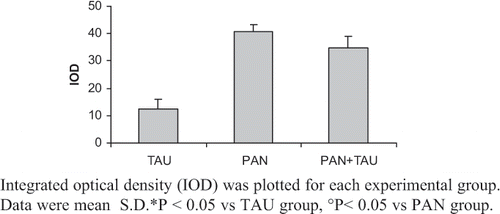
TABLE 1 24 h-Urine analysis after Puromycin and Taurine administration in rats
TABLE 2 Semiquantitative analysis of interstitial collagen
216 Availability of Essential Antidotes in Catalonia Hospitals (Spain)
Aguilar R (1), Soy D (2), Nogué S (3). 1. Pharmacy Department, Dr. Josep Trueta Hospital, Girona, Spain; 2. Pharmacy Department, Clinic Hospital, Barcelona, Spain; 3. Toxicology Unit, Clinic Hospital, Barcelona, Spain.
Objective: Certain drugs are considered as essential for the treatment of serious acute poisoning, as its efficacy cannot be replaced by supportive care nor decontaminating resources. These drugs should be available in hospitals, depending on their complexity. The aim of the study was to know about qualitative and quantitative availability of essential antidotes (IA) in Emergency Departments (ED) and Pharmacy Services (PS) of: Basic General Hospitals (Level I), Reference Hospitals (Level II) and High Technology Hospitals (Level III) from Catalonia. Methods: Descriptive study. A questionnaire was sent to hospitals from May to July 2005. Eight drugs were considered as IA, and its availability assessed according to the hospital level: 1) for all hospitals: N-acetyl-cysteine (NAC), hydroxocobalamine, ethanol (IV) and magnesium sulphate; 2) for level II-III hospitals: methylene blue and deferoxamine; 3) for level III hospitals: digoxin Immune Fab and antivenin snake. Qualitative availability (QLA) was defined, for each group, as the proportion of hospitals with IA in stock. Quantitative availability (QTA) was defined, for each drug, as the total dose needed to treat one 70 kg adult for 12, 24 or 48 hours in level I, II or III hospitals, respectively. Results: Twelve hospitals participated in the study (4 at each level). Availability in ED and PS is shown in . Conclusions: Some hospitals lack of antidotes considered as essential. The available dose of antidote is, in many cases, insufficient to treat an acute poisoning for a certain period of time. In general, it is necessary to improve the stocking of essential antidotes in Catalonian hospitals.
TABLE 1 Availability of IA in different hospital level
217 Regulatory Status of Antidotes: An Italian Perspective
Butera R, Locatelli C, Petrolini V, Lonati D, Giampreti A, Manzo L. Pavia Poison Center, IRCCS Maugeri Foundation and University of Pavia, Pavia, Italy.
The therapeutic armamanetarium we use to counteract acute poisoning is a unique mixture of histories: some antidotes were identified on the basis of traditional remedies, others represent highly sophisticated therapies; moreover, for many years the therapy of acute poisoning has been based on empirical observations. If the scientific community is moving to a more rigorous approach, this puzzled situation continues to be reflected in the regulatory status of antidotes. Objective: To describe regulatory status of antidotes in Italy. Methods: An extensive list of molecules used or proposed as antidotes was obtained integrating available lists reported in relevant international professional, technical and regulatory documents. The review of recent medical literature and updated reference textbooks allowed to identify antidotes currently used in clinical practice. Drugs available and/or licensed to the Italian market for antidotal use were identified analyzing the national directory of pharmaceutical products, according to ATC code (V03AB, V03AC and V03AF) and/or labelled uses as described in the medicinal products leaflets. Results: A total of 149 molecules were identified. In the toxicological literature and reference textbooks 122 antidotes were mentioned: among these, 64 antidotes are currently used in clinical practice with the support of scientific evidences, while 58 molecules are obsolete antidotes or drugs routinely used as symptomatic treatment. In Italy, 67 molecules with ATC code and/or labelled therapeutic uses consistent with antidotal use are available: 28 molecules are antidotes currently used in clinical practice, but only 14 of them are approved by National Health Authority for the use in the management of poisoning cases (information available in the drug leaflet is complete and correct in 5 cases only); a further 27 molecules are available to the market and approved for antidotal use even if they are obsolete and/or not supported by toxicological literature (). The other 36 antidotes used in clinical practice include Italian medicinal products used off-label, antidotes imported from foreign counties where they are markets, and galenical preparations. Conclusion: The described situation underlines the factual need of off-label use of antidotes in the management of poisoned patients; by contrast, approved antidotes are often outdated drugs: both situations may have relevant consequences in terms of medical responsibility. Antidotes use is usually restricted to hospital setting, with few exceptions; cheap antidotes are too cheap to be developed, expensive ones are rarely used once developed: for these reasons, the interest of pharmaceutical companies in clinical toxicology have been quite low for many years. The recent development of EU regulation on orphan drugs offers new opportunities in the area of antidotes.
FIG. 1
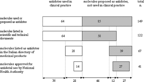
218 Availability of Antidotes in Medical Ambulances of Catalonia, Spain
Aguilar R (1), Nogué S (2), Soy D (3). 1. Pharmacy Department, Dr. Josep Trueta Hospital, Girona, Spain; 2. Toxicology Unit, Clinic Hospital, Barcelona, Spain; 3. Pharmacy Department, Clinic Hospital, Barcelona, Spain.
Objective: Availability of antidotes is, sometimes, limited by demographic, geographic and economic factors. Swift to act can be essential for any acutely poisoned patients. Medical ambulances (MA) play an important role, especially, in situations that require the treatment with antidotes classified by the International Programme on Chemical Safety (IPCS) as those needed immediately (within 30 minutes). Legislation about what antidotes must to be present in MA is not regulated in Catalonia. The aim of the study was to assess: 1) qualitative availability (QLA) and 2) quantitative availability (QTA) of antidotes in MA. Methods: Descriptive and transversal study carried out with a questionnaire from May to June 2005. The QLA was evaluated by means of a list of 67 drugs. We considered 30 drugs as recommended presence in MA. The QTA was defined (for each drug) as the total dose needed to treat one 70 kg adult patient for 6 hours. Results: Only 15 out of the 30 recommended antidotes were present in MA. Present antidotes were: adrenaline, atropine, sodium bicarbonate, biperiden, activated charcoal, dexclorfeniramine, diazepam, fenitoin, flumazenil, calcium gluconate (IV), dextrose 50% (IV), haloperidol, hydroxocobalamin, Mg sulphate (IV) and naloxone. Absent antidotes were: folinic acid, albumin, starch, apomorphine (IV), BAL, EDTA, ethanol, glucagon, calcium gluconate (topic), insulin, ipeca syrup, Mg sulphate (oral), noradrenaline, pyridoxine and protamine sulphate. Present antidotes achieved the required stock in all cases. Conclusion: the qualitative availability of antidotes in MA is inadequate. Only 50% of all recommended antidotes were present but, if so, their quantitative availability was adequate. Between absent antidotes stand out those that, in Catalonia, only are available as compounded formulations.
TABLE 1 Availability of IA in different hospital level (NA = not applicable)
219 Status Epilepticus Following Pediatric Ingestion of Thuja Essential Oil
Friesen MS (1), Phillips B (2). 1. BC Drug and Poison Information Centre, Faculty of Pharmaceutical Sciences, University of BC, Vancouver; 2. BC Children's Hospital, Vancouver, Canada.
Objective: Thuja consists of terpenoid alkaloids alpha-thujone and beta-thujone. Alpha-thujone is the active toxic constituent in wormwood oil and absinthe liqueur and is a known pro-convulsant. The mechanism responsible involves modulation of the gamma-aminobutyric acid (GABA) type A receptor similar to the classic GABA type A receptor antagonist, picrotoxin. There is one previous report of an adult male ingesting 10 mL of wormwood oil that resulted in seizures and acute renal failure. Reports of thuja oil ingestion are lacking. We present a case of thuja oil ingestion resulting in status epilepticus (SE). Case Report: A 2-year-old female ingested up to 15 mL of thuja 0.1% essential oil. Two seizures, each lasting 5 minutes, occurred within 20 minutes of ingestion. The child was actively seizing when EHS arrived 30 minutes after the exposure. An oropharyngeal airway could not be placed due to jaw clenching and persisting seizures. Oxygen was administered via bag-valve-mask en route to hospital. On presentation to hospital 50 minutes post-exposure, the child was in SE, with apneic spells, and was minimally responsive with GCS 7, HR 150 beats/min, BP 112/54 mmHg, and temperature 36.6C. Venous blood gas: pH 7.08, pCO2 84 mmHg, pO2 61 mmHg, bicarbonate 24 millimol/L, base excess -9. Electrolytes and renal function were normal, random glucose was 13.1 millimol/L. Complete blood count was normal except for elevated white blood cells. Protein, albumin, and liver function tests were all within normal limits. Treatment included intravenous lorazepam (0.125 mg) and phenytoin (18 mg/kg). By 1.5 hours post-exposure there was response to painful stimuli, arterial blood gas was normal, and seizures had resolved although there was still some residual extensor posturing. By 3 hours post-exposure, the child was sedated but rousable. Following 15 hours of hospitalization the patient was discharged with no apparent adverse sequelae. Thuja essential oil had been recommended to the father as a naturopathic topical anti-fungal nail treatment. Conclusion: Ingestion of thuja essential oil may result in rapid seizure onset and progress to SE. Treatment should include optimal doses of GABA type A receptor agonists such as benzodiazepines and barbiturates. References: Weisbord SD, Soule JB, Kimmel PL. Poison on line – acute renal failure caused by oil of wormwood purchased through the Internet. New Engl J Med 1997; 337:825–827. Höld KM, Sirisoma NS, Ikeda T, et al. Alpha-thujone (the active component of absinthe): gamma-aminobutyric acid type A receptor modulation and metabolic detoxification. Proc Natl Acad Sci 2000; 97:3826–2831.
220 Death After Accidental Ingestion of a Calcium Chloride Solution from a Humidity Absorber: A Case Report
Demarque R (1), Mostin M (1), Vanhove M (2), Banyihishako L (3). 1. National Poison Centre, Brussels, Belgium; 2. Service des urgences/soins intensifs, CHIREC site de Braine l’Alleud, Belgium; 3. Laboratoire de Biologie Clinique, Hôpital Saint Joseph, Gilly, Belgium.
Objective: Calcium chloride is a hygroscopic calcium salt which is used in humidity absorber devices. It is known for its irritant properties. We report an unusual case of severe hypercalcemia following accidental ingestion of a calcium chloride solution. Case Report: A thirsty 74-year-old man with antecedents of Chronic Obstructive Pulmonary Disease mistakenly ingests a glassful of calcium chloride solution collected from a humidity absorber device. The solution was stored in a mineral water bottle. The product information sheet recommends keeping the solution in a plastic container for further use as an herbicide or a deicing agent. Half-an-hour after ingestion, the patient complains of vomiting, diarrhea and abdominal pain. He becomes dyspneic and he is admitted at the emergency department one hour after the onset of the symptoms. Physical examination reveals a GCS 15/15, blood pressure 70/32, heart rate 140/min regular; wheezing and bilateral crackles at lung bases; abdominal tenderness. Laboratory evaluation shows an acidosis pH 7.09, BE -13.4 mmol/L, lactate 4.7 mEq/L, pCO2 49.3 mmHg, pO2 70.9 mmHg, saturation of oxygen 86.5%. The calcemia is 27.1 mg/dL (6.8 mmol/L). Renal function is normal. The patient is transferred to the intensive care unit. He becomes unconscious and is intubated and ventilated. Despite intensive hydration and diuresis, he dies from intractable cardiac arrest 4 hours after ingestion. An autopsy is not performed. No factor enhancing calcium absorption is found. A calcium concentration of 220 g/L was measured in the solution collected from a humidity absorber similar of that involved in the accident. Conclusion: The risk of hypercalcemia should be taken into account when evaluating a patient with a history of calcium chloride ingestion. Safety data sheets and major textbooks insist on the irritant properties of calcium chloride but do not consider the risk of hypercalcemia after ingestion. In 1995, at the North US Congress of Clinical toxicology, Slattery and colleagues presented a poster on two cases of hypercalcemia after accidental ingestion of a calcium chloride solution in similar circumstances. We were unable to retrieve other case reports. Reference: Slattery A, King WD, Nichols M, Fowler J. Hypercalcemia following DAMP-RID™ ingestion. J Toxicol Clin Toxicol 1995; 33:487.
221 Pharmacokinetic – Pharmacodynamic (PK – PD) Relationships in Acute Ethanol Poisonings
Mégarbane B (1,2), Bloch V (1,2), Debray M (2), Résière D (1), Karyo S (1), Abdel-Wahab A (1), Baud F (1,2). 1. 1Réanimation Médicale et Toxicologique, Hôpital Lariboisière, Paris, France; 2. INSERM U705, Hôpital Fernand Widal, Paris, France.
Objectives: The interest of PK-PD relationships is well-known in pharmacology but it was less investigated in toxicology. Ethanol-induced coma is a frequent cause of admission in Emergency Room and Intensive Care Unit (ICU). Our objective was to prospectively evaluate the correlation between the coma depth and the plasma ethanol concentration in acutely poisoned patients. Methods: Parameters were collected hourly in all successive patients admitted to our ICU for an acute ethanol poisoning, in the absence of other significant positive toxicological screening. Coma depth was assessed using the Glasgow Coma Scale (GCS). Plasma ethanol concentrations were measured using an enzymatic assay. We performed a descriptive analysis (median [10%-90% percentiles]). Non-linear regression was used for modeling PK-PD relationships (WinNonlin® software). This study was approved by the Ethic Committee of the French Society of Intensive Care Medicine. Results: PK-PD relationships were studied in 14 successive patients (9F/5M, age: 35 years [22–67], SAPS II: 45 [30–72]). Among these patients, 4/14 were chronic alcoholic (gGT: 140 UI/l [85–160]), 5/14 had a psychiatric past history, and 6/14 used regularly psychotropic drugs. The GCS at the time of hospital admission was 3 in all poisoned patients and the plasma ethanol concentration was 84.7 mmol/l [55.3–138.0]. ICU hospitalization was short (1 day [1–2]). Poisoning was complicated with an aspiration pneumonia (3/14) or a cardiovascular collapse (1/14). Regarding ethanol, we describe a Michaelis-Menten plasma kinetics (Vmax: 5.1 mmol/l/h [3.5–5.6] et Km: 0.60 mmol/l [3.105–4.2]). During the course of poisoning, PK-PD correlation between coma depth (E, Emax = 15, EO = 3) and ethanol concentration (C) well fitted the sigmoidal Emax model E = Emax.Cn/[C50n + Cn] + E0 (Hill coefficient (n): 15.7 [5.2–50.0] and C50: 61.8 mmol/l [39.3–88.9]). A maximal toxic effect (GCS of 3) was associated with a wide range of plasma ethanol concentrations, indicating clearly the saturation of all drug receptors in these situations of high dose ingestion. The high values of the Hill coefficient showed that a small decrease in plasma concentrations near the C50 was associated with a dramatic improvement in the level of consciousness. We showed the existence in acutely ethanol-poisoned patients, of a real acute PD tolerance to ethanol, in men as well as in women, independently from the existence of chronic ethanol intoxication. Conclusions: TK-TD relationships can be helpful to measure the acute PD tolerance in ethanol poisonings.
222 Ethylene Glycol Poisoning Masquerading as Mesenteric Ischemia because of a Falsely Elevated Lactate
Kirrane BM (1), Wiener SW (2), Hoffman RS (1), Nelson LS (1). 1. New York University/Bellevue Hospital Center, New York, NY, USA; 2. State University of New York-Downstate Medical Center, New York, NY, USA.
Objective: Lactate analysis is rapidly available in most clinical settings, and is an important tool in determining the etiology of an anion-gap acidosis. Lactate is most commonly measured by enzymatic based analyzers, and can be performed on both serum and whole blood samples. Since ethylene glycol (EG) levels are not readily available in most hospital laboratories, physicians must rely on surrogate markers to make a timely diagnosis. For example, a lactate level that largely explains an anion gap is often used to exclude EG poisoning. Unfortunately, there may be a false elevation of the lactate level in the setting of EG poisoning, as glycolate may interfere with the enzymatic assay. We report a case of EG poisoning resulting in an exploratory laparotomy due to a misdiagnosis of mesenteric ischemia based on an anion gap acidosis with an elevated lactate and a misread abdominal CT scan. Case Report: A 58 year-old woman was found unresponsive and intubated immediately on arrival to the ED. She had a history of diabetes mellitus, depression and a positive PPD. She was taking INH, trazadone, clonazepam and paroxetine. Her vital signs were: BP, 150/90 mmHg; P, 110 beats/min; R, 20 breaths/min; T, 37.3 C. A post-intubation arterial blood gas revealed: pH, 6.89; pCO2, 2.0 kPa; pO2, 30.1 kPa, HCO3, 3 mEq/L; and lactate>15 mEq/L. Her WBC count was 30 × 10 E9/L and the remainder of her CBC was normal. Her serum chemistry was remarkable only for a K 2.9 mEq/L and a measured serum osmolality was 305 mOsm/L. Serum for paracetamol, salicylates and ethanol were negative. The patient was treated with intravenous fluid and sodium thiosulfate, but her acid-base status remained unchanged. Fomepizole was administered, and a repeat venous blood gas showed mild improvement: pH, 7.15; pCO2, 3.6 kPa; pO2, 10 kPa, HCO3, 9.4 mEq/L; and lactate 12.8 mEq/L. An abdominal CT, performed because of the persistent metabolic acidosis and lactate elevation, and was preliminarily interpreted as showing bowel thickening and pneumotosis coli. She underwent an exploratory laparotomy that was negative. Shortly thereafter, a serum lactate drawn on a serum tube was 3.1 mEq/L. A serum EG concentration that was drawn on admission returned at this time revealing a level of 18.7 mmol/L. Conclusion: Failure to consider ethylene glycol as a cause of a severely elevated lactate level may lead to misdiagnosis.
223 Survival Despite Intravenous Self – Injection of a Potentially Fatal Dose of Aniline Hydrochloride
Chochrad D (1), Maes V (2), Lheureux PER (3). 1. Emergency Department, Hôpitaux IRIS Sud, Brussels, Belgium; 2. Laboratory of Analytical Toxicology, Academisch Ziekenhuis VUB, Brussels, Belgium; 3. Department of Emergency Medicine, Acute Poisoning Unit, CUB – Hôpital Erasme, Brussels, Belgium.
Background: Aniline is a colourless aromatic liquid, widely used as a chemical intermediate or solvent or in the manufacture of synthetic dyes. Aniline hydrochloride consists of white hygroscopic crystals that darken when exposed to air and light. Acute aniline intoxication is a very rarely reported condition, especially when it results from an intentional poisoning in adults, in particular by self-injection. A PubMed search disclosed no similar reported case. The main toxic effect of aniline is methaemoglobinemia and methaemoglobin levels above 60% are usually considered life-threatening. Case Report: A 28-year-old man without previous medical history except for severe depressive disorders (daily treatment: venlafaxine 75 mg and a flupentixol 0.5 mg / melitracen 10 mg combination) and several suicidal attempts self-injected intravenously a 2 g dose of aniline hydrochloride mixed with deionised water in a syringe at 11:30 pm. Multiple sites of injection were used to complete the self-administration. The products were found in the laboratory of a museum where the patient was employed as nightwatchman. His colleagues called an ambulance when they found him the next morning at 5:00 am. On arrival at the hospital the patient was conscious and fully able to explain what he did. His arterial pressure was 130/60 mmHg and the heart rate was regular at 140/min. He was deeply cyanotic and tachypneic with a respiratory rate of 24/min. Examination was otherwise unremarkable except for needle tracks on the forearms and a pinpoint zone of skin necrosis around a recent site of injection. EKG showed sinus tachycardia. The blood gas analysis was pH 7.47, pCO2 25.7 mmHg, pO2 301.7 mmHg (high concentration oxygen mask). The methaemoglobin level was determined by cooxymetry at 69.4%. The lactate level was 7.4 mmol/L, reflecting severe tissular hypoxia. Other biological values were unremarkable, except for blood glucose (183 mg/dL) and troponin T (0.06 ng/mL; N < 0.03). Treatment consisted of oxygen supplementation through a high concentration mask, infusion of 5% dextrose and slow IV injection of 150 mg (2 mg/kg) methylene blue (methylthioninium chloride). The condition rapidly improved as shown by the evolution of the methaemoglobin and lactate levels (). Neither further increase in troponin level, nor biological sign of haemolysis or organ dysfunction was observed, so that the patient was discharged on the same day to the psychiatry department. The presence aniline and the metabolite acetanilide in urine was confirmed by GC-MS. Conclusion: This observation shows that rapid recovery is possible in serious acute aniline poisoning provided tissular oxygenation is promptly restored by generous oxygen supplements and proper antidotal treatment with methylene blue.
FIG. 1 Evolution of methaemoglobin and lactate levels
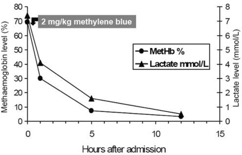
224 A Case of Intravenous Mercury Emboli Removed by Interventional Radiology
Rogers JJ (1), Brent J (2), Nutting C (3). 1. Rocky Mountain Poison and Drug Center/Denver Health, Denver, Colorado, USA; 2. Toxicology Associates/NewFields, University of Colorado Health Sciences Center, Denver, Colorado, USA; 3. Porter Memorial Hospital, Denver, Colorado, USA.
Background: The manifestations of elemental mercury toxicity are highly dependent on route of exposure. Exposure to elemental mercury via intravenous (IV) injection is uncommon, yet well documented. Previously published options for management of IV mercury injection include local excision and chelation. We report a case of elemental mercury injection treated by embolectomy using interventional radiography (IR). Case Report: This is a suicidal 41-year-old male, who injected approximately 1.5 cc of elemental mercury into his right antecubital fossa. He described a “twinge” of right-sided chest pain and was then asymptomatic. In the emergency department his exam was unremarkable with normal vital signs. Initial x-ray of the extremity showed several small pinpoint spots of mercury at the injection site, throughout the proximal extremity, and a 4 × 4 mm bolus lodged behind a valve in the bascilic vein. The chest x-ray showed a few small emboli (<2 mm) in the pulmonary vasculature. Under ultrasound and fluoroscopic guidance in the IR suite, a guidewire was steered through the mercury deposit and a 6 french sheath was placed into the vein. A Fogarty balloon was then advanced over the wire and inflated centrally to prevent additional embolization. A 6 french Excisor thrombectomy device was used to remove a majority of the intravascular mercury. The remaining mercury was cleared with a 5-french suction thrombectomy catheter and a Possis Angio-Jet device. The patient remained asymptomatic with normal renal function. He received 13 days of oral succimer treatment (10 mg/kg q8 hrs X 5 days then BID). Prior to embolectomy, blood mercury concentration was 3.7 mg/L (normal < 10 mg/L). On day two, while receiving succimer, his measured blood concentration peaked at 32.5 mg/L and by post exposure day 24 was it 9.8 mg/L. 24 hr urine mercury levels on days 2 (on succimer) and 24 (off succimer) were 24.2 mg/L and 8.6 mg/L, respectively (normal < 20 mg/L). The patient was discharged from the hospital on day 15 without signs or symptoms of mercury toxicity and was asymptomatic on follow up 24 days post exposure. Conclusion: Previous reports of IV mercury exposure have suggested that patients may have elevated blood mercury concentrations and that removal is often difficult (Torres-Alanis 1997, Dell’Omo 1997). We describe the intravascular removal of a mercury deposit with normal post-injection blood and urine mercury concentrations and no signs or symptoms of mercury poisoning 24 days after a large exposure. This intervention should be considered in cases with a large deposit of intravascular mercury.
225 Suspected Munchausen's Syndrome Presenting as Recurrent Severe Coagulopathy Following Rodenticide Ingestion
Fox BJ, Spreitzer PJ, Kelly MG, Mueller PD. Orlando Regional Medical Center, Orlando, Florida, USA.
Background: Coagulopathy after intentional ingestion of brodifacoum-containing rodenticides is prolonged, and reportedly persists long after the brodifacoum is no longer detectable in the serum. We present an unusual case of recurrent severe anticoagulation and a sporadically measurable serum brodifacoum level leading to the suspicion of Munchausen's syndrome. Case Report: A 30-year-old woman with a prior history of peptic ulcer disease presented with epistaxis and hemoptysis, stating that she had overdosed on D-Con rat poison 4 months prior in a suicide attempt. She reported she was previously asymptomatic on oral vitamin K therapy and denied repeated ingestion. Her INR was>10 and her PT was>120 seconds. Her coagulopathy resolved with FFP and vitamin K, however the patient left the hospital against medical advice two days later. She was again hospitalized two weeks after her initial presentation with a coagulopathy that was successfully treated with vitamin K. Warfarin, brodifacoum, and difenacoum levels obtained were non-detectable. The patient was subsequently hospitalized an additional 8 times over the ensuing 6 months for severe coagulopathy. Notably, a brodifacoum level obtained 3 months after her initial presentation was 27 ng/mL. Complications related to her coagulopathy included GI bleeding from peptic ulcers, anemia from recurrent bleeding requiring multiple blood transfusions, exploratory laparotomy with right salpingo-oophorectomy secondary to hemorrhagic luteal cyst, postoperative enterocutaneous fistula, and central line sepsis from postoperative total parenteral nutrition. She has received a total of 33 units of FFP since her initial presentation. Consultation with psychiatry on one occasion diagnosed major depressive disorder, however the patient has refused further psychiatric intervention. Discussion: Plasma half life of brodifacoum in humans has been reported to range from 16 to 36 days, and the duration of anticoagulant action from 51 to 240 days. However, review of the cases with the longest duration of anticoagulation suggests repeated ingestion. One series (1), consisting of 3 cases of single brodifacoum ingestion, revealed a prolonged but gradual decline in PT responsive to therapy with vitamin K and FFP. These cases never showed the marked, recurrent peaks in PT demonstrated in our patient. Conclusion: The pattern of recurrent severe coagulopathy with a sporadic positive serum brodifacoum level led us to suspect Munchausen's syndrome in this patient. References: 1. Weitzel JN, Sadowski JA, Furie BC, et al. Surreptitious ingestion of a long acting vitamin K antagonist/rodenticide, brodifacoum: clinical and metabolic studies of three cases. Blood 1990; 76:2555–2559.
226 A Study of Acute Corrosive Ingestions in Bulgaria for the Period 2003–2005
Stankova E (1), Loukova A (2), Tzekov H (3), Galov H (4). 1. Toxicology Clinic, Emergency Medicine Institute “Pirogov,” Sofia, Bulgaria; 2. Psychiatry sector, Emergency Medicine Institute “Pirogov,” Sofia, Bulgaria; 3. First Clinic of Surgery, Emergency Medicine Institute “Pirogov,” Sofia, Bulgaria; 4. Clinic of Imaging Diagnostics, MHAT “St. Anna,” Sofia, Bulgaria.
Objective: To present the results of a 3-year clinico-epidemiological study of characteristics and severity of acute corrosive ingestions in EMI “Pirogov” Bulgaria. Attention is paid to motivation of patients for committing suicide by means of corrosives. Presented is the approved natrional protocol for handling patients with this pathology. Methods: The study includes 113 adult patients with acute corrosive ingestions, hospitalised in the Toxicology Clinic, Emergency Medicine Institute “Pirogov,” Sofia, Bulgaria for the period 2003–2005. The patients are followed-up with regard to general condition, local damage, psychiatric state, concomitant diseases, and complications. The methods used include: clinical observation and examination, together with laboratory, statistical, imaging and psychiatric methods. Results: 113 patients between the ages of 19 and 87 with acute corrosive ingestions out of 4512 poisoned patients have been observed (corrosive ingestions represent 2.5% of all poisonings). 55 were male (48.7%) and 58 female (51.3%). Accidental poisonings occurred in 38 of the cases (33.6%), while intentional ingestions for committing self-poisoning were registered in 75 patients (66.4%). Alkaline agents are used by 86% of the patients, and acid substances by 14% of them. The severity of poisonings varied from moderate to extremely severe. We observe significant correlation between type of corrosives and severity and between severity and patient's motivation. The motivation in different age groups was also studied. Different complications were seen in 80% of the cases – severe bleeding, perforation, fistula or/and stricture formation. Four patients (3.6%) died in the acute phase of poisoning. Mean hospital stay in toxicology clinic was 16 ± 3 days. 26 of the patients have undergone different surgery in later phases of poisoning and 18 of them have recovered completely. Conclusions: Acute corrosive ingestions are rare, but cause severe pathology. Timely diagnosing and establishing the extent of damage is important for the successful treatment of acute corrosive poisonings. The severity and complex character of the injuries necessitate the implementation of active monitoring with good coordination between different specialists and a multidisciplinary approach to any patient with acute corrosive ingestion.
227 Soft Tissue Lesion After Injection of Hydrocarbons
Deters M (1), Schoen H (2), Hentschel H (1). 1. Poisons Information Centre Erfrut, Erfurt, Germany; 2. Klinik für Pädiatrie, Kliniken Erlabrunn, Erlabrunn, Germany.
Objective: While there is an experience with the pulmonary and central nervous system manifestations of inhaled and ingested hydrocarbon toxicity, very few reports of soft tissue petroleum distillate injection are published. Therefore we present an illustrative case. Case Report: A 17-year-old male abuser of cannabis, amphetamine, methamphetamine, and cocaine injected himself lubricating oil into the subcutaneous soft tissue of both forearms and the right knee after drug consumption. After experiencing three days of pain whilst walking he presented to the emergency department. Physical examination showed an abscess in both forearms (the whole left forearm up to elbow and the right forearm at a length of 15 cm) and in the right knee (with a extension of 10 cm). Body temperature was 38.2°C. Laboratory findings were Hb 7.9 mmol/L, Hct 0.38, thrombocytes 251 G/L, and leucocytes 12.2 G/L. In urine amphetamine, cannabinoides, and methamphetamine were detected. At the day of presentation, the abscesses were opened and the necrotic areas with early phlegmonous inflammation were excised. Coagulase-negative staphylococcus aureus was found in the detritus by microscopic preparation. The patient received cefotiam and metronidazole intravenously over 10 days and was analgesia and sedation. Wound treatment was performed by wet wound dressing and lavage. However, wound revision of the left elbow and the right knee with debridement of further necrotic material was necessary after 2 and 4 days. Eleven days after admission a final wound debridement was made. The borders of wound of the left forearm remained dehiscent but were without inflammation. The patient was discharged after 17 days for further psychological treatment. Conclusions: Symptoms and time course observed in our case report are in accordance with the few case reports in the literature (1,2). Soft tissue injection of hydrocarbons can result in severe phlegmonous inflammation with necrosis and can afford large wound debridement. Results can be complicated by healing by second intention. The optimum time of excision is controversial. References: 1. Rush MD, Schoenfeld CN, Watson WW. Skin necrosis and venous thrombosis from subcutaneous injection of charcoal lighter fluid (naphtha). Am J Emerg Med 1998; 16:508–511. 2. Shusterman EM, Williams SR, Childers BJ. Soft tissue injection of hydrocarbons: a case report and review of the literature. J Emerg Med 1999; 17:63–65.
228 Mortality Following an Aldicarb Overdose: A Departure from Classic Teaching
Schwaner RA, Hoffman RS, Nelson LS, Rao RB. New York City Poison Control Center, New York, USA.
Objective: It is often stated that compared to organophosphate poisoning, carbamate poisoning is relatively mild.(1–3) This is based on its relatively poor CNS penetration and the spontaneous reversibility of the carbamate-cholinesterase bond. As such, many authors also suggest that the use of oximes is unnecessary. We present a case of fatal carbamate poisoning to highlight both its potential severity, and the safe use of pralidoxime. Case Report: A 44-year-old woman with a history of depression was witnessed to be normal ten minutes before being found on the floor, foaming at the mouth. Upon arrival, EMS noted that she was asystolic and standard ACLS was instituted. She was intubated and received a total of 5 mg of epinephrine IV, 3 mg of atropine IV, 160 mg of lidocaine IV and three defibrillations prior to hospital arrival. In the ER, she was in sinus tachycardia with a blood pressure of 95/60 mm Hg, a pulse of 130/min, spontaneous respirations of 16/min and a temperature of 37.6 C. She received intravenous fluids, norepinephrine and activated charcoal. Because of her rapid collapse an empiric dose of sodium thiocyanate (12.5 gm) was also given IV. Subsequently, when her AST was reported as 2176 U/L, IV NAC was administered. A urine toxicology screen for drugs of abuse was negative. Approximately ninety minutes after arrival, she became bradycardic and hypoxic. Atropine, 2 mg IV, resulted in improved oxygenation and rapid atrial fibrillation developed. At this point, her daughter arrived with a cup containing a rodenticide called tres pasitos, known to contain the carbamate aldicarb.4 Retrospectively, her improvement during the resuscitations was likely related to the multiple doses of atropine. A continuous infusion of pralidoxime was initiated at 500 mg/hr, and no subsequent atropine was required. The following day she experienced a bradycardic arrest without other cholinergic signs. She was pronounced dead twenty-five hours after being found unresponsive. Conclusion: Despite current teachings, carbamate poisoning may be lethal and should be treated aggressively. References: 1. Erdman AR. Pesticides. In: Dart RC, ed. Medical Toxicology. 3rd ed. New York, New York: Elsevier, 1988:1475–1496. 2. Carlton FB, Simpson WM, and Haddad LM. The Organophosphates and Other Insecticides. In: Haddad LM, Shannon MW, Winchester JF, eds. Clinical Management of Poisoning and Drug Overdose. 3rd ed. Philadelphia, Pennsylvania: Saunders, 1998:836–845. 3. Woo OF. Carbamate Insecticides. In: Olson KR, ed. Poisoning and Drug Overdose. 2nd ed. Norwalk, Connecticut: Appleton & Lange, 1994:118–119. 4. Nelson LS, Perrone J, Deroos F, et al. Aldicarb poisoning by an illicit rodenticide imported into the United States: Tres Pasitos. J Toxicol Clin Toxicol 2001; 39:447–452.
229 Contact Dermatitis Caused by Smoke from Cashew Nut Shell Oil Burning in a Lithraea Molleoides Sensitized Man and Local Skin Memory
Vieira RJ. Sate University of Campinas (UNICAMP), Campinas, Brazil.
Objective: To present an extensive cashew (Anaccardium occidentals, family Anacardiaceae) nut oil contact dermatitis in a Lithrarea molleoides (Anacardiaeae) sensitized man, the passage of the dermal lesion to another area of skin through contact and the reactivation of a cured Lithraea molleoides skin lesion (1,2) as a local skin memory (3) Case Report: A 68-year-old man, was exposed to the smoke from burning oil from cashew nut shell as he opened the oven in which 25 cashew seeds were being baked. The oil from the shells dropped over the oven flame and was burning. As he was wearing trunks and a long-sleeved shirt, the most exposed area were the anterior parts of his legs, wrists, hands and, in minor degree, face and neck. On the second day he noticed some erythema and small papule with pluritus in the area of his legs, wrists, hands and in other two areas (one in the internal face of the left arm and the other in the inferior part of the axilla), where there had been skin lesion from Lithraea molleoides exposition three months before, but cured. In the night between the 2nd and 3rd day, the patient slept intentionally with both arms and wrists lesion on the correspondent antero-lateral faces of the thorax. This contact resulted in similar skin lesions on both involved areas of the thorax. The skin lesions got worse and kept growing up to the 7th day, with erythema, infiltration, small papules, small vesicals and intense edema. The patient's weight increased from 85 to 90 kg in the first week, without corticotherapy. The urine and blood exams were within normal limits, except for a 10% eosinophily on hemogram. On the 7th day a skin biopsy showed edema, spongyoses, eosinophily, infiltration and small vesicals. Conclusion: A case of contact dermatitis caused by smoke from cashew nut shell oil burning in a Lithraea molleoides sensitized man is presented. The exposition reactivated a previously cured dermal lesion from Lithraea molleoides showing a local skin memory. The dermal lesion caused by smoke from cashew oil passed to normal skin by contact as a stamp-like skin lesion. References: 1. Alé SI, Ferreira F, González G, et al. Allergic contact dermatitis caused by Lithraea molleoides and Lithraea brasiliensis: identification and characterization of the responsible allergens. US Journal Contact Dermatitis 1997; 8:144–149. 2. Diógenes MJN, Morais SM, et al. Contact dermatitis among cashew nut workers. Contact Dermatitis 1996; 35:114–115. 3. Moed H, Boorsma DM, et al. Increased CCL27-CCR10 expression in allergic contact dermatitis: implications for local skin memory. The Journal of Pathology 2004; 204:39–46.
230 Life-Threatening Methoxyflurane Ingestion
Halcomb SE, Hoffman RS, Nelson LS. New York City Poison Center, New York, USA.
Objective: Methoxyflurane is a volatile halogenated anesthetic. Seven years after its introduction, a case series of 16 patients reported renal abnormalities resulting from methoxyflurane administration. The toxicity was due to the urinary excretion of inorganic fluoride and tubular accumulation of oxalate, both products of methoxyflurane metabolism. This discovery led to its removal from the US market. Ingestion may cause anesthesia by GI absorption or by oropharyngeal volatilization and subsequent inhalation. Patients who ingest volatile anesthetic products may often require aggressive supportive care because of respiratory depression and hypotension. Some physicians collect old medications for historical purposes. Occasionally patients who have access to these medications will overdose on them. We report a life-threatening ingestion of methoxyflurane that occurred in a woman who kept this anesthetic as part of her father's collection. Case Report: A 50-year-old woman with a history of depression presented to the ER after ingesting 30 mL of methoxyflurane in a suicide attempt. While being transported a short distance to the hospital, the patient experienced a brief episode of apnea. Her husband performed CPR, but avoided mouth-to-mouth-resuscitation since the patient smelled strongly of the anesthetic. On arrival to the ED, the patient was apneic and comatose with a BP of 105/64 mmHg, a pulse of 79/min and a temperature of 36.8°C. She was intubated and the staff reported smelling a strong odor. She recovered neurologically intact with supportive care and was extubated two days later. Methoxyflurane is the most potent inhalational anesthetic with an MAC of 0.16%. In this case, the patient appeared to off-gas the product, which could present an occupational hazard to transporters in enclosed spaces or hospital personnel attempting resuscitation. Her husband, a dentist, was familiar enough with anesthesia to avoid using mouth-to-mouth resuscitation. Conclusion: Methoxyflurane ingestion may result in apnea, although the route of absorption is unclear. Supportive care and monitoring of renal function appear to be sufficient in the management of these patients.
231 Oxidative Stress in Subjects with Asbestos Exposure
Pelclová D (1), Fenclová Z (1), Kacer P (2), Kuzma M (3), Lebedová J (1), Klusácková P (1), Navrátil T (4). 1. Toxicological Information Centre, Department of Occupational Medicine, Charles University, Prague, Czech Republic, 2. Department of Organic Technology, Institute of Chemical Technology, Prague, Czech Republic; 3. Laboratory of Molecular Structure, Institute of Microbiology, Prague, Czech Republic; 4. Heyrovský Institute of Physical Chemistry, Prague, Czech Republic.
Objective: The aim of this study was to determine oxidative stress and airways inflammation markers in subjects with previous asbestos exposure. The formation of oxidants in macrophages and lung epithelial cells is supposed to be the key issue in the toxic effect of asbestos. Oxidative stress is probably a crucial event in the initiation of functional changes in fibroblasts contributing to pulmonary and pleural fibrosis. F2-isoprostanes are produced by the radical-catalyzed lipid peroxidation of arachidonic acid and the determination of 8-isoprostane in biological fluids is valuable for quantification of oxidant stress in vivo. On the other hand, leukotrien B4 points to the inflammation in the airways. Exhaled breath condensate reflects the composition of bronchoalveolar extra cellular lining fluid and enables non-invasive measurements of several parameters directly in the environment of the airways. Methods: Fourty-four persons were examined, mean age 68 years, previous exposure to asbestos 24 years on average. Control group was represented by 30 age- and gender-matched controls, without occupational history of asbestos. Lung functions were measured; 8-isoprostane was analyzed by HPLC/MS, and leukotrien B4 by SPE. Cotinine in urine was detected by GC/MS to verify the smoking status. Student t-test and correlation coefficient were used for statistical comparison of the groups. Results: Mean level of 8-isoprostane in persons exposed previously to asbestos and controls was 72 ± 12 vs. 51 ± 8 pg/ml (p < 0.01). Increased levels of 8-isoprostane were seen both in subjects with interstitial fibrosis and with pleural changes. Mean level of leukotriene B4 was 38 ± 9, v.s.34 ± 6 pg/ml. Conclusion: This is the first study to determine 8-isoprostane and leukotriene B4 in subjects with asbestos exposure. The level of 8-isoprostane in exhaled breath condensate was significantly increased in asbestos-exposed subjects; it seems sensitive as it was elevated also in patients with pleural changes without severe lung fibrosis. This finding is biologically plausible because asbestos fibres persist in the lungs for decades. Normal level of leukotrien B4, which is usually increased in chronic bronchitis and in smokers, excludes the interference of airways inflammation in the patients. References: Mossman BT: Role of reactive oxygen and nitrogen species (ROS/RNS) in lung injury and diseases. Free Rad Biol Med 2003; 9:1115–1116. Cracowski J-L et al.: Isoprostanes as a biomarker of lipid peroxidation: physiology, pharmacology and clinical implications. Trends in Pharmacol Sciences 2002; 23:360–363. Acknowledgement: IGA 8107-3/2004.
232 Protein Adducts of Industrial Chemicals in Biological Monitoring: Preparation of the Reference Materials
Mráz J, Stránský V, Kicová R, Nohová H. Centre of Occupational Health, National Institute of Public Health, Prague, Czech Republic.
Background: Some reactive chemicals absorbed in the organism or reactive intermediates produced by the metabolism form stable adducts with proteins in vivo. In the industrial toxicology, these adducts are determined for the biological monitoring purposes. Major attention is focused on the adducts with the blood protein globin. Globin adducts persist in the body over the whole lifetime of the erythrocytes (4 months in humans), therefore, they are particularly useful as biomarkers of the integrated internal dose of the chemicals. Determination of the adducts presents a methodical problem as analytical standards of the modified globins are not commercially available. Objective: To prepare and characterize batches of globin modified with selected alkylating agents to be used as reference materials. Methods: Full human blood (25 ml) was incubated with 1 mM alkylating agents. Small aliquots were taken at regular intervals for determination of the parent agent, to calculate its removal rate. After 24 h, the erythrocytes were separated and globin was isolated. The alkyl adducts at the N-terminal valine of globin were determined using modified Edman degradation procedure to produce specific 1-alkyl-5-isopropyl-3-pentafluorophenyl-2-thiohydantoins, followed by gas chromatography/mass spectrometry. Calibration was carried out using alkyl-peptide standards. Results: Half-times of the tested agents (1 mM) in blood and the levels of the alkyl adducts at the N-terminal valine in globin are shown in the table. Conclusion: The obtained data will be used to prepare batches of globin modified with the alkyl adducts at various requested concentrations. Acknowledgement: The study was supported by the Internal Grant Agency of the Czech Ministry of Health, grant NJ/7387-3/2003.
TABLE 1
233 Severe Keratoconjunctivitis after Accidental Ingestion of Dibutylphthalate
Martens M (1), Mostin M (1), Duchesne B (2). 1. National Poison centre Brussels Belgium; 2. Department of Ophthalmology, CHU Sart-Tilman, University of Liege, Belgium.
Objective: Keratoconjunctivitis after oral chemical exposure without direct eye contact is unusual. We report a case of severe keratitis and signs of pancreatitis after accidental ingestion of 15 ml Dibutylphthalate in a 68-year-old man. Case Report: A 68-year-old man with antecedents of hypertension, dyslipidemia, and multiple heart infarcts mistakenly swallowed his medication with Dibutylphthalate (DBP) instead of water. The product was brought home from his workplace in a water bottle. A few hours after ingestion he complained of hypoacousia, tinnitus, and irritation of both eyes. A day later, he was admitted to the hospital with epigastric and right hypochondrial pain. On admission, the epigastric and right hypochondrial regio were tender to palpation without guarding or rebound. Laboratory examinations were remarkable for hyperleucocytosis (15.550 white cells/mm3) with neutrophilia and a slight elevation of liver and pancreatic enzymes: gamma-GT 62 IU/l, SGPT 37 IU/l amylases 378 IU/l and lipases 764 IU/l. Coagulation tests, ionogram, and renal function tests were normal. An abdominal scan showed a slight oedema of the pancreas. Ophthalmologic examination showed photophobia, slight bilateral conjunctivitis, diminution of the cornea transparency without severity. An eye examination performed on the second day of hospitalization disclosed complete desepithelialisation of the cornea. The abdominal pain resolved after treatment with analgesics and antispasmodics and pancreatic enzymes normalized after a few days. The patient was discharged after five days and followed up by the ophthalmologist. After three weeks, his vision was still slightly disturbed by the persistence of a punctual superficial keratitis of the right eye. Conclusions: Our case report is very similar to the observation Cagianut published in 1954. Phthalate esters are widely used as plasticizers in the industry. There are also found in consumer products like glow sticks and luminous necklaces. Accidental ingestion of small quantities by young children is quite common. Acute toxicity of phthalate esters is usually considered to be low although there is little data available. We suggest that all cases of dibutylphthalate ingestion should be carefully observed for occurrence of ocular symptoms. Reference: Cagianut B, Keratitis erosiva und Nephritis toxica nach Einnahme von Dibutylphthalat Schweiz Med Wochenschr 1954; 84(44):1243–1244.
234 Obesity Associated with Chronic Exposure to Chorpyrifos in Rats
Meggs WJ, Brewer KL. Brody School of Medicine at East Carolina University, Greenville, NC, USA.
Objective: The worldwide obesity epidemic could be related to chronic exposures to environmental chemicals. Many environmental chemicals, including organophosphate insecticides, are known to disrupt endocrine and neurological systems, so a connection between obesity and exposures is possible. The objective of this work was to expose rats to low levels of the organophosphate insecticide chlorpyrifos and monitor for toxic effects, including weight gain. Methods: Subjects were female Long-Evans rats about 6 months of age. Research design was a placebo controlled study. Intervention was a subcutaneous injection of chorpyrifos, 5 mg/kg, or an equal volume of vehicle. Injections were given daily for 4 months. Chlorpyrifos was obtained from SigmaAldrich (St. Louis) and dissolved in DMSO then diluted with normal saline just prior to injection, for a final concentration of 4.0 mg/mL. Subjects were observed for 30 minutes after injection for signs of acute cholinergic or other toxicity, with atropine and pralidoxime available for treatment as needed. Weights were recorded at baseline, 2 months, 3 months, and 4 months. Weights in the two groups were compared using ANOVA. Results: No signs of acute cholinergic toxicity were observed after injection of either group. Rats in the exposed group were significantly heavier than those in the control group by 2 months after exposure was initiated (335.7 ± 16.7 g vs. 318.6 ± 15.8 g; p = 0.034). This difference increased at 3 months (350.1 ± 16.4 g vs. 322.3 ± 21.3 g p = 0.006) and 4 months (374.4 ± 22.2 g vs. 340.2 ± 25.2 g p = 0.006). Conclusions: Chronic exposure to the organophosphate insecticide chlorpyrifos caused statistically significant weight gain in young rats. Further research is needed to determine the mechanism of this weight gain, and whether or not exposure to other pesticides and environmental chemicals have similar effects.
235 Analysis of the Renal Damage Following Ethylene Glycol Poisoning
Krenová M, Pelclová D. Toxicological Information Centre, Charles University, General University Hospital, Prague, Czech Republic.
Objective: To evaluate retrospectively the renal impairment and permanent consequences following ethylene glycol (EG) ingestion in a 5-year study in the Czech Republic. Methods: Data from medical reports concerning clinical course of patients with EG intoxication reported to the Czech Toxicological Information Centre and toxicological laboratories between 2000 and 2004 were analysed. Results: Medical records of the 206 cases of EG intoxication were analysed. 139 patients (24 children, 115 adults) developed only mild symptoms of EG intoxication without any renal damage. Twenty-four adults (18 males, 6 females) died due to EG intoxication. Nineteen patients developed acute renal failure with the mean maximal serum creatinine level 396 micromol/l. The most common immediate cause of death was cardiopulmonary or metabolic failure. Eight autopsies were performed. In five histological findings calcium oxalate crystals were found predominantly in the proximal tubules, besides that vacuolar degeneration and acute tubular necrosis was present. In three patients also the signs of renal atherosclerosis were found, one of them was treated for hypertension. Forty-three patients (34 males, 9 females) developed signs of nephrotoxicity. All were treated by ethanol and haemodialysis. In fourteen patients renal function normalized until the discharge from the hospital. Their mean maximal serum creatinine level was 371 micromol/l, mean maximum serum EG level 1.00 g/l. In twenty-nine patients the renal function did not normalise at discharge, mean maximal serum creatinine level reached 523 micromol/l; mean maximal serum EG level l.58 g/l. Eighteen patients were followed-up, 11 did not comply with it. In five patients the renal function completely recovered during six months, in two patients until twelve, and in one patient until seventeen months after discharge from the hospital. In five patients renal parameters were altered (mean serum creatinine level 123 micromol/l) six months after the discharge from the hospital but they did not comply with further follow-up. Two of them had history of renal damage. In five patients renal damage persisted (mean serum creatinine level 137 micromol/l) twelve months after discharge from the hospital. One of them had also history of renal damage before EG ingestion. Mean time delay between EG ingestion and admission to the hospital was 19 days, versus 11 days, and mean pH 6.988 versus 7.086 in patients with more persistent renal impairment comparing with patients whose renal function recovered until 12 months. Conclusion: Recovery of kidney function following EG intoxication is influenced by the dose ingested (serum EG level), time delay between EG ingestion and admission to the hospital, and history of renal disease. Follow-up of the patients continues.
236 Chemical Skin Burn of a Two-Year-Old Boy Exposed to a Paint Remover Containing Formic Acid and Dichloromethane
Sydow A (1), Schiffmann H (2), Bardeck MA (3), Desel H (1). 1. GIZ-Nord Poisons Centre, University Hospital, Göttingen, Germany; 2. Department of Pediatrics, University of Göttingen, Göttingen, Germany; 3. Department of Pediatric Burns, Children's Hospital Park Schönfeld, Kassel, Germany.
Objective: Labelling of dangerous products as “irritating” or “corrosive” according to the European Union's legislation usually indicates the maximal degree of skin damage correctly. However, for some products labelling is misleading. Poisons centres and clinical toxicologists must be aware of this problem. Case Report: A two-year-old healthy boy intended to drink from an almost empty 10 liter container of a paint remover for professional use in his parents’ carpenter workshop. According to label and safety data sheet the product contained 3.6% formic acid and 85% dichloromethane. It was correctly labelled as “irritating for eye and skin.” The boy tipped it over, thus contaminating the chest, the front side of arms and legs, lips, parts of the throat, nose and neck, but not the eyes. Only minutes after exposure clothes have been removed and the skin was carefully decontaminated using a shower. Within the next 24 hours in the hospital skin irritation developed increasingly on 40 percent of the body surface, but neither signs of metabolic acidosis nor any toxic organ damage have been be observed. A subtoxic concentration of formic acid but no dichloromethane could be detected in urine by toxicological analysis. The formic acid concentration in the product was determined to 5.6% (due to evaporation of dichloromethane), dichloromethane concentration was as indicated by the manufacturer. The patient was transferred to a specialized treatment unit for children with skin burns. Histologic analysis of a skin sample from day 2 showed epidermic necrosis but no damage of the dermis. Within 3 months’ treatment at the hospital the patient needed two split skin transplants. No severe complications developed during the treatment period and one year of follow up. Conclusion: Simultaneous dermal exposures to 5.6% formic acid and dichloromethane have caused severe skin corrosion although the product was labelled as irritant for eye and skin only (correct according to EU legislation). Synergistic toxic effects of the ingredients (and maybe the absence of water) may have caused the severe symptoms.
237 Alcohol-Based Hand Cleansers – Enquiries to a Poisons Centre Before and After Their Widespread Introduction into Hospitals
Tizzard Z. Guy's Poisons Unit, Guy's and St Thomas’ NHS Foundation Trust, London, UK.
Objective: In order to reduce infection rates in National Health Service (NHS) hospitals in the UK, by April 2005 alcohol-based hand cleansers were put in place in hospitals at the entrances to wards, at bedsides and were to be carried by staff throughout the NHS. In anticipation of wider use and availability of such products, prospective surveillance of enquiries to our Poisons Unit regarding these products was instigated. Methods: Telephone enquiries to the Unit regarding alcohol-based hand cleansers during 2004 and 2005 were identified at the time of the enquiry and a written follow-up questionnaire requesting further information was then sent to the enquirer, if sufficient details were available, by post the next day. The data recorded at the time of the enquiry and any data from returned follow-up questionnaires was collated and analysed. A comparison of enquiries from the 6-month period before the widespread introduction of these products into hospitals with enquiries from the 6-month period following their introduction was undertaken. Results: In the six months before the introduction of alcohol based hand cleansers in hospitals (October 2004–March 2005) we received 15 enquiries. These enquiries account for 0.06% of the total enquiries for that period (26,193). Seven enquiries were regarding ingestion and 8 were eye exposure. 10 were adults and 5 were children. Thirteen were accidental (including 5 occupational accidents) and 2 were intentional abuse. Eight were symptomatic, 6 were asymptomatic; 1 was not stated. In the six months after the introduction of alcohol based hand cleansers in hospitals (April 2005–September 2005) we received 32 enquiries. These enquiries account for 0.16% of the total enquiries for that period (20,047). This is a percentage increase of 200%, and has occurred despite a drop in telephone enquiry numbers overall, which decreased by 23.5%. Twenty-three enquiries were regarding ingestion and 9 were eye exposure. 27 were adults and 4 were children; 1 was not stated. Seventeen were accidental (including 6 occupational accidents), 12 were intentional and 3 were not stated. Seventeen were symptomatic, 10 were asymptomatic; 5 were not stated. Conclusion: Poisoning due to alcohol-based hand cleansers remains uncommon although enquires regarding such products have increased since their widespread introduction in April 2005. Exposures are rarely serious, however, the potential for self-harm or abuse with these products and subsequent serious clinical effects remains. Their increased use is likely to result in more adverse effects, particularly from eye exposures, occurring occupationally in hospitals. Poisons information services are well placed to monitor exposures relating to changes in working practice.
238 Cesium Chloride Toxicity from Alternative Cancer Treatment: Case Series
Geib A (1), Martin TG (2), Burns-Ewald M (1). 1. Children's Hospital Boston, MA, USA; 2. University of Washington, Seattle, WA, USA.
Objective: To describe cardiotoxicity resulting from chronic ingestion of an alternative cancer treatment. Methods: Cases of suspected cesium chloride (CsCl) poisoning were pulled from the records of two poison centers. Results: Three cases are reported in . Two patients were adults and one <18 years. All admitted to ingesting CsCl. Two had serum Cs measured. All patients had torsades de pointes and two had QTc duration of>600 msec. The adult patients were hypokalemic. Serum potassium was not recorded in the child. All three patients were treated with cardioversion. The pediatric patient received esmolol and propranolol to prevent apparently rate-related torsades. She also received hemodialysis twice with an unknown effect on serum Cs. The adult patients received electrolyte replacement. Prussian blue was considered in one patient but never administered. All patients were discharged from the hospital. Discussion: Cs blocks voltage-sensitive inward potassium channels (1–4) and is associated with hypokalemia, QTc prolongation, and torsades de pointes. (5–7) Prolongation of the QTc can last for days or even weeks despite electrolyte replenishment(5,7). Treatment consists of discontinuing exposure, restoring electrolyte balance, and antiarrhythmic medication (5,6). Prussian blue (ferric ferrocyanate) has been used for treatment of internal radiocesium contamination, but never reported in stable Cs poisoning. Cs is dialyzable but hemodialysis has never been reported as a treatment for toxicity(8). Clinicians should continue to warn patients of harmful effects of unproven, alternative treatments. References: 1. Gay LA, Stanfield P.R. Cs+ Causes a Voltage Dependent Block of Inward K+ Current in Resting Skeletal Muscle Fibers. Nature 1977; 267:169. 2. Levine JH, Spear, J.F., Guarnieri, T., et al. Cesium chloride-induces long QT syndrome: demonstration of afterdepolarizations and triggered activity in vivo. Circulation 1985; 72:1092–1103. 3. Patterson E, Szabo B, Scherlag BJ, Lazzara R. Early and Delayed Afterdepolarizations Associated with Cesium Chloride-Induced Arrhythmias in the Dog. J Cardiovasc Pharmacol 1990; 15:323–331. 4. Nayebpour M and Nattel S. Pharmacologic Response of Cesium-Induced Ventricular Tachyarrhythmias in Anesthetized Dogs. J Cardiovasc Pharmacol 1990; 15:552–561. 5. Dalal AK, Harding JD, Verdino RJ. Acquired Long QT Syndrome and Monomorphic Ventricular Tachycardia After Alternative Treatment with Cesium Chloride for Brain Cancer. Mayo Clinic Proceedings 2004; 79:1065–1069. 6. Lyon AW and Mayhew WJ. Cesium Toxicity: A Case of Self-Treatment by Alternate Therapy Gone Awry. Therapeutic Drug Monitoring 2003; 25:114–116. 7. Saliba W, Erdogan O, Niebauer M. Polymorphic Ventricular Tachycardia in a Woman Taking Cesium Chloride. Pacing & Clinical Electrophysiology 2001; 24:515–517. 8. Krachler M, Scharfetter H, Wirnsberger GH. Exchange of alkali trace elements in hemodialysis Patients: a comparison with Na(+) and K(+). Nephron 1999; 83:226–236.
TABLE 1 Cesium cases
239 Olbas Oil and Respiratory Arrest in a Child
Crandon KL, Thompson JP. National Poisons Information Service (Cardiff Centre), Llandough Hospital, Penarth, UK.
Background: Olbas Oil contains a mixture of essential oils including eucalyptus, levomenthol, cajuput peppermint and clove oil. It is used via inhalation for a variety of minor ailments including bronchial and nasal congestion or to bring symptomatic relief of muscular pain and stiffness including backache, sciatica and rheumatic pain via local application. Inhalation of the vapours can be achieved by sprinkling several drops onto a handkerchief and placed near the patient or by mixing a few drops in hot water. For muscular relief it is applied lightly and massaged into the affected area. Olbas Oil comes in 10 ml and 28 ml dropper bottles. Case Report: A six-month-old child was admitted to hospital shortly after an accidental therapeutic error whereby Olbas Oil was instilled intranasally by mistake. On admission to A&E the child had a respiratory arrest from which he was successfully resuscitated. The child was subsequently admitted to hospital for overnight observation. During this time the child was asymptomatic and suffered no further complications. He was discharged the following day. Further investigation into this case found the child had been suffering a cold and had been prescribed saline drops for intranasal application. At the time of the exposure the two bottles had become confused and the Olbas Oil instilled instead of the saline drops intended. It was felt that this may have been due to the bottles being of a similar size and shape. Discussion: Severe adverse reactions to Olbas Oil are unusual, but not unheard of. Wyllie and Alexander (1994) reported a similar case of nasal instillation in a 4-month-old child which led to immediate respiratory distress and agitation, followed by corneal scarring and conjunctivitis. Essential oils may also be highly toxic via ingestion. Conclusion: As Olbas Oil has the potential to cause serious adverse effects when applied incorrectly, it is important that users should be aware of potential adverse effects. The severity of the reactions observed raise the question as to whether the packaging of Olbas Oil should be improved to make accidental administration via intranasal application or eye contact less likely. All instructions for the correct use of Olbas Oil are currently carried on the outer box and bottle label and are therefore relatively limited. As no patient information leaflet is currently included the potential dangers of Olbas Oil misuse may be under appreciated. Improved labeling and public awareness together with a bottle redesign may help prevent further unnecessary accidents. Reference: Wyllie JP, Alexander FW. Nasal instillation of ‘Olbas Oil’ in an infant. Archives of Diseases in Childhood 1994; 70:357–358.
240 Clinical Implications of Methidathion Toxicokinetics
Zoppellari R, Ferri E, Lamborghini S, Osti D, Petrini S, Pinamonti A, Guberti A. Department of Anesthesia and Critical Care, S. Anna Hospital, Ferrara, Italy.
Objective: A relationship between toxicokinetic data of three dimethyl-organophosphate methidathion poisoning cases and clinical implications is proposed. Case Report 1: After ingestion of about 9.5 g of methidathion, a 50-year-old woman was ventilated for 5 days and treated with atropine titrated to control bronchorrhea and bronchospasm and pralidoxime administration (2 g bolus followed by 2 mg/kg/h). During the following 9 days the doses of antidotes were gradually reduced. Three gastric lavages, made at 1.5, 18 and 43 hours postingestion, removed 4,100, 6.53 and 0.01 mg of methidathion, respectively. The estimated absorbed dose was 5.4 g (68 mg/kg). Methidathion was present in plasma and gastric fluid until day 9. Plasma peak level was 761 microg/L at 3 hours postingestion. Case Report 2: A 50-year-old man ingested about 6.2 g of methidathion. He was treated with repeated gastric lavages (the first removed 61 mg of methidathion) and ventilation for 16 days. Atropine and pralidoxime (2 g bolus followed by 1 g/day) were administered until day 10, when methidathion was absent in plasma, gastric fluid and fat biopsy. The estimated absorbed dose was 90 mg/kg and the plasma peak level was 1675 microg/L, 12 hours postingestion. Case Report 3: A 61-year-old man self injected 26 mg/kg of methidathion. He was treated with mechanical ventilation for 22 days, atropine and pralidoxime (30 mg/kg followed by 2 mg/kg/h). The cholinergic signs lasted 18 days and antidotes were discontinued on day 20, when plasma methidathion level was 5 microg/L. Plasma peak level was 242 microg/L at 40 hours postinjection. On day 13 and 25 methidathion levels in fat were respectively 162 and 42 microg/L. Conclusion: The injection of a relatively low amount resulted in prolonged toxic effects. After ingestion, high loads of absorbed methidation caused early poisoning features. A prompt gastric lavage may be of benefit. In the parenteral poisoning, the severe cholinergic syndrome appeared on day 2, when plasma level was the highest. A level higher than 50 microg/L was associated with the most serious phases of poisonings. The prolonged mechanical ventilation was associated with the persistence of methidathion, accumulated in the fat, and with the occurence of pneumonia. The patients were treated with prolonged infusion of pralidoxime because significant levels of organophosphate might inhibit the newly synthetized acetylcholinesterase. The knowledge of body organophosphate level was a guide to prolonged antidotal therapy.
241 Outbreak of Respiratory Distress after Exposure to Textile Proofing Spray with Fluorpolymer – Effective Toxicovigilance Through a Poison Center
Gregersen PA (1), Klixbüll U (2), Skanning PG (1), Jacobsen P (3). 1. Department of Anaesthesiology, H:S Bispebjerg Hospital, Copenhagen; 2. National Preparedness Agency, Chemical analytical laboratory; 3. Danish Poison Center, H:S Bispebjerg Hospital, Copenhagen, Denmark.
Objective: To report on an outbreak with 16 cases of respiratory distress after exposure to textile proofing spray. Case Series: Respiratory problems after exposure to aerosols of proofing agents are well known (1–4). It has been described how particularly the fluor-carbon component of the agent causes toxic pneumonia and collapse of the alveoli (3,4). Due to advances in the manufacture of these sprays the number of new cases has been very low in recent years. However, outbreaks have occasionally been reported, some due to a specific chemical component (4,5). During the summer of 2005, the Danish Poison Information received a number of calls concerning respiratory symptoms after exposure to a particular waterproofing agent, (). The 16 patients were aged 3–64 years, 9 were females. Several of the patients showed significant morbidity with complaints including dyspnoea, cough, chest pain, nausea and general malaise. Oxygen saturations ranged from 80%–100%. Eight subjects needed oxygen-therapy, 4 patients presented diffuse pulmonary infiltrates. When the Danish Poison Information recognized that an outbreak took place the National authorities were involved and a ban on the product was issued. Later of it was revealed that one component of the product, a fluor polymer, might have been changed in the spring 2005. Presently the chemical contents of both the old and new spray cans are being analyzed. Conclusion: Aerosols composed of solvents and proofing agents are potentially harmful to the airways. Small changes in the composition of the product seem to have changed the hazard associated with this product. The poison centers potential for accurate and real time toxicovigilance has been demonstrated by this outbreak. References: 1. Centers for Disease Control and Prevention. Acute respiratory illness linked to use of aerosol leather conditioner – Oregon, December 1992. Morb Mortal Wkly Rep 1993; 41:965–967. 2. Burkhart KK et al. Pulmonary toxicity following exposure to an aerosolized leather protector. J Toxicol Clin Toxicol 1996; 34:21–24. 3. Wallace GM, Brown PH. Horse rug lung: toxic pneumonitis due to fluorocarbon inhalation. Occup Environ Med 2005; 62:414–416. 4. Vernez DS et al. Characterizing emission and breathing-zone concentrations following exposure cases to fluororesin-based waterproofing spray mists. J Occup Environ Hyg 2004; 1:582–592. 5. Groot R, Vries I de, Meulenbelt J. Sudden increase of acute respiratory illness after using a spray product to waterproofing clothing and shoes. Abstract, EAPCCT XXIV international congress, Strasbourg, 2004.
FIG. 1 Respiratory cases after exposure to a textile proofing agent in 2005
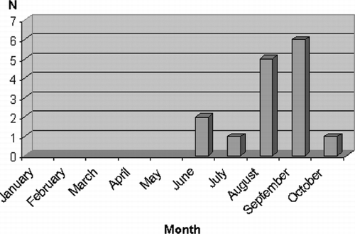
242 A Case of Trichloroethylene-Induced Ventricular Sustained Tachycardia
Macovei R, Voicu VA, Danescu I, Tudosie M. ICU II Toxicology, Emergency Clinical Hospital, Bucharest, Romania.
Background: Trichloroethylene is a solvent for degreasing metallic parts. Ingestion of trichloroethylene affects nervous system through solvent effect on lipoprotein neuronal membrane components with excitation, dizziness, stupor, coma and peripheral nervous and cranial lesions through neurotoxic metabolite dichloroacethylene. On cardiovascular system trichloroethylene sensitives myocardium to cathecolamines, producing a decrease of myocardial contractility, electrical aberrant conduction, vasodilatation, and hypotension. Gastrointestinal absorption is rapidly and substantial, producing severe central nervous depression. Treatment is unspecific and consists in sustaining of vital functions (1). We report a case of trichloroethylene-induced ventricular sustained polymorphous bidirectional tachycardia. Case Report: A 28-year-old male was admitted into the hospital 12 hours after a suicidal attempt through ingestion of 200 ml trichloroethylene. On admittance: critical status, profound coma, myosis, facial hyperemia, intubated and ventilated with hemodynamic instability. An ECG revealed monster aspect with difficult interpretable complexes, suggesting ventricular sustained tachycardia (). Endoscopic exam: erythematous gastroduodenitis. Laboratory: metabolic acidosis, rhabdomyolysis, leucocytosis. He underwent intensive care treatment with ventilator support, antiarrhythmic association (lidocaine, amiodarone, beta-blockers) in continuous adjustable doses, fluids replacement, hydroelectrolytical and acidobasic balancing, anticoagulants, forced diuresis, gastric protectors and cerebral anabolysants. Evolution was favorable, the patient became hemodynamic stable and the ECG progressively normalized in 72 hours. The laboratory data normalized. The patient recovered consciousness and mechanical ventilation was discontinued after 4 days. The patient was released from hospital after 14 days, with total recovery of the symptoms, without neurological sequelae and normal ECG aspect. Conclusion: Acute poisoning with trichloroethylene is very rare, but very severe, with a great lethal potential through cardiac arrhythmias and respiratory and central nervous depression. This report demonstrates the acute cardiovascular toxicity of trichloroethylene, reversible under antiarrhythmics and sustaining vital functions. Reference: 1. Agency for Toxic Substances and Disease Registry (ATSDR). Toxicological Profile for trichloroethylene. Atlanta, GA: US Department of Health and Human Services, Public Health Service, 1997.
FIG. 1
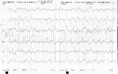
243 Liver Transplantation in a Ceramic Worker after Acute Hepatitis and Severe Lead Poisoning
Bacis G (1), Marini B (2), Farina ML (1). 1. Bergamo Poison Control Center, Ospedali Riuniti, Bergamo, Italy; 2. Infectious Diseases Unit, Ospedali Riuniti, Bergamo, Italy.
Introduction: Ceramic workers are potentially exposed to lead from ceramic glazes. Red lead (lead tetroxide) was one of the most used glaze in the past. We describe a severe lead poisoning after acute hepatitis followed by liver transplantation and a severe Guillain-Barré neuropathy. Case Report: A 47-year-old male ceramist, with chronic hepatitis C, diabetes and alcoholic abuse, used a small amount of red lead for preparing a ceramic product. After 5 days the patient had fever and sore throat treated only with propolis; in the following days nausea, vomiting, arthralgia, legs’ paresthesia, abdominal pain, jaundice and confusional state were the symptoms observed. After his admission to our hospital, the laboratory controls showed an increase of transaminases (ALT 2,519–AST 2,422) and hyperbilirubinemia (total 9.2, direct 5.5 mg/dL), elevated LDH (3,938 U/L) and alcaline phosphastase (153 U/L), hyperglycemia (289 mg/dL), elevated urinary copper (700 mcg/24 h), mild C-reactive protein increase (2.2 mg/dL) and mild anemia (Hb 12.7 g/dL). Virological exam showed high level of HCV-RNA (115.719 U/mL). A liver biopsy showed diffuse coagulative necrosis, moderate inflammation reaction with Kupfer cells hyperplasia and focal siderosis. An electromyography showed a significative demyelinating polyneuropathy. The severe abdominal pain, after exclusion of porphyria, made possible the hypotheses of lead poisoning. Blood lead was found very high (184 mcg/mL) with high urinary lead level (19,400 mcg/L; 23,280 mcg/24 h). Chelation with calcium disodium edetate was immediately started (2 g/day for 5 days). During the chelation the urinary lead level increased (25,300 mcg/L; 78,430 mcg/24 h) and the blood lead was reduced (134 mcg/mL). The only alteration observed in the renal function was the increase of urinary beta2-microglobulin (1,741 ng/mL). Ten days after the first chelation the blood lead was greatly reduced (62 mcg/mL) and also urinary elimination (4,170 mcg/mL; 11,676 mcg/24 h), but a significative redistribution from tissue stores was observed with an increase of the blood level to 80 mcg/mL and the chelation treatment was repeated for two times. In the next two weeks the patient developed a severe hepatic failure with hyperbilirubinemia (total 46.8, direct 42.0 mg/dL), prolonged PT-INR (1.78), hypoalbuminemia (2.5 g/dL) and high ferritin levels (4,512 ng/mL), an initial renal damage (creatinine clerance 46.1 mL/min), anemia (7.3 g/dL), elevated erythrocyte protoporphyrin (155 mcg/mL) and low delta-aminolevulinate dehydratase (1.5 U/L). The lead levels found in tissues were: 470 mcg/g in the liver, 126 mcg/g in bone and 19.3 mcg/g in pubis hairs. An estimation of the total body amount of lead was about 4 to 5 grams. In the following month, after liver transplantation and tacrolimus/ciclosporine treatment for the prophylaxis of rejection, the polyneuropathy became very severe with a complete flaccid paralysis and a cerebrospinal fluid compatible with a Guillain-Barré syndrome treated with steroids, plasmapheresis, immunoglobulins and mechanical ventilation. The patient died eleven months after the hospital admission. Conclusion: In our patient we could not determine if it was an acute lead poisoning with severe hepatitis (a very rare effect) or a huge lead redistribution from liver after hepatic necrosis of unknown origin.
244 Lung Injury due to Trichloroethylene Aspiration
Botti P, Dannaoui B, Gambassi F, Ieri A, Missanelli A, Peruzzi S, Pistelli A. Toxicology Unit and Poison Control Centre, Azienda Ospedaliera Careggi, Firenze, Italy.
Background: Trichloroethylene (TCE), as other halogenated hydrocarbons, is a liquid, highly volatile, lipophilic compound. Widely used as industrial solvent and domestic dry cleaning agent, it is very well absorbed from both gastrointestinal and respiratory tract, besides intact skin. It is also known as a substance of abuse due to its anaesthetic and dysforic effects. TCE is metabolized in the liver by CYP2E1 isoenzyme of cytochrome P450 oxidative system, thus producing an intermediate reactive metabolite (epoxide free radical). It is excreted unchanged by the lungs and as inactive metabolite (trichloroacetic acid) in the urine. Whatever the absorption route, TCE toxicity on CNS (coma due to reversible interaction with neuronal membrane), liver (epoxide-induced acute hepatitis) and cardiovascular system (arrhythmias due to sensitization of β1-catecholamine receptors) is very well known. Lung injuries following TCE aspiration are less reported. A recent case is here reported. Case Report: A 25-year old male, fire eater in his free time, arrived at the Emergency Department of Florence Hospital, 48 hours after having accidentally inhaled a small amount of TCE: soon after the event he had presented vomit and right thoracic pain. On admission he reported intense abdominal pain in right hypochondriac region, showed great discomfort and reduced ventilation with intense hypophonesis at right-pulmonary base level on chest clinical evaluation. Blood tests showed leucocytosis (WBC 19,300/mm3), arterial PaO2 was 61.8 mmHg with 94.4% saturation. Trichloroacetic acid urine excretion was 25 mg/g creat (n.v: <2.5 mg/g creat). Chest X-rays showed increased parenchymal density at lower right lung level. Despite administration of antidotal therapy (N-acetylcysteine) and antibiotic prophylaxis, the patient's conditions worsened during the following days, with the onset of fever, productive cough with blood-stained expectoration and increased platelet count. Further chest x-ray films and CT showed the progressive deterioration of the pulmonary lesion, with pleuric involvement and bronchoscopy-evident purulent exudate (bacterioscopically negative) leaking from the right-middle lobar bronchus. The whole clinical pattern did not show any substantial change through the third week, when the patient started to improve and was released from the hospital in good conditions on day 47. Conclusions: Bronchial aspiration of trichloroethylene can induce insidious, severe and slowly resolving pulmonary lesions. These are characterized by interstitial inflammation, polymorphonuclear exudation, intra-alveolar haemorrhage and oedema, bronchial and bronchiolar necrosis and vascular thrombosis. The cause of these lesions is the direct toxicity of TCE to pulmonary tissue and the disruption of lipid surfactant layer. These lesions seem to be very frequent (up to 90%) after direct contact of the bronchoalveolar mucosa with liquid TCE, but not with gaseous TCE. Patients with TCE ingestion should be thoroughly investigated in order to exclude TCE aspiration.
245 Complete Recovery after Repeated Suicidal Ethylene Glycol Ingestion
Krenová M. Toxicological Information Centre, Charles University, General University Hospital, Prague, Czech Republic.
Objective: In the Czech Republic, suicidal ethylene glycol (EG) ingestion is one of the most severe causes of poisoning. We report a patient who ingested four times massive dose of antifreeze as a suicide attempt. Case Report: A 30-year-old man attempted repeatedly in the years 2002–2005 suicide by ingesting various amounts of antifreeze solutions containing EG. The psychiatric examination diagnosed a severe depressive disorder. In December 2002 the patient was admitted in coma, algid, with peripheral cyanosis, miosis, and irregular pulse. He had to be ventilated. Severe metabolic acidosis (lactate 20 mmol/l) developed. EG intoxication was confirmed (serum EG level 1.025 g/l) and the therapy was started (natrium bicarbonate, ethanol, haemodialysis). During the next days the patient developed ARDS, acute cardiovascular and renal failure and coagulative impairment. After intensive treatment he recovered. In February 2003, this patient drank EG (1000 ml) and two beers, after he vomited twice. On admission twelve hours after ingestion (serum EG level 0.257 g/l) he had no symptoms of intoxication. He received ethanol iv Only mild metabolic acidosis (pH = 7.365) developed during the next two days. In May 2003 the patient arrived to the hospital six hours after ingestion of 500 ml of EG (serum EG level 0.282 g/l). Metabolic acidosis (pH = 7.176) developed, serum creatinine (up to 168 micromol/l) and serum osmolality (up to 315 mOsm/l) were elevated. He received natrium bicarbonate, ethanol i.v. and underwent haemodialysis. After three days he was displaced to a psychiatric department. In May 2005 the patient was found comatose in the street. The laboratory monitoring on admission showed severe metabolic acidosis (pH = 6.899), elevated serum creatinine level (166 micromol/l), osmolality (367 mOsm/l), and serum EG level 2.877 g/l. The therapy (natrium bicarbonate, ethanol iv) was performed. The haemodialysis was started, in the course of which the patient improved. Only serum creatinine (301 micromol/l) and urea (8.17 mmol/l) level increased with the maximum in the sixth day of hospitalisation, but both normalized before discharge. Conclusion: EG in the antifreeze products remains quite common and available agent for suicide attempts. To our knowledge this is the first case report describe a repeated ingestion of large amount of EG by one patient and documents his complete recovery.
246 Renal Impairment Following Low Dose Intravenous and Oral Diquat Administration
Hunt K (1), Thomas SHL (1,2). 1. Newcastle Hospitals NHS Trust, Newcastle upon Tyne, UK; 2. School of Clinical and Laboratory Sciences, University of Newcastle, Newcastle upon Tyne, UK.
Objective: Diquat is a dipyridyl herbicide and plant growth regulator structurally similar to Paraquat. It is used in high concentration in products available to agricultural industry, while dilute solutions are available to the general public. There have been several reported cases of diquat ingestion. Here we present a case involving ingestion and intravenous administration of a small amount of diquat resulting in significant renal impairment. Case History: A 52-year-old male presented 28 hours after ingesting 250 mls of “Weedol Gun” (1 g/L diquat) and injecting a further 40 mls intravenously. The delay to presentation resulted from steps taken to avoid detection and the need to transfer the patient from another hospital. He initially experienced nausea, abdominal cramps and diarrhoea. On admission he had normal vital signs other than a slight tachycardia. There was some erethema in the pharynx and around the injection site in the arm. A chest X-ray and electrocardiogram were normal. A urinary dithionite test was negative. The patient was initially admitted to ITU and treated with continuous veno-venous haemofiltration, although this was discontinued after clinical toxicology review. The patient developed mild transient pyrexia without evidence of infection. Evidence of impairment of renal function was present on admission (creatinine 180 micromol/L) and this improved over several weeks. Conclusions: Diquat ingestion may be associated with local burns, gastrointestinal disturbance, liver and renal dysfunction and (uncommonly) cardiac arrhythmias, coma, convulsions and blood dyscrasias. Lung toxicity is very rare. A dose of>6 g is considered life threatening (1). Since diquat is poorly absorbed from the gut, the intravenous dose required for significant toxicity will be smaller. According to the history obtained, this patient ingested 240 mg and injected 40 mg diquat but clinically important effects were associated with this small dose, including significant renal impairment. This may result from direct diquat toxicity, but the possibility of a contribution from hypovolaemia as a result of diarrhoea and/or gut sequestration of fluid cannot be excluded. This case illustrates that in the rare event of parenteral administration of diquat, clinically important effects may occur with small doses. Reference: 1. Jones GM, Vale JA. Mechanisms of toxicity, clinical features and management of diquat poisoning: a review. Clinical Toxicology 2000; 38:123–128.
247 Exposures to Limescale Removers and pH Measurement
Rícarová B (1), Kotasová I (1), S`enholdová Z (1), Pelclová D (1), Rakovcová H (1), Navrátil T (2). 1. Toxicological Information Centre, Department of Occupational Medicine, General University Hospital; 2. J. Heyrovský Institute of Physical Chemistry, Prague, Czech Republic.
Objective: Study of important factors to evaluate severity of limescale remover ingestion. Methods: Data on kettle limescale remover ingestion in years 2000–2005 were collected from the database from the Czech Toxicological Information Centre (TIC). Data compiled from phone calls and medical reports. The pH dependence on the time interval of the reaction in the kettle was analyzed for five most common limescale removers. The influence of consumables (tea, coffee) on the pH of the citric acid solution was studied; pH was measured by meter inlab, Fischer Scientific. Results: Between 2000 and 2005 TIC answered 301 inquiries (about 50 per year) following limescale remover ingestion. Ninety-one percent of questions concerned diluted remover; only 9% dealt with intake of concentrated remover. At the time of the call (within 1–2 hours after ingestion) 62.5% of patients reported no symptoms, 32.2% reported burning in oral cavity or retrosternally and, in 5.3% the symptoms were not recorded. Information obtained during 38 calls throughout 2005 (1.11.05–21.11.05) generated detailed data from medical reports, in 3 cases concentrated remover was ingested. No symptoms during observation had 68.4% patients, mild symptoms were recorded in 31.6%. In 9 cases otorhinolaryngologic examination and in 4 cases oesophagoscopy was performed but revealed no pathologic findings or only grade I injury (hyperaemia). Results of pH measurements are shown in ; pH of tea and coffee prepared from remover containing citric acid increased by 0.7 compared to pure boiled remover. Conclusion: Reported accidents had no serious course. The results of pH-measurements of the most common removers do not correspond with objective findings (expected corrosive damage typical for such a low pH value was not found). The reactions between acids and scale and by the consumables buffering capacity as in coffee, tea, soup etc. could explain why the mucous membrane are not seriously injured after contact with removers. The influence of other factors on final pH, e.g. neutralization capacity of consumables prepared from boiled water containing a limescale remover should be further studied.
TABLE 1 pH measurements
248 Poisoning by Low – Level Mercury Chronic Exposure in Dental Workers?
Schach V (1), Jahanbakht S (1), Livardjani F (1), Jaeger A (2), Haïkel Y (3). 1. AD Scientifique, Strasbourg, France; 2. Réanimation Médicale, Hôpital de Hautepierre, Université Louis Pasteur Strasbourg; 3. Faculté de Chirurgie Dentaire, HUS, 67091 Strasbourg, France.
Objective: To investigate mercury contamination and its possible effects on health in volunteers occupationally exposed to mercury vapor in dental practice: dental surgeons and dental assistants. Methods: Clinical examination and measurement of 24 hours urinary mercury elimination were performed in each volunteer. Apart from urinary mercury analysis, data collected included history of exposure, symptoms and results of clinical examination which was especially focused on the neurological system for search of signs of possible mercury toxicity. Neurological examination included motor, cerebellar, discriminative sensitivity and cognitive functions. All data were collected by the laboratory and rendered anonymous for statistical processing. Results: 135 volunteers were included in the study: 79 dental surgeons and 54 dental assistants. 123 cases could be analysed, all data having been collected. Urinary mercury elimination was mean 1.96 μg/g creatinine (range: 0.35–6.52). Mercury elimination was independent of age, sex, time of working in dental practice and number of dental procedures performed (setting or removal of amalgams). There was no difference between dental surgeons and assistants. Symptoms included: decrease in cognitive functions (27 cases), anxiety (24 cases), intentional tremor (6 cases), abnormal spiral test (21 cases), decrease in cognitive functions (27 cases). 49 cases had a positive Romberg test which, however, was strongly investigator dependent. No correlations were found between the different symptoms and the level of urinary mercury or the time of occupational exposure. Conclusion: This study does not bear evidence for a relation between the dentists’ weak mercury exposure and effects on their health, especially neuropsychological or neurological symptoms. This study may gain considerable strength by using a paired control population not exposed to possible neurotoxic substances, such as mercury. A prospective protocol has been written out for the medical and occupational follow-up of dental workers possibly exposed to mercury.
249 Aggressive Surgical Treatment for a Large, Intentional Ingestion of Sodium Hypochlorite Associated with Minor Toxicity
Ginsburg BY, Hoffman RS, Nelson LS. Department of Emergency Medicine, New York University School of Medicine, New York, NY, USA.
Objective: Adverse effects secondary to ingestion of household bleach (3–6% sodium hypochlorite) are usually limited to superficial mucosal burns. This benign effect, in comparison to other caustic substances, has been demonstrated in several studies. Reports of strictures are limited to poorly documented cases in which co-ingestion was often not excluded. However, most ingestions of household bleach are unintentional and of small volume. Large intentional ingestions, in which there is prolonged contact time, may be a cause for greater concern regarding significant esophagogastric injury. We report a case of overly aggressive surgical treatment, following a sub-optimal evaluation, for a large, intentional ingestion of household bleach that was associated with only minor toxicity. Case Report: A 22-year-old man with schizophrenia presented to the emergency department following a witnessed ingestion of “half a bottle” of household bleach. On arrival, he was lethargic with vital signs: BP, 143/81 mm Hg; HR, 132/min; RR, 30/min; temperature, 37.1 degrees C; and oxygen saturation, 100% on room air. His physical examination was unremarkable. Arterial blood gas analysis included pH, 7.49; pCO2, 3.2 kPa; and lactate, 3.0 mEq/L. Additional laboratory results included Na, 141 mEq/L; K, 3.0 mEq/L; Cl, 104 mEq/L; bicarbonate, 21 mEq/L; BUN, 7.9 mmol/L; creatinine, 124 micromol/L; glucose, 8.7 mmol/L; AST, 85 U/L; ALT, 47 U/L; and a normal complete blood count. The patient was intubated because of tachypnea, and a post-intubation chest radiograph was normal. The patient had an episode of hematemesis prompting concern for esophagogastric injury. Computed tomography of the chest and abdomen revealed minimal pneumomediastinum, presumed to be secondary to a small esophageal perforation. However, the perforation was not visualized and there was no leak of oral contrast. An esophagogastrectomy was performed without prior endoscopy or laparoscopy. Pathologic evaluation revealed only small areas of mucosal erosion in the esophagus, esophagogastric junction, and stomach, without any perforation. Conclusion: Although there is the possibility of significant toxicity following a large ingestion, this case confirms that ingestions of household bleach are not typically associated with significant mucosal injury. In this case, the etiology of pneumomediastinum might have been rupture of a pulmonary bleb during vomiting or intubation, or even a small tear at the esophagogastric junction that rapidly sealed spontaneously. Esophagogastrectomy could have been avoided if endoscopy and/or laparoscopy had been performed. A thorough evaluation for esophagogastric injury, including endoscopy and/or laparoscopy as indicated, should be made prior to aggressive surgical intervention in patients with large ingestions of household bleach.
250 A Train with a Smell
Groot R de (1), Vries I de (1,2), Meulenbelt J (1,2). 1. National Poisons Information Centre, RIVM, The Netherlands; 2. Dept of Intensive Care and Clinical Toxicology, University Medical Centre Utrecht, The Netherlands.
Introduction: One of the tasks of Poisons Centres is to perform risk analyses and provide information in case of chemical accidents and disasters. Generally, these events cause a lot of concern about potential health effects among first responders and the general public. It is essential to manage these incidents adequately in the acute phase to prevent the occurring of a large disaster with many casualties. Furthermore, it is extremely important to establish the nature of a chemical incident in order to deal with the phenomenon of attribution of health effects to the incident in the aftermath of the event. Biological measures such as blood analyses can be an important tool. This case report is a typical example of such an incident. Case Report: In November 2004, a cargo train is brought to a standstill after some bystanders noticed a pungent smell on its passing. The nearby railway station is evacuated, railway traffic is halted and the cargo, consisting of highly flammable Methyl tert-Butyl Ether (MTBE), is investigated. In the absence of any leakage the measures are recalled and the green sign is given to the railway traffic. In the following hour, several bystanders and 8 policemen who assisted in the evacuation, reported symptoms of nausea, throat irritation and headache although they had not observed the pungent smell. The Poisons Centre reported that the complaints could have been caused by a minor exposure to MTBE. Closer investigation of the cargo train now revealed a ‘not entirely’ screwed butterfly nut on one of the lids. It is thought that during movement of the train MTBE could have splashed out. Although a causal relationship seems likely, the local officials remain sceptic. Extensive air samples near the cargo train were taken, which were all negative for MTBE. Evaporation of the splashed MTBE long before the samples were taken, can be an explanation for the negative results. Because of the confusion and health effects reported, even though there was no medical indication, blood samples of the policemen were drawn for further investigation to assess a possible exposure. Both MTBE and the metabolite tert-Butyl Alcohol (TBA) were present in significant amounts confirming the exposure. Conclusion: For forensic reasons, taking blood samples can be useful to confirm exposure, though there is no direct medical indication for it. There is a task for clinical toxicologists and Poisons Centres to provide guidance on how to act here.
251 Effects of Organophosphates Insecticides on Oxyhemoglobin Dissociation Curve
Pignataro A, Lanza V. Anesthesia, Buccheri La Ferla, Palermo, Italy.
Introduction: We describe the effects of organophosphates (OP) intoxication on oxyhemoglobin dissociation curve (ODC) expressed by P50, in two patients admitted in ICU with multiple organ failure. Case Series: A 57-years-old male accidentally ingested parathion contained in a bottle that he mistook with wine. He showed coma, dyspnea, cyanosis, bronchorrhea, bradycardia and hypotension. Treatment consisted of artificial ventilation, gastrointestinal decontamination and specific antidotes (atropine and pralidoxime). He was discharged, completely recovered, from intensive care unit after 24 days. Case 2 A 32-year-old male, who adsorbed malathion during the work: psychomotor agitation, tachypnea, sweating, bronchorrhea, hypertension and tachycardia on arrival to emergency area. Arterial blood gases revealed hypoxia and hypocapnia. Mechanical ventilation, cutaneous and ocular decontamination and antidotal therapy were administered. The patient was discharged without major complication after fourteen days. Methods: Blood samples every day until discharge were collected for the determination of P50 and plasma cholinesterase in both of patients. The P50 was calculated using the 2-point tonometric method and Hill's equation by a microtonometer (BMS2-MK2 Radiometer) (1,2). Results: The values of P50 showed a decrease in the first days after exposure to organophosphate with progressive normalization until discharge, correlated with the trend of plasma cholinesterase levels. Discussion: The effects of OP on oxygenation are mediated by an increase of the haemoglobin affinity for the oxygen with less release to the tissues; these effects show the same trend of the cholinesterase levels. OP insecticides might interferes with the enzyme glyceraldehyde 3-phosphate dehydrogenase (G3PD) (3), another component of the red blood cell membrane, altering oxygen binding to hemoglobin. Another study, instead, have shown that phosalone shift ODC to right and decrease the affinity of haemoglobin for oxygen (4). Conclusion: Other studies in vivo and in vitro need to confirm these mechanism on tissue hypoxia caused by severe OP poisoning. References: 1. Lanza V. Une méthode rapide et simple pour l’estimation de la position de la courbe de dissociation de l’oxyhémoglobine. Ann Fr Anesth Réanim 1989; 8:382–384, 2. Lanza V, Mercadante S, Pignataro A. Effects of Halothane, Enflurane, and Nitrous Oxide on Oxyhemoglobin Affinity. Anesthesiology 1988; 68:591–594. 3. Sharlai N, Benitez L, Ranney HM. Binding of 2,3-DPG by spectrin and its effect on oxygen affinity of hemoglobin. Am J Physiol 1978; 234:C36-C40, 3. Reddy SJ, Reddj BV, Ramamurthi R. Impact of chronic phosalone toxicity on Bohr factor and oxygen equilibrium curves of rat. Biochem Int 1992; 26:171–179.
252 Arsenic-Induced Torsades de Pointes in Multiple Myeloma
Halcomb SE (1), Howland MA (1,2), Hoffman RS (1), Nelson LS (1), Barbey JT (3), Rao RB (1). 1. New York City Poison Control Center, New York, USA; 2. St. Johns University College of Pharmacy, New York, USA; 3. Georgetown University, Washington DC, USA.
Objective: Arsenic trioxide (AsO3) is used as a chemotherapeutic agent for acute promyelocytic leukemia. It prolongs the QT and causes torsades de pointes (TdP). AsO3 has also recently been used for refractory multiple myeloma because it inhibits myleoma cell proliferation by depolarizing the mitochondrial transmembrane potential. This ultimately leads to caspase activation and apoptosis of myeloma cells. When AsO3 is used for multiple myeloma it may also may prolong the QT and increase the risk of TdP. Although a recent study showed that QT prolongation occurred with a half-life of 6+/-2 days in patients receiving arsenic infusions, the onset and duration of TdP remains poorly defined. We report a case of recurrent TdP nine days after discontinuation of AsO3. Case Report: A 77-year-old woman with a history of multiple myeloma and was started on a 60-dose regimen of AsO3 (0.15 mg/kg/dose). She was also being treated for depression with citalopram. During her hospitalization, she developed altered mental status and a right-sided CVA. The AsO3 was stopped and she was transferred to another facility. Nine days later she developed TdP. She was shocked with 200J and returned to a normal sinus rhythm. She subsequently experienced 11 more episodes of TdP, each of which responded to 200 J of electricity. She received 3 grams of MgSO4, was bolused twice with 50 mg lidocaine followed by a continuous infusion at 1 mg/min. She also received 100 mg of metoprolol. An ECG on the day of TdP showed a QTc of 562 ms, and a repeat ECG after being shocked had a QTc of 600 ms. At that time, her vital signs were: BP, 130/80 mmHg and a pulse of 72/min. She was afebrile and had normal respirations. Her laboratory studies were remarkable for magnesium of 0.74 mmol/L and potassium of 3.2 mEq/L. Her potassium was repleted. The patient's medication list was reviewed and the only other medication with QT prolonging effects was citalopram, which was discontinued after the arrhythmia. The patient had no further episodes of TdP. Conclusion: As arsenicals become increasingly used in hematologic malignancies, it is important to monitor patients for arsenic toxicity. Further research is needed to evaluate arsenic-drug interactions and the optimal treatment of TdP in this setting. Clinicians need to be aware of this risk and discontinue other medications that prolong the QT when arsenic therapy is given.
253 Severe Hyperkalemia After Ingestion of KCL Liquid Suspension in a Patient with Normal Renal Function
Wiegand T, Smollin C. University of California and California Poison Control System, San Francisco, USA.
Background: Total body potassium is approximately 3,500 mmol of which only 65 mmol (less than two percent) is extracellular. The typical US diet contains 50 to 150 mmol potassium/day of which approximately 85–90% is excreted in the urine with the rest excreted in the feces (1). It is suggested that hyperkalemia can occur with an exogenous potassium burden of 2.5 mmol/kg orally (2). Normal compensation for increased potassium load is for the body to increase urinary potassium excretion and shift the potassium intracellular. Rarely are these mechanisms overcome. Hyperkalemia after ingestion of potassium tablets has been previously reported, however it is rare (3,4). We present a case in which a patient ingested a massive amount of KCl liquid suspension with delayed presentation to health care facility. Serial EKG's are included in the poster presentation. Treatment strategies for hyperkalemia are also reviewed. Case Report: A 28-year-old woman with history of bulimia ingested 420 mL of a 20% KCl suspension (total 1120 mEq). Ten hours later the patient presented to the ED with complaint of severe muscle cramping. Initial vitals included: HR 74, BP 120/80, and RR 18. The EKG showed a wide complex rhythm, absent P waves with QRS prolongation to 208 msec. The initial potassium was 7.1. CaCl2 was immediately given. Despite aggressive treatment to lower the potassium including use of: nebulized albuterol, insulin and dextrose infusion, sodium bicarbonate, furosemide, oral and retention enema kayexalate, the patient's potassium continued to rise. Hemodialysis against a 0 potassium bath for 4 hours subsequently lowered the potassium to normal and the patient had no further sequelae. Conclusion: We report here a case of severe hyperkalemia resulting from the acute ingestion of 1120 mEq KCl liquid suspension in a healthy young woman without preexisting renal disease. Despite the body's inherent compensatory mechanisms for hyperkalemia, this dose produced refractory hyperkalemia with cardiac toxicity. In this patient hemodialysis rapidly and effectively corrected the serum potassium levels. References: 1. Bradberry S, Vale JA., Disturbances of Potassium Homeostasis in Poisoning. J Toxicol Clin Toxicol. 1995; 33:295–310. 2. Saxena K. Clinical Features and Management of Poisoning Due to Potassium Chloride. Med Toxicol Adverse Drug Exp 1989; 4:429–443. 3. Su M, Stork C, Ravuri S et al., Sustained-Release Potassium Chloride Overdose. J Toxic Clin Toxic 2001; 39:641–648. 4. Frako DL, Banitt PF. Abuse of potassium by a patient with bulimia nervosa. US Journal of Psychiatry 1991; 148:682.
254 GHB Urine Concentrations in Humans After Single-Dose Administration of XYREM®
Haller CA (1), Thai Dung (2), Dyer JE (3), Benowitz NL (1). 1. Department of Medicine, University of California, San Francisco (UCSF), San Francisco, CA, USA; 2. Amgen, Thousand Oaks, CA, USA; 3. School of Pharmacy, UCSF, San Francisco, CA, USA.
Objective: Gamma hydroxybutyric acid (GHB) is used as a recreational drug and as an illicit agent in drug-facilitated sexual assault, but also has limited therapeutic uses in some countries to treat alcoholism and narcolepsy. Detection of GHB in urine is important for forensic testing, and could be of clinical benefit in overdose management if assays for rapid testing were available. The duration of detection in urine after ingestion of GHB is reported to be approximately 12 hours, but this has not been well characterized in relation to dose or other factors such as race, sex, or co-ingested drugs (1). Methods: Urine GHB levels were measured by gas chromatography/mass spectrometry in timed urine collections from 16 healthy volunteers (9 women) administered single doses of 50 mg/kg Xyrem® alone, and combined with 0.6 g/kg ethanol. Results: At a proposed cut-off of 10 mg/L to distinguish endogenous versus exogenous GHB levels (2), 12.5% of samples collected from 3–6 hours, 81.3% of samples collected from 6–12 hours, and 100% of urine specimens collected from 12–24 hours after dosing were below this threshold concentration. GHB levels were higher in the first (0–3 hour) urine collection with GHB alone (mean 203.6 mg/L) versus GHB + ethanol (mean 132.6 mg/L) (p = 0.039), but were not significantly different in subsequent collections (). Caucasians had a lower mean GHB level (69.7 mg/L) than Asians (236.8 mg/L) or people of other races (202.7 mg/L) in the 3–6 hour urine collection (p = 0.033). There were no sex differences in urine GHB levels. Conclusions: At modest doses, the duration of detection of GHB in urine is less than 12 hours. Co-ingestion of ethanol reduces GHB urine levels in the first 3 hours. Urine concentrations of GHB showed significant inter-individual variability, with race differences but not sex differences observed. References: 1. Brenneisen R, Elsohly MA, Murphy TP, Passarelli J, Russmann S, Salamone SJ, Watsone DE. Pharmacokinetics and excretion of gamma-hydroxybutyrate (GHB) in healthy subjects. J Anal Toxicol 2004; 28:625–630. 2. Yeatman DT, Reid K. A study of urinary endogeneous gamma-hydroxybutyrate (GHB) levels. J Anal Tox 2003; 27:40–42.
FIG. 1 GHB concentrations in serial times urine collections in 16 humans administered 50 mg/kg GHB alone (solid diamonds), and with 0.6 mg/kg ethanol (open triangles). Horizontal lines represent mean values
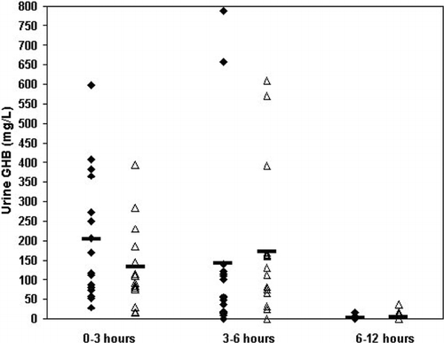
255 Clenbuterol-Associated Myocardial Injury: A Case Series
Kirrane BM (1), Johnson-Arbor K (2), Richardson WH (3), Tarabar AF (4), Ginsburg BY (1), Halcomb SE (1), McGee M (5), Stajic M (5), Hoffman RS (1). 1. New York City Poison Control Center, NY, USA, 2. Dept. of Emergency Medicine, University of Connecticut/Hartford Hospital, Hartford, USA; 3. Palmetto Poison Center, South Carolina College of Pharmacy, University of S. Carolina, Columbia, USA; 4. Section of Emergency Medicine, Yale University, New Haven, USA, 5. Office of the Chief Medical Examiner, Dept. of Health, New York, USA.
Objective: Clenbuterol is a potent, long-acting beta-2-adrenergic agonist primarily used for asthma and veterinary indications. It is also used illicitly by body builders for its sympathomimetic, lipolytic, and anabolic effects. Clenbuterol increases myocardial oxygen demand through positive inotropic and chronotropic effects, raising concerns for potential myocardial injury. Although two isolated cases of myocardial infarction are associated with oral clenbuterol use, they are complicated by both chronicity of use and the presence of additional substances (1,2). An outbreak of clenbuterol-tainted heroin in the US provided a unique opportunity to observe a disproportionately high rate of cardiac injury resulting from acute exposure. We report six cases of myocardial injury temporally associated with clenbuterol use. Methods: During January-October 2005, cases of potential exposure to tainted heroin reported to regional poison centers (RPC) were collected. Patients were identified based on a provisional case definition as described in the MMWR (3). In each case, therapy and testing were determined by the primary physicians in consultation with the RPC. Cases where cardiac enzymes were documented are included in this analysis. Results: Five US Poison Control Centers (CT, NC, NJ, NY, SC) reported a total of 34 patients who met the case definition for heroin-related clenbuterol toxicity. Chest pain, and/or ECG changes consistent with ischemia occurred in 19/34. Unfortunately, cardiac markers were only documented in 14/34. Of these, 6 patients had elevated troponin I levels consistent with myocardial injury. Clenbuterol was confirmed in the urine and/or blood in 5/6. All patients with evidence of myocardial injury were male, between 22 and 44 years old. Initial complaints included chest pain (6 patients), palpitations (1 patient), dizziness (1 patient), abdominal pain (1 patient), nausea (2 patient) and vomiting (2 patient). All patients were initially tachycardiac (mean pulse 134 per min), with a mean BP of 119/64 mm Hg. Only 2/6 patients had evidence of recent cocaine use. Troponin levels ranged from 0.12 ng/mL to 30 ng/mL, and were all elevated by hospital-based normal values. Conclusion: Acute exposures to clenbuterol may result in myocardial injury. Since cardiac markers were not obtained in every patient, it is possible that the incidence of injury is higher than reported here. References: 1. Goldstein DR, et al. Clenbuterol and anabolic steroids: A previously unreported cause of myocardial infarction with normal coronary arteriograms. South Med J 1998; 91:780–784. 2. Kierzkowska B, et al. Myocardial infarction in a 17-year-old body builder using clenbuterol. Circ J 2005; 69:1144–1146. 3. CDC. Atypical reactions associated with heroin use-Five states, January–April 2005. MMWR 2005; 54:793–796.
256 Pulmonary Edema Associated with Clenbuterol Exposure
Schechter EM (1), Hoffman RS (2), McGee M (3), Stajic M (3), Tarabar AF (1). 1. Yale University, Section of Emergency Medicine, New Haven, CT, USA; 2. New York City Poison Control Center, New York City, NY, USA; 3. New York City Office of the Chief Medical Examiner, NY, USA.
Objective: Clenbuterol is a long acting beta-2-adrenergic agonist used outside the US for treating pulmonary disorders and within the US in veterinary medicine. Due to its promotion of muscle growth and lipolysis (beta-3 effects), clenbuterol is abused by bodybuilders and the animal production industry. Recently there have been reports of clenbuterol contamination of heroin and cocaine, resulting in prolonged tachycardia, hypokalemia, hypophosphatemia, and myocardial injury. We report a case of clenbuterol toxicity causing pulmonary edema and respiratory failure. Case Report: The patient was a 33-year-old man who inhaled an unknown white powder and developed immediate onset of chest pain, palpitations, tremors, headache, nausea and vomiting. He presented to the ED where significant initial vital signs were sinus tachycardia at 146 bpm; wide pulse pressure: 116/27 mm Hg; respiratory rate 20/min and room air oxygen saturation of 99%. He was diaphoretic with a fine tremor and enlarged, reactive pupils. EKG showed 1 mm ST depressions laterally. A working diagnosis of cocaine toxicity was made, and he was treated with 5 mg of IV lorazepam and 3 L of normal saline without significant change in vital signs. Laboratory results revealed hypokalemia of 1.9 mmol/L (normal 3.5–5.0 mmol/L), hypophosphatemia 0.4 mg/dL (3.1–4.5 mg/dL), hyperglycemia 230 mg/dL (70–105 mg/dL), lactate 7.2 mmol/L (normal < 2 mmol/L) and anion gap 18. The patient was admitted to ICU and treated with benzodiazepines and haloperidol for agitation, chest pain, and persistent tachycardia. Six hours after presentation he desaturated to 64% on 100% oxygen. He was intubated for respiratory failure and CXR showed bilateral pulmonary edema. Ultrasound revealed bilateral loculated pleural effusions. Cardiac markers peaked on day 2: CK 8880 U/L (normal 24–185 U/L), CK-MB 57 ng/mL (normal <5 ng/mL), TroponinI 1.18 ng/mL (normal <0.4 ng/mL). He was extubated on hospital day 4 and discharged 9 days after initial presentation. His toxicology screen was negative for cocaine. His serum and urine analysis confirmed the presence of clenbuterol: 6 ng/mL and 874 ng/mL respectively. Conclusion: Pulmonary edema is a known complication of IV beta-2-adrenergic agonists with a reported incidence of 5% in the obstetric literature. The etiology is unknown but likely multifactorial and can include cardiogenic mechanisms such as fluid overload and catecholamine induced myocardial necrosis, and non-cardiogenic mechanisms such as direct toxicity and increased pulmonary vessel permeability. This is the first documented case of pulmonary edema following inhalational beta-2-adrenergic agonist exposure. Based on the recent epidemic of clenbuterol exposure, clinicians should be aware of the possibility of pulmonary edema and consequent respiratory failure. References: 1. Hoffman RJ, Hoffman RS, Freyberg CL, et al. Clenbuterol ingestion causing prolonged tachycardia, hypokalemia, and hypophosphatemia with confirmation by quantitative levels. Clinical Toxicology 2001; 39:339–344. 2. Hoffman RS, Burkhart K, Chan G, et al. Multistate outbreak of clenbuterol contaminated heroin and cocaine. J Toxicol Clin Toxicol 2005; 43:599–630. 3. De La Chapelle A, Benoit S, Bouregba M, et al. The treatment of severe pulmonary edema induced by beta adrenergic agonist tocolytic therapy with continuous positive airway pressure delivered by face mask. Anesthesia & Analgesia 2002; 94:1593–1594.
257 Escitalopram and Cocaine Use: A Case Series and Recreational Misadventure
Paoloni DR, Hernon CH, Babu KM, Ganetsky M, Boyer EW. Division of Toxicology, Dept of Emergency Medicine, University of Massachusetts, Worcester, USA.
Objective: We report a case series of patients who developed acute agitation after intentionally using escitalopram to prevent the typical negative symptoms following binge cocaine use. While many prescription medications have been employed to modify the effects of recreational drugs, the use of escitalopram for this purpose has not been previously reported. Case Series: Case 1: A 20-year-old male presented acutely agitated via EMS. The patient had been smoking cocaine at a party the previous night. His heart rate was 110 bpm, blood pressure was 140/90 mmHg and temperature was 100.5 °F. He was severely agitated, and diaphoretic. He required endotracheal intubation after intravenous lorazepam did not adequately control his agitation. An electrocardiogram demonstrated normal EKG intervals. Case 2: The patient's 17-year-old sister and 18-year-old girlfriend arrived to visit him shortly afterwards. Within an hour of his sister's arrival, she became progressively more agitated, and developed frank hallucinosis. She was not tachycardic or hypertensive, but did have a temperature of 100.4 °F. She also required intubation after lorazepam failed to control her agitation. Case 3: The third partygoer, patient #1's girlfriend, although agitated, had milder symptoms than the siblings. She was able to answer questions and required less benzodiazepines for sedation. She remained afebrile, with normal vital signs and normal EKG intervals. The patient was admitted for 24 hour observation. The first two cases were both extubated by 6 hours after admission. Comprehensive urine toxicologic testing of all three patients revealed the presence of cocaine metabolites, escitalopram and nicotine. On further questioning, these patients revealed that they had intentionally ingested the girlfriend's escitalopram in order to avoid “coming down hard and crashing” after their cocaine use. Conclusion: There are sporadic references in the literature of pharmaceuticals being used in combination with recreational drugs for purposes of “harm reduction” or to modify desired or unwanted effects (1,2). After the initial increased endogenous serotonergic activity associated with cocaine use, there is a decline in serotonergic tone that contributes to symptoms described as “coming down” or “crashing.” In this case series, patients used escitalopram, a highly selective serotonin reuptake inhibitor, to alleviate the malaise commonly described after smoking cocaine. It is unclear whether the symptoms displayed by the patients in this case series can be attributed to their escitalopram exposure. However, the concomitant use of cocaine and escitalopram does place recreational drug users at higher risk of developing serotonin syndrome. Toxicologists must be vigilant for the health hazards posed by use of pharmaceutical coingestants among recreational drugs users. References: 1. Brush DE, Bird SB, Boyer EW. Monooxamine Oxidase Inhibitor Poisoning Resulting from Internet Misinformation on Illicit Substances. J Toxicol Clin Toxicol 2004; 32:191–195. 2. Vuori E, et al. Death Following Ingestion of MDMA (ecstasy) and Meclobemide. Addiction 2003; 98:365–368.
258 Pyoderma Gangrenosum Following Cocaine Use
Climent B (1), Sanchez J (2), Roche E (2), Garcia D (1), Oliver V (2), Garcia-Melgares ML (2). 1. Clinical Toxicology Unit, Hospital General Universitario, Valencia, Spain; 2. Department of Dermatology, Hospital General Universitario, Valencia, Spain.
Objective: Pyoderma gangrenosum is a noninfectious neutrophilic dermatosis that usually starts with sterile pustules wich rapidly progress to painful ulcers of variable depth and size with undermined violaceus borders. In 17 to 74% of cases, Pyoderma gangrenosum is associated with an underlying disease, most commonly inflammatory bowel disease, rheumatological or hematological disease or malignancy. Diagnosis of Pyoderma gangrenosum is based on a history of underlying disease, typical clinical presentation and histopathology, and exclusion of other diseases that would lead to a similar appearance. In the literature have been described a few cases of cutaneous lesions produced by cocaine abuse but this two cases are the first reported of Pyoderma gangrenosum following cocaine use. Case Report: Two male patients, a 30- and 37-year-old presented with multiple painful lesions located on the trunk and face beginning as a tender papulopustule with surrounding erythematous induration that progressed to central shallow and ulceration. The ulcer has a purulent base with irregular undermined and overhanging violaceous borders which extends centrifugally. The histopathology of the lesions were concordant with Pyoderma gangrenosum. Explorations realized ruled out systemic disease, but urinary tests revealed cocaine positive. Later they admitted a nasal use of cocaine during 2 and 10 years respectively. Case 1 was initially treated with classical therapies for Pyoderma gangrenosum (oral and intralesional corticoids, topic tacrolimus, oral ciclosporyn, metotrexate) without improvement. Finally he was treated with infliximab and abstaining from cocaine that lead to healing the lesions. The second patient consulted later, and was directly treated with infliximab and abstaining from cocaine with healing of lesions since he was two months on treatment. Conclusion: Cocaine has been found to induce ischaemic changes mostly involving the heart or the central nervious system in drug users. Most events are attributed to transient cocaine-induced vasospams. In the literature had been reported several cases of vasculitis secondary to cocaine use but to our knowledge, this two cases are the first cases of Pyoderma gangrenosum following cocaine use. By the other hand our clinical experience in the management of this phenomenon show us that the treatment with intravenous infliximab and abstaining from cocaine conduced to scarring of the lesions. References: 1. Wollina U. Clinical mangement of Pyoderma gangrenosum. Am J Clin Dermatol 2002; 3:149–158. 2. Hofbauer GF, Hafner J, Trueb RM. Urticarial vasculitis following cocaine use. Br J Dermatol 1999; 141:600–601. 3. Brust JMC. Vasculitis owing to substance abuse. Neurol Clin 1997; 15:945–957.
259 Clenbuterol Toxicity Masquerading as Strychnine Poisoning
Kirrane BM (1), Kilbury LL (2), Stajic M (3), Mc Gee M (3), Nelson LS (1), Hoffman RS (1), Rao RB (1). 1. New York City Poison Center, NY, USA; 2. Department of Medicine, St. Barnabas Hosptial, Bronx, USA; 3. Office of the Chief Medical Examiner, Department of Health, New York, USA.
Objective: Many substances are used to adulterate street drugs. Strychnine, a competitive inhibitor of glycine at the chloride channel, is a reported contaminant of heroin that produces diffuse muscle spasms mimicking tonic-clonic seizures. Clenbuterol, a long acting beta adrenergic agonist, was identified in 2005 as an adulterant of heroin in the eastern United States. Acute toxicity is similar to that of other beta-2-adrenergic agonists including palpitations, tachycardia, tremor, anxiety, agitation, and vomiting. Electrolyte abnormalities, such as hyperglycemia and hypokalemia, and lactic acidosis are also described. We present a case of clenbuterol toxicity in an injection drug user, who presented with agitation, muscle spasms and hyperreflexia who was initially suspected to have strychnine poisoning. Case Report: A 47-year-old man injected heroin he had purchased from a new dealer. Shortly thereafter, he developed muscle cramping that progressed in severity prompting an ED visit 16 hours after exposure. He had severe pain, anxiety and diaphoresis, and was arching his back upon arrival. His vital signs were: BP, 140/84 mmHg; P, 105 beats per min; R, 28 per min T, 37.3 C. His head, neck, chest and abdominal examinations were unremarkable. A neurological examination was limited because of patient agitation, but no focal motor deficits were noted. Initial laboratories included: Na, 142 mEq/L; K, 3.3 mEq/L; Cl, 105 mEq/L; HCO3, 27 mEq/L; BUN, 3.2 mmol/L; Cr 70.4 mcmol/L and glucose 7.2 mmol/L. His CK was 5539 U/L, but troponin was negative. He was intubated and sedated with benzodiazepines and propofol. After sedation, the patient was noted to have intermittent spasms of his lower extremities, hyperreflexia and clonus. Because of the presentation of muscle spasms after heroin use, both strychnine poisoning and tetanus were strongly suspected, and presumptive treatment for tetanus was begun. He remained intubated for 8 days, and made a complete recovery. His urine and blood were negative for strychnine (performed by GC/MS), and tetanus was excluded by the rapid clinical response. However, testing for clenbuterol revealed: urine 206 ng/ml, blood 3.5 ng/ml and cerebral spinal fluid 1.9 ng/ml/(all performed by LC/MS). Conclusion: Clenbuterol toxicity can present with atypical symptoms, such as muscle spasms and agitation. Clenbuterol can also be detected in the CSF.
260 Twenty-Four-Hour Holter ECG and Arterial Blood Pressure Monitoring in Alcoholics Acutely Poisoned with Ethanol or with Withdrawal Syndrome
Lasinska V, Groszek B, Pach J. Department of Clinical Toxicology, Jagiellonian University Medical College, Kraków, Poland.
Objective: The aim of the study was to evaluate the differences in blood pressure, arrhythmias and the heart conduction disturbances in alcoholics acutely poisoned with ethanol or with withdrawal syndrome. Methods: The study group consisted of 89 males between 18 to 40 years of age (mean = 38.6 ± 7.49) with at least 5-year dependency (12.44 ± 5.59 years). The first group comprised of 59 patients admitted with withdrawal syndrome (CIWA 15–35 points, mean 19 points, blood ethanol 0 g/L), the second group comprised of 30 patients acutely poisoned with ethanol (ethanol blood concentration> 2.5 g/L). The control group consisted of 32 males, occasional drinkers, at the age between 18–50 years (mean 34.7 ± 5.98). A written consent of participation in the investigations was obtained from all the patients. In all patients 24-hour blood pressure monitoring and 24-hour ECG were performed twice – on admission and after symptoms resolving. Results: On admission, in alcoholics, compared to control group, significantly higher 24-hour systolic blood pressure (139.34 ± 10.22 v. 130.65 ± 10.65 mm Hg; p < 0.001) and 24-hour diastolic blood pressure (85.67 ± 9.29 v. 75.53 ± 11.11 mm Hg; p < 0.001) were stated; there was no significant difference between two alcoholics groups. After symptoms resolving, systolic (135.36 ± 8.69 mm Hg v. 130,65 ± 10,65 mm Hg; p < 0.05) and diastolic (82.11 ± 11.15 v. 75.53 ± 11,11 mm Hg; p < 0.05) pressure was lower, but still significantly higher than in control group. In 24-hour ECG monitoring on admission significantly higher number of nocturnal tachycardia events was noted in alcoholics group compared to the control group (10.59 v. 1.87; p < 0.05). In alcoholics with withdrawal symptoms events of irregular rhythms (615 v. 13; p < 0.001) were significantly more frequent, single supraventricular extrasystoles (7565 v. 940: p < 0.001), and bigeminy (345 v. 7; p < 0.001) events than in acutely poisoned. Bradycardia, both nocturnal (10 v. 67; p < 0.001) and diurnal (13 [min = 49 beats/min] v. 28 [50beats/min]; p < 0.001) events, single ventricular extrasystoles (8875 v. 9204; p < 0.01) occurred significantly more often in alcoholics poisoned with ethanol. In the second examination the considerably higher number of single supraventricular extrasystoles maintained despite remission of the withdrawal symptoms. The remarkably higher number of tachycardia and bradycardia in general, in comparison to the acute poisoned alcoholics persist despite withdrawal symptoms resolving. Conclusions: In both alcoholics groups higher systolic and diastolic pressure were stated, compared to the control group, after remission of symptoms. In alcoholics with withdrawal symptoms events of irregular rhythms and supraventricular extrasystoles were more frequent, which maintained after symptoms remission.
261 Increase in Cases of Cocaine Use Seen in the Emergency Department
Munné P, Nogué S, Sanjurjo E, Montori E, Rovira E, Sanchez M 1. Clinical Toxicology Unit, Hospital Clínic, Barcelona, Spain; 2. Internal Medicine Service, Hospital Clínic, Barcelona, Spain; 3. Emergency Department, Hospital Clínic, Barcelona, Spain.
Objective: In the last 5 years, surveys carried out in Spain have shown a change in the pattern of substance abuse, characterized by a significant increase in cocaine users. The objective of this study was to determine if this rise has been accompanied by an increase in hospital emergencies associated with an overdose or adverse reaction to cocaine consumption and to describe the clinical characteristics of these patients. Method: Three-year (2002–2004) retrospective study of admissions to the Emergency Department (ED) of a university hospital. Patients reporting cocaine use during the previous 24 hours were included. Epidemiological, clinical and toxicological data were collected for all patients and the evolution was recorded. The role of cocaine consumption as a direct cause of admission to the ED was analysed. Results: A total of 745 patients were included: average age 31 years, 68% males. Distribution by year was: 223 cases in 2002, 232 cases in 2003 and 290 cases in 2004. Fifty-three per cent of cases attended the ED at the weekend and 53% of these occurred between 00.00 and 12.00 hours. The main substances associated with cocaine use were: ethyl alcohol (38%), opiates (14%), cannabis (13%), and amphetamines and derivatives (9%). Cocaine was directly responsible for consulting the ED in 70% of cases. Cocaine was consumed by the nasal (sniffing) route (82%), pulmonary route (9%) and intravenous route (8%). The main reasons for consultation were anxiety or agitation (48%) and thoracic pain or palpitations (25%). Eleven per cent of cases required hospital admission (19 cases in the ICU) and there were three deaths. Conclusions: The increase in the number of cocaine users in recent years has been associated with an increase in consultations in the ED generated by cocaine. The characteristic profile of the cocaine user is a 30-year-old man who uses cocaine at the weekend, frequently with other substances, including alcohol, cannabis or opiates. The main reasons for attending the ED are generally due to the adrenergic effects of cocaine. Although cocaine overdose has a low mortality (0.4%), cocaine use causes morbidity and sometimes requires hospital admission.
262 An Epidemiologic Study of Opioid Dependent Subjects Who Were Volunteered for Opioid Detoxification in Iran, 2005
Afshari R (1), Shafaeeyan H (2), Afshari R (3). 1. Medical Treatment Deputy Department, Shiraz University of Medical Sciences, Shiraz; 2. Shafaeeyan Clinic, Shiraz; 3. Medical Toxicology Centre, Imam Reza (p) Hospital Mashhad University of Medical Sciences, Iran.
Background: In site of heavy regulation, opioid substance abuses are common in Iran (1,2) and may reach millions (Loyd A, The Times, 16.07.05). Therefore, a wide spread program of abstinence therapies exists. Methods: The files of all volunteers for detoxification from 21 March 2005 to 20 September 2005, in Shafaeeyan clinic, Shiraz (137 cases) were screened retrospectively. Results: Out of these cases 92.9% were male, 66.9% married, 34.9 (0.9) years old and started illicit drugs at 24.8 (0.8) y. They had a positive history of cigarette smoking (82.3%). They were using raw opium (79.9%), diamorphine (5.5%), illicit vials contained buprenorphine (3.5%), cannabis (0.8%) and mixed (13.8%). Their motive for applying was dominantly related to psychological (50.7%) and familial (24.9%) problems. Just 2.1% of subjects revealed concurrent ethanol use (ethanol is an illegal substance in Iran). Among them, 48.2% were smoking, 27.7% ingesting, 4.4% injecting, 2.1% snoring opioids, and 17.6% were current mixed abusers. Opioids were used predominantly 2–3 times a day in all subgroups. History of poly-substance use existed in 68.6%, which was significantly more common in younger group (p < 0.001). Diamorphine and bupronorphine were also more common in younger group (p < 0.001). Subjects mainly started illicit drugs with opium (81.9%) and cannabis (10.2%) when they were on average 25.5 (0.9) and 19.0 (0.7) years of age. Of these cases, 65.7% had previous attempts of withdrawal, which dominantly lasted one week to three months. Current age and age at which they started illicit drugs were positively correlated (r = 0.791, P = < 0.001) as shown in the Figure. Conclusion: Young male who smoke raw opium, and under psychological problems was the mode of subjects. Unlike the Western World3, the gateway to illicit drugs seems to be opium rather than cannabis, and also it is cigarette smoking related and alcohol independent. It seems that in this country an opium dependence epidemiologic transition is emerging in which younger subjects are more in favor of diamorphine and bupronorphine, start drugs in younger age, and are more interested in poly-substances abuse. References: 1. Afshari R, et al. J Toxicol Clin Toxicol 2004; 42:965–975. 2. Ahmadi J, et al. J Clin Nurs 2004; 13:60–64. 3. Otten EJ. Marijuana. In: Goldfrank's Toxicologic Emergencies, 2002:1054–1059.
FIG. 1
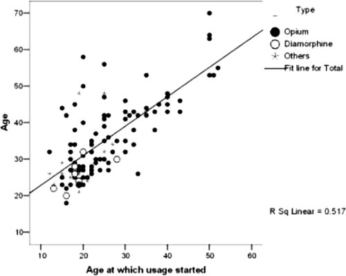
263 Atypical Reactions in Heroin Users – Western Massachusetts
Geib A (1), Babu KM (2), Ganetsky M (2), Boyer EW (2), Burns-Ewald M (1). 1. Children's Hospital Boston, Massachusetts, USA; 2. University of Massachusetts Medical School, Worcester, Massachusetts, USA.
Introduction: Clusters of recreational drug users with atypical toxicity raise the suspicion of adulteration. In early 2005, 26 patients in five states were observed to have atypical reactions to heroin use; laboratory analysis confirmed the presence of clenbuterol, a beta agonist banned in the United States, contaminating the heroin (1). Case Series: We report a series (n = 5) of heroin users presenting to western Massachusetts emergency departments in November 2005 with tachycardia, hypokalemia, hyperglycemia, and hypophosphatemia (). These cases met clinical criteria for heroin-related clenbuterol toxicity (1). One patient (Patient 4) experienced chest pain with elevation of serum troponin I; he received an esmolol infusion for rate control that was discontinued after hypotension worsened. Most patients were treated with benzodiazepines for agitation and tachycardia. All were lost to follow up by the poison center. Testing of an environmental sample likewise was lost to follow up. Discussion: Clenbuterol is a beta-2 agonist with anabolic activity (2). Its use in the US is highly restricted to treatment of airway obstruction in nonfood animals, but has been illegally imported. Toxicity has been reported from consumption of tainted livestock products and bodybuilding supplements (3,4). Toxicity has been treated with beta-blockers, benzodiazepines and judicious potassium supplementation (5.6). Clinicians should be aware of the signs of clenbuterol toxicity, as clenbuterol contamination may represent an emerging trend in illegal heroin distribution in the US. References: 1. Centers for Disease Control. Atypical reactions associated with heroin use—Five states, January – April 2005. MMWR 2005; 54:793–796. 2. Choo JJ, Horan, MA, Little RA, et al. Anabolic effects of clenbuterol on skeletal muscle are mediated by beta 2-adrenoceptor activation. Am J Physiol 1992; 263:E50–56. 3. Hoffman RJ, Hoffman RS, Freyburg CL, et al. Clenbuterol ingestion causing prolonged tachycardia, hypokalemia, and hypophosphatemia with confirmation by quantitative levels. J Toxicol Clin Toxicol 2001; 39:339–344. 4. Barbosa J. Cruz C, Martins J, et al. Food poisoning by clenbuterol in Portugal. Food Addit Contam 2005; 22:563–566. 5. Ramos F, Silveira I, Silva JM, et al. Proposed guidelines for clenbuterol food poisoning. Am J Med 2004; 117:362. 6. Chodorowski Z, Sein Anand J. Acute poisoning with clenbuterol – a case report. Przegl Lek 1997; 54:763–764.
TABLE 1 Patient characteristics
264 Choreoathetoid Movements After Excessive Cocaine Consumption, Effect of Biperidene
Pfab R, Eyer F, Jetzinger E, Zilker T. Toxikologische Abt., 2. Med. Klinik, Klinikum rechts der Isar, Muenchen, Germany.
Case report: After excessive consumption of self reported 7 grams cocaine over three days without other drugs or alcohol, a 24-year-old female experienced sudden involuntary onset choreoathetoid movements while driving her car. Since she was unable to use the steering wheel, she called the paramedics who admitted her to our toxicological department. On arrival, she had stable vital signs, no tachycardia nor hypertension, she was oriented and cooperative. She had involuntary choreoathtoid-ballistic movements of her arms and legs, and her speech was slurred. She was unable to hold or drink a glass of water. Drug screening of her urine only revealed cocaine-metabolites, no other drugs nor alcohol. Treatment with 5 mg biperidene iv resolved the movement-disorder. However, after 7 hours recurrent choreoathetosis necessitated 5 mg biperidene iv again, which had to be repeated after 5 hours a third time. Fourteen hours after admission the patient slept 19 hours and awakened completely recovered. Discussion: Choreotathetoid movements are a known, but rare effect of cocaine consumption. However, this is to our knowledge the first report about the treatment of cocaine induced choreoathetosis with biperidene.
265 Identification of a New Piperazine Related Compound in Festive Parties
Arditti J (1), Bourdon JH (1), Evrard I (2), de Haro L (1), Gilli R (1). 1. Centra Antipoison, Hôpital Salvator, Marseilles, France; 2. OFDT, Saint Denis, France.
Method: Since the beginning of 2005, several pills collected in France during festive parties and sold as Ecstasy containing MDMA were analysed by different laboratories of the SINTES programme. Results: The gas chromatography mass spectrometry analysis confirmed the presence of mCPP (1-(3-chlorophenyl) piperazine). mCPP is a serotonin agonist (5-HT2C, 5-HT1A, 5-HT1B and 5-HT1D receptors) and is one of the pharmacologically active metabolite of two antidepressants (trazodone and nefazodone). Several seizures were recently declared by French customs and the use of this piperazine-related compound was reported in numerous European Union countries (Sweden, Netherlands, Austria, Latvia, Norway and Belgium). Conclusion: As there is great suspicions about the existence of mCPP illegal market quickly growing in Europe, the European Monitoring Centre for Drugs and Drug Addiction (EMCDDA) and Europol are jointly elaborating an European report concerning this substance.
266 Outcome of Pregnancy After Maternal Treatment with Antipsychotic Drugs
Newham J (1), Thomas SHL (2), MacRitchie K (1), Young AH (1), McElhatton PR (2). 1. School of Neurology, Neurobiology & Psychiatry, University of Newcastle upon Tyne, UK; 2. National Teratology Information Service, RDTC, UK.
Objective: Pregnancy outcome is poorer in psychotic women, but many of the drug treatments are associated with fetotoxicity. This on-going prospective case series aims to collect data and assess the potential fetotoxic effects of antipsychotics during pregnancy. Method: Using standardised procedures, NTIS has provided fetal risk assessment and collected outcome data on 52 pregnancies exposed to monotherapy with neuroleptics (27) and atypicals (25). Results: Results are shown in . The majority (90–95%) of liveborn infants were healthy; 92% were born at term, none were small for dates (<2.5 Kg). Although the mean birth weight was within the expected range (3.4 kg,) 7/14 infants exposed to atypicals weighed >3.4 Kg compared with 4/16 exposed to neuroleptics. Neuroleptics: ninety percent (18/20) of liveborns had no malformations. Two infants had common minor malformations (10% vs. 2–3% expected) of unknown aetiology and three had neonatal withdrawal symptoms. The incidence of miscarriage (4/27, 15% vs. 10–20%) and elective termination (3/27, 11% vs. 23%) was within the expected range. Atypicals: Ninety-five percent (19/20) of liveborns had no malformations. One infant had malformations of unknown aetiology and withdrawal symptoms. One other infant had neonatal withdrawal symptoms. The incidence of miscarriage (2/25, 8% vs. 10–20%) and elective terminations (3/25, 12% vs. 23%) was within the expected range. Conclusions: The majority of liveborn infants were normal. The malformation incidence (3/52, 7% vs. 1/40, 2–3%) was higher than expected, but the small numbers preclude the drawing of reliable conclusions. No pattern of malformations was observed and no causal relationship could be established. Further data are required before any firm conclusions can be drawn regarding the safety of antipyschotics, particularly atypicals in pregnancy.
TABLE 1 Outcome of pregnancy following maternal monotherapy with antipsychotic drugs
267 Outcome of Pregnancy Following Maternal Treatment with Conazoles
McElhatton PR. National Teratology Information Service, RDTC, Newcastle upon Tyne, UK.
Objective: Fluconazole and itraconazole are triazole antifungal agents used orally or parenterally to treat mycotic infections. A single oral dose of fluconazole 150–200 mg is used to treat vaginal candidiasis. Higher doses (200–800 mg/d) are used chronically in severe systemic illnesses and have been associated with congenital malformations (CM) similar to Antley-Bixler syndrome but no causal relationship has been established (1,2). Itraconazole does not seem to pose the same risk of fetotoxicity (3). This ongoing prospective case series aims to collect data and assess the potential fetotoxic effects of these triazoles during pregnancy. Method: Using standardised procedures, NTIS has provided prospective fetal risk assessment and collected outcome data in 108 pregnancies exposed to these drugs mainly in the first trimester. Results: The results are shown in . Twenty-seven (63%) pregnant women were on fluconazole monotherapy and 34 (69%) in the itraconazole group. The majority (93%) of liveborn babies were healthy with no malformations, 95% were born at term and only one was small for dates (<2.5 Kg). The overall incidence of malformations was 7% vs 2–3% expected, but 4 of these were common minor malformations. The incidence of miscarriage, elective termination and sex ratio was within the expected range. The mean birth was within the range expected (about 3.4 Kg); 3.35 Kg (2.6–4.5 Kg) in the fluconazole group and 3.47 Kg (2.6–4.8 Kg) for itraconazole. Conclusions: The majority of liveborn infants were normal. The malformation incidence of 6–9% was higher than expected, but 4 were minor and no causal relationship could be established. The small numbers preclude the drawing of reliable conclusions. Further data are required before any firm conclusions can be drawn regarding the safety of triazole antifungals in pregnancy. References: 1. Mastroiacovo P, Mazzone T, Botto LD, Serafini MA, Finardi A, Caramelli L, Fusco D. Prospective assessment of pregnancy outcomes after first-trimester exposure to fluconazole. Am J Obstet Gynecol 1996; 175:1645–1650. 2. Lopez-Rangel E, Van Allen MI: Prenatal exposure to fluconazole: an identifiable dysmorphic phenotype. Birth Defects Res Part A Clin Mol Teratol 2004; 70:261. 3. Jick SS. Pregnancy outcomes after maternal exposure to fluconazole. Pharmacotherapy 1999; 19:221–222.
TABLE 1 Outcome of pregnancy following maternal treatment with triazoles
268 Multiple Bilateral Ear Malformations Associated with Prenatal Isotretinoin Exposure
Pistelli A, Caramelli L, Ieri A, Occupati B, Damiani G, Smorlesi C. Toxicology Unit, Perinatal Toxicology and Poison Control Centre, AOU Careggi, Firenze, Italy.
Background: Isotretinoin has been extensively used for the treatment of severe nodulo-cystic acne since the early 1980s. As other vitamin A derivatives, isotretinoin is a well known teratogen in both rodents and primate models. Birth control methods are strongly suggested and pregnancy should be avoided for a mean period of 3 months after discontinuing the drug. Case report: A 37-years-old woman consuming 75 mcg/day levothyroxin for hypothyroidism has started isotretinoin therapy (30 mg/day) to treat a severe case of acne while taking an oral contraceptive. Conception could not be avoided 6 weeks after starting this therapy and both drugs were withdrawn at the 4th and 2nd post-conceptional week respectively. No other pharmaceutical (apart from folic acid), environmental, professional, infectious and recreational factors were encountered during the pregnancy which proceeded apparently uneventful. The patient had already delivered 5 normal babies and referred 3 miscarriages which were not investigated in particular. When she called the Florence Teratology Information Service, during her 6th week of pregnancy, she was suggested to undergo a second level ultrasound at week 22 to better investigate the vitamin A target structures. Unfortunately we have not been able to retrieve the answer of the exam. A baby girl was spontaneously delivered at week 40. Her weight was 3720 grams, length 51 cm and head circumference 35 cm. Apgar score was 8, 9. A bilateral anotia with mandibular hypoplasia was present. The newborn was thoroughly studied for other possible anomalies which were ruled out. The brain CT scan revealed severe anomalies at bilateral/middle ear level. An external mobile acoustic vibrating device was tailored and applied since the age of 6 months. The girl is now 2-years-old and is suffering from a mild developmental neurobehavioural impairment. Her hearing function seems to be satisfactory and she's now able to articulate simple words and to identify domestic items with their exact meaning in most times. Conclusions: As previously reported in the literature, we confirm the vitamin A-derivative teratogenicity. In this case the effect of the drug was particularly focused on the inner and external ear in the absence of any other apparent involvement. So far, a definitive clinical prognosis of the patient is difficult to assess due to the involvement of still developing systems. A further rehabilitative acoustic programme has been scheduled and a permanent acoustic device will be implanted in the ear inner structures together with external plastic surgery.