Abstract
Background. The American Association of Poison Control Centers (AAPCC; http://www.aapcc.org) maintains the national database of information logged by the country's 61 Poison Control Centers (PCCs). Case records in this database are from self-reported calls: they reflect only information provided when the public or healthcare professionals report an actual or potential exposure to a substance (e.g., an ingestion, inhalation, or topical exposure.), or request information/educational materials. Exposures do not necessarily represent a poisoning or overdose. The AAPCC is not able to completely verify the accuracy of every report made to member centers. Additional exposures may go unreported to PCCs, and data referenced from the AAPCC should not be construed to represent the complete incidence of national exposures to any substance(s). U.S. Poison Centers make possible the compilation and reporting of this report through their staffs' meticulous documentation of each case using standardized definitions and compatible computer systems. The 61 participating poison centers in 2005 are:1
Regional Poison Control Center, Birmingham, AL
Alabama Poison Center, Tuscaloosa, AL
Arizona Poison and Drug Information Center, Tucson, AZ;
Banner Poison Control Center, Phoenix, AZ
Arkansas Poison and Drug Information Center, Little Rock, AK
California Poison Control System–Fresno/Madera Division, CA
California Poison Control System–Sacramento Division, CA
California Poison Control System–San Diego Division, CA
California Poison Control System–San Francisco Division, CA
Rocky Mountain Poison and Drug Center, Denver, CO
Connecticut Poison Control Center, Farmington, CT
National Capital Poison Center, Washington, DC
Florida Poison Information Center, Tampa, FL
Florida Poison Information Center, Jacksonville, FL;
Florida Poison Information Center, Miami, FL
Georgia Poison Center, Atlanta, GA
Illinois Poison Center, Chicago, IL
Indiana Poison Center, Indianapolis, IN
Iowa Statewide Poison Control Center, Sioux City, IA
Mid-America Poison Control Center, Kansas City, KA
Kentucky Regional Poison Center, Louisville, KY
Louisiana Drug and Poison Information Center, Monroe, LA
Northern New England Poison Center, Portland, ME
Maryland Poison Center, Baltimore, MD
Regional Center for Poison Control and Prevention Serving Massachusetts and Rhode Island, Boston, MA
Children's Hospital of Michigan Regional Poison Control Center, Detroit, MI
DeVos Children's Hospital Regional Poison Center, Grand Rapids, MI
Hennepin Regional Poison Center, Minneapolis, MN
Mississippi Regional Poison Control Center, Jackson, MS
Missouri Regional Poison Center, St Louis, MO
Nebraska Regional Poison Center, Omaha, NE
New Jersey Poison Information and Education System, Newark, NJ
New Mexico Poison and Drug Information Center, Albuquerque, NM
New York City Poison Control Center, New York, NY
Long Island Regional Poison and Drug Information Center, Mineola, NY
Ruth A. Lawrence Poison and Drug Information Center, Rochester, NY
Upstate (formerly Central) New York Poison Center, Syracuse, NY
Western New York Poison Center, Buffalo, NY
Carolinas Poison Center, Charlotte, NC
Cincinnati Drug and Poison Information Center, Cincinnati, OH
Central Ohio Poison Center, Columbus, OH
Greater Cleveland Poison Control Center, Cleveland, OH
Oklahoma Poison Control Center, Oklahoma City, OK
Oregon Poison Center, Portland, OR
Pittsburgh Poison Center, Pittsburgh, PA
The Poison Control Center, Philadelphia, PA;
Puerto Rico Poison Center, San Juan, PR
Palmetto Poison Center, Columbia, SC
Tennessee Poison Center, Nashville, TN
Central Texas Poison Center, Temple, TX
North Texas Poison Center, Dallas, TX
Southeast Texas Poison Center, Galveston, TX
Texas Panhandle Poison Center, Amarillo, TX
West Texas Regional Poison Center, El Paso, TX
South Texas Poison Center, San Antonio, TX
Utah Poison Control Center, Salt Lake City, UT
Virginia Poison Center, Richmond, VA
Blue Ridge Poison Center, Charlottesville, VA
Washington Poison Center, Seattle, WA
West Virginia Poison Center, Charleston, WV
Wisconsin Poison Center, Milwaukee, WI
INTRODUCTION
The American Association of Poison Control Centers (AAPCC) is a not-for-profit nongovernmental association representing the United States' 61 Poison Control Centers (PCCs) and their staffs. The AAPCC compiles the information reported by the regional PCCs into its national database. These data are used to identify hazards early, focus prevention education, guide clinical research, direct training, and detect chemical/bioterrorism incidents. AAPCC data have prompted product reformulations, repackaging, recalls, and bans; are used to support regulatory actions; and contribute to post-marketing surveillance on newly released drugs and products.
From its inception in 1983, the AAPCC's number of poisonings and exposures reported by the country's PCCs has grown dramatically, with increases in the number of participating poison centers, population served by those centers, and reported human exposures () (Citation1–22).
TABLE 1A Growth of the AAPCC Toxic Exposure Surveillance System (TESS®) database
Database Fluidity
Information in the AAPCC's database is dynamic, with follow-up calls and updated information allowing for changes in coding of some cases over time. The information reported in this article reflects only those cases classified as:
exposure calls (non-administrative, non-information calls; the caller was concerned about an exposure to a substance)
having occurred in humans (no animal species)
where the call status has been deemed closed (the PCC has determined no further information is available or no further follow-up/recommendations will be made). Most calls are closed within the first few hours; some calls about patients admitted to hospitals remain open for weeks or months depending on the particulars of a case.
Database Record Count – Exposures Reported in Humans
The cumulative AAPCC database now contains over 49 million case records of which 41.08 million represent human exposure cases. This report includes 2,424,180 human exposure cases reported to all 61 participating PCCs during 2005. While an additional 2,093 calls were classified as open at the time of preparation of this report, all prior Annual Data Reports have looked only at closed human exposure calls and for appropriate comparison this report does the same.
Trends in Reported Poisonings/Exposures
The data do not directly identify a trend in the overall incidence of poisonings in the United States because the percentage of actual exposures and poisonings reported to PCCs is unknown ().
FIG. 1. Daily count of exposures in humans as reported calls made to U.S. Poison Control Centers and transmitted to the AAPCC from 2000–2005. Not all PCCs record that a call regarding an animal has occurred if the caller is immediately referred to the ASPCA hotline.
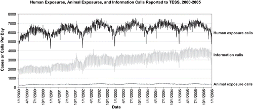
Although this report focuses on the human exposure cases reported to Poison Control Centers in 2005, the database also contains data on animal exposures (), human confirmed nonexposures (Citation7,983), animal confirmed nonexposures (375), and information calls (Citation1,400,904) ().
TABLE 1B Non-human exposures by animal type
TABLE 1C Distribution of information calls
An additional 4,688 duplicate reports (reported to more than 1 participating poison center) were excluded. This total of 3,825,084 exposure cases and information calls reported to PCCs in 2005 does not reflect the full extent of poison center effort, such as prevention and education.
In addition, 3,976,586 million follow-up calls were placed by PCCs in 2005 to provide further patient guidance, confirm compliance with recommendations, and gather final outcome data. Follow-ups were done in 44.9% of human exposure cases. One follow-up call was made in 22.2% of human exposure cases, and multiple follow-up calls (range 2–125) were placed in 21.8% of cases.
Information (Non-exposure) Calls to Poison Centers
Data from 1,400,904 information calls reported to PCCs in 2005 was transmitted to the AAPCC database, including 376,040 calls coded in optional reporting categories such as administrative, immediate referral, and prevention/safety/education (). Information calls are not required to be recorded by PCCs and may be reported inconsistently. Overall, the volume of information calls handled by U.S. PCCs increased 9.5% from 2004 to 2005.
The most frequent information call was for drug identification, comprising of 848,082 calls to PCCs during the year. Of these, 129,825 (15.3%) could not be identified over the telephone. Of the drug identification calls, 78.2% were received from the public, 8.6% from health professionals, and 12.4% from law enforcement. Forty-nine percent of drug identification requests involved drugs sometimes involved in abuse; however, these cases were categorized based on the abuse potential, generally without knowledge of whether abuse was actually intended.
Drug information calls (176,782 calls) comprised 12.6% of all information calls. Of these, 19.2% were questions about drug-drug interactions, 15.7% were questions about therapeutic use and indications, and 10.6% were questions about adverse effects. Environmental inquiries comprised 2.4% of all information calls. Of these environmental inquiries, 20.2% related to cleanup of mercury thermometers and 13.0% involved pesticides.
Poison information comprised 7.0% of information calls, with 12.3% of these information calls involving food poisoning or food preparation practices and 9.4% involving plant toxicity.
CHARACTERIZATION OF PARTICIPATING POISON CONTROL CENTERS 2005
All 61 participating centers submitted data to the AAPCC for all of 2005. Fifty-six centers (92%) were fully certified by the AAPCC at the end of 2005.
The annual human exposure case volume by center ranged from 11,478 to 113,740 (mean 40,852) for centers. The entire population of the 50 states, the District of Columbia and Puerto Rico (296.4 million people (Citation23)) was served by PCCs in 2005.
The average number of human poison exposure consultations handled per day by all U.S. poison centers was 6,642. Higher volumes were observed in the warmer months, with a mean of 6,965 consultations per day in June compared with 6,015 per day in December. On average, ignoring time of day and seasonal fluctuations, U.S. PCCs received one call concerning a suspected or actual human poisoning/exposure every 13 seconds.
Due to variations in poison center penetrance (number of calls made to a PCC per 1,000 population served), it is difficult to extrapolate the number of actual poisonings occurring annually in the United States using AAPCC data alone. Using U.S. census data, the number of human exposure cases reported to any poison center per 1,000 population was calculated by caller state. The minimum penetrance of calls from a state per 1,000 population was 3.4. The maximum number of calls from a state per 1,000 population was 24.3. Mean penetrance across states, the District of Columbia and Puerto Rico was 8.7 and the median was 8.3. If all centers had reached the penetrance level of 24.3 reported exposures in humans per 1,000 population as reported for 1 state, 7.2 million exposures in humans would have been reported to PCCs in 2005. Using the average penetrance of 8.7 calls per 1,000 population, 2.6 million calls would have been reported.
Management of Calls – Specialized Poison Emergency Providers
Calls received at U.S. PCCs are managed by healthcare professionals who have received additional training in managing poisoning emergencies. Poison Center operation as well as clinical education and instruction are directed by Managing Directors (most are PharmDs and RNs with American Board of Applied Toxicology (ABAT) board certification). Medical direction is provided by Medical Directors who are board certified medical toxicologists (MD or DO). At some poison centers, the Managing and Medical Director positions are held by the same person.
Specialists in Poison Information (SPIs) are primarily PharmDs, RNs and RPhs. They work under the supervision of a Certified Specialist in Poison Information (CSPI). SPIs must log a minimum of 2,000 calls at a poison control center to become eligible to take the certifying exam for specialists in poison information.
Poison Information Providers (PIPs) are allied healthcare professionals-in-training. They handle information-type and non-medical (non-hospital) calls and work under the supervision of at least one Certified Specialist in Poison Information (CSPI). Non-medical calls are those which do not require management recommendations to another allied healthcare professional.
U.S. PCCs employ the full-time equivalent of 75 PIPs and 635 SPIs (of whom more than 75% are CSPIs) (Citation24).
REVIEW OF 2005 HUMAN EXPOSURE DATA
No changes to the data collection format were implemented in 2005. Prior revisions had occurred in 1984, 1985, 1993, 2000, 2001, and 2002. Data reported after January 1, 2000, allow an unlimited number of substances for each case, a factor that should be considered when comparing substance data with prior years.
Exposure Site
Of the 2,424,180 human exposures reported in 2005, 92.7% occurred at a residence (). Exposures occurred in the workplace in 2.1% of cases, schools (1.4%), health care facilities (0.3%), and restaurants or food services (0.3%). Poison center peak call volumes were from 4 to 11 p.m., although call frequency remained consistently high between 8 a.m. and midnight, with 89.7% of calls logged during this 16-hour period.
TABLE 2 Site of call and site of exposure, human exposure cases
Age and Gender Distribution
The age and gender distribution of human poison exposure victims is outlined in . Children younger than three years were involved in 38.1% of cases, and 50.9% occurred in children younger than six years. A male predominance is found among recorded cases involving children younger than 13 years, but this gender distribution is reversed in teenagers and adults, with women comprising the majority of reported poison exposure victims.
TABLE 3 Age and gender distribution of human exposure cases
Exposures in Pregnancy
Of all poison exposures captured, 8,636 occurred in pregnant women. Of those with known pregnancy duration, exposures reported in patients reported as being pregnant, 32% occurred in the first trimester, 33% in the second trimester, and 26% in the third trimester. In 8.2% of cases (199,127 cases), multiple patients were implicated in the poison exposure episode (i.e., cases were coded as being related to another case, as in siblings sharing a household product, or multiple patients inhaling vapors at a hazardous material spill).
Fatalities ( and )
Fatalities differed from the total exposure data set in several ways. presents the age and sex distribution for the 1,261 reported fatalities. Although children younger than six years were involved in the majority of poisoning reports, they comprised just 1.9% (Citation24) of the recorded and verified fatalities. Fifty-six percent of poisoning fatalities occurred in 20- to 49-year-old individuals. is a log of each of the 1,261 fatalities reported to PCCs.
TABLE 4 Distribution of age and gender for 1,261 fatalities
TABLE 21 Summary of fatal exposures reported to TESS in 2005
A single substance was implicated in 91.3% of reported human exposures, and 8.7% of patients were exposed to two or more drugs or products (). In contrast, 640 (50.8%) of fatal case reports noted exposure to two or more drugs or products.
TABLE 5 Number of substances involved in human exposure cases
Chronicity
The overwhelming majority of human exposures were acute (91.5%), compared to just 51.0% of reported poisoning-related fatalities (643 of 1,261). Chronic exposures comprised 1.9% of all poison exposure reports, and acute-on-chronic exposures comprised 5.8% (chronic exposures were defined as continuous or repeated exposures occurring over a period exceeding eight hours).
Reason for Exposure
Specialists in Poison Information (ISPIs) coded the reasons for exposure reported by callers to PCCs according to the following definitions:
Unintentional general: All unintentional exposures not otherwise defined as follows.
Environmental: Any passive, nonoccupational exposure that results from contamination of air, water, or soil. Environmental exposures are usually caused by manmade contaminants.
Occupational: An exposure that occurs as a direct result of the person being on the job or in the workplace.
Therapeutic error: An unintentional deviation from a proper therapeutic regimen that results in the wrong dose, incorrect route of administration, administration to the wrong person, or administration of the wrong substance. Only exposures to medications or products used as medications are included. Drug interactions resulting from unintentional administration of drugs or foods which are known to interact are also included.
Unintentional misuse: Unintentional improper or incorrect use of a nonpharmaceutical substance. Unintentional misuse differs from intentional misuse in that the exposure was unplanned or not foreseen by the patient.
Bite/sting: All animal bites and stings, with or without envenomation, are included.
Food poisoning: Suspected or confirmed food poisoning; ingestion of food contaminated with microorganisms is included.
Unintentional unknown: An exposure determined to be unintentional, but the exact reason is unknown.
Suspected suicidal: An exposure resulting from the inappropriate use of a substance for reasons that are suspected to be self-destructive or manipulative.
Intentional misuse: An exposure resulting from the intentional improper or incorrect use of a substance for reasons other than the pursuit of a psychotropic or euphoric effect.
Intentional abuse: An exposure resulting from the intentional improper or incorrect use of a substance where the victim was likely attempting to achieve a euphoric or psychotropic effect. All recreational use of substances for any effect is included.
Intentional unknown: An exposure that is determined to be intentional, but the specific motive is unknown.
Contaminant/tampering: The patient is an unintentional victim of a substance that has been adulterated (either maliciously or unintentionally) by the introduction of an undesirable substance.
Malicious: This category is used to capture patients who are victims of another person's intent to harm them.
Withdrawal: Effect related to decline in blood concentration of a pharmaceutical or other substance after discontinuing therapeutic use or abuse of that substance.
Adverse reaction: An adverse event occurring with normal, prescribed, labeled, or recommended use of the product, as opposed to overdose, misuse, or abuse. Included are cases with an unwanted effect because of an allergic, hypersensitive, or idiosyncratic response to the active ingredients, inactive ingredients, or excipients. Concomitant use of a contraindicated medication or food is excluded and coded instead as a therapeutic error.
The vast majority (83.8%) of poison exposures were unintentional; suicidal intent was present in 8.1% of cases (). Therapeutic errors accounted for 9.9% of exposures (241,033 cases), with unintentional nonpharmaceutical product misuse comprising another 4.2% of exposures. The types of therapeutic errors observed in each age group are delineated in . Thirty-two percent of therapeutic errors involved double-dosing. Dispensing cup errors were seen in 5,466 cases, 10-fold dosing errors in 1,369 cases, iatrogenic or dispensing errors in 5,022 cases, and errors resulting from exposure to multiple products with common ingredients in 7,081 cases.
TABLE 6A Reason for human exposure cases
TABLE 6B Scenarios for therapeutic errors
Unintentional poisonings outnumbered intentional poisonings in all age groups (). In contrast, of the 1,261 human poisoning fatalities reported, 89.6% of adolescent deaths and 76.6% of adult deaths (older than 19 years) were intentional ().
TABLE 7 Distribution of reason for exposure by age
TABLE 8 Distribution of reason for exposure and age for 1,261 fatalities
Route of Exposure
Ingestion was the route of exposure in 76.7% of cases (), followed in frequency by dermal, inhalation, and ocular routes. For the 1,261 fatalities, ingestion, inhalation, and parenteral were the predominant exposure routes.
TABLE 9 Distribution of route of exposure for human exposure cases and 1,261 fatalities
Clinical Effects
The AAPCC database allows for the coding of up to 131 clinical effects (signs, symptoms, or laboratory abnormalities) per case. Clinical effects were coded in 882,083 (36.4%) of cases (18.9% had 1 effect, 9.6% had 2 effects, 4.9% had 3 effects, 1.8% had 4 effects, 0.6% had 5 effects, and 0.6% had >5 effects coded). Of 1,641,600 total clinical effects coded, 80.2% were deemed related to the exposure(s), 8.9% were considered not related, and 10.9% were coded as unknown if related.
Case Management Site
The majority of cases reported to PCCs were managed in a non–health care facility (75.5%), usually at the site of exposure, the patient's own residence (). This includes the 2.0% of cases that were referred to a health care facility but refused to go. Treatment in a health care facility was rendered in 22.8% of cases.
TABLE 10 Management site of human exposure sites
The percentage of patients treated in a health care facility varied considerably with age. Only 10.5% of children younger than six years and only 13.5% of children between six and 12 years were managed in a health care facility, compared with 48.5% of teenagers (13–19 years) and 37.1% of adults (age>19 years).
Of cases managed in a health care facility, 51.4% were treated and released without admission, 14.5% were admitted for critical care, and 8.0% were admitted for noncritical care.
Where treatment was provided in a health care facility, 37.2% of the patients were referred by the PCC, and 62.8% were already in or en route to the health care facility when the poison center was contacted.
displays the medical outcome of the human poison exposure cases distributed by age, showing a greater rate of severe outcomes in the older age groups. compares medical outcome and reason for exposure and shows a greater frequency of serious outcomes in intentional exposures. demonstrates an increasing duration of the clinical effects observed with more severe outcomes.
TABLE 11 Medical outcome of human exposure cases by patient age
TABLE 12 Distribution of medical outcome by reason for exposure in human exposure cases
TABLE 13 Duration of clinical effects by medical outcome
Medical outcome categories were as follows:
No effect: The patient did not develop any signs or symptoms as a result of the exposure.
Minor effect: The patient developed some signs or symptoms as a result of the exposure, but they were minimally bothersome and generally resolved rapidly with no residual disability or disfigurement. A minor effect is often limited to the skin or mucus membranes (e.g., self-limited gastrointestinal symptoms, drowsiness, skin irritation, first-degree dermal burn, sinus tachycardia without hypotension, and transient cough).
Moderate effect: The patient exhibited signs or symptoms as a result of the exposure that were more pronounced, more prolonged, or more systemic in nature than minor symptoms. Usually, some form of treatment is indicated. Symptoms were not life-threatening, and the patient had no residual disability or disfigurement (e.g., corneal abrasion, acid-base disturbance, high fever, disorientation, hypotension that is rapidly responsive to treatment, and isolated brief seizures that respond readily to treatment).
Major effect: The patient exhibited signs or symptoms as a result of the exposure that were life-threatening or resulted in significant residual disability or disfigurement (e.g., repeated seizures or status epilepticus, respiratory compromise requiring intubation, ventricular tachycardia with hypotension, cardiac or respiratory arrest, esophageal stricture, and disseminated intravascular coagulation).
Death: The patient died as a result of the exposure or as a direct complication of the exposure. Only those deaths that were probably or undoubtedly related to the exposure are coded here.
Not followed, judged as nontoxic exposure: No follow-up calls were made to determine the outcome of the exposure because the substance implicated was nontoxic, the amount implicated was insignificant, or the route of exposure was unlikely to result in a clinical effect.
Not followed, minimal clinical effects possible: No follow-up calls were made to determine the patient's outcome because the exposure was likely to result in only minimal toxicity of a trivial nature (the patient was expected to experience no more than a minor effect).
Unable to follow, judged as a potentially toxic exposure: The patient was lost to follow-up, refused follow-up, or was not followed, but the exposure was significant and may have resulted in a moderate, major, or fatal outcome.
Unrelated effect: The exposure was probably not responsible for the effect.
Confirmed nonexposure: This outcome option was coded to designate cases where there was reliable and objective evidence that an exposure initially believed to have occurred actually never occurred (e.g., all missing pills are later located). All cases coded as confirmed nonexposure are excluded from this report.
and outline the use of decontamination procedures, specific antidotes, and measures to enhance elimination in the treatment for patients reported in this database. These must be interpreted as minimum frequencies because of the limitations of telephone data gathering.
TABLE 14 Decontamination and therapeutic interventions
TABLE 15 Therapy provided in human exposure cases (frequency, divided by patient age groups)
demonstrates the continuing decline in the use of ipecac-induced emesis in the treatment of poisoning. Ipecac was administered in only 3,027 (0.12%) human poison exposures in 2005. A 35.6% decrease in ipecac syrup use in 2005 compared with 2004 was observed, likely as a result of ipecac use guidelines issued in late 2003. At that time, a joint Guidelines Consensus Panel formed by the American Association of Poison Control Centers, American College of Medical Toxicology, and American Academy of Clinical Toxicology issued a guideline which concluded that the circumstances in which ipecac syrup is the appropriate or desired method of gastric decontamination are rare (Citation25). In a separate report, the American Academy of Pediatrics concluded not only that ipecac should no longer be used routinely as a home treatment strategy, but also recommended disposal of ipecac currently in homes (Citation26).
TABLE 16 Decontamination trends
presents the most common substance categories involved in human exposures, listed by frequency of exposure. and present similar data for children and adults, respectively, and show the considerable differences between pediatric and adult poison exposures.
TABLE 17A Substances most frequently involved in human exposures
TABLE 17B Substances most frequently involved in pediatric exposures (children younger than 6 years)
TABLE 17C Substances most frequently involved in adult exposures (>19 years)
TABLE 18 Categories associated with largest number of reported deaths
Table 19 shows little variation over the past two decades in the percentage of cases reported to the AAPCC's national database that are fatal poisonings, and in the percentage of reported fatalities as a result of suicide. A breakdown of plant exposures is provided for those most commonly implicated ().
TABLE 19 Twenty-one year comparisons of fatality data
TABLE 20 Frequency of plant exposures by plant type
Fatalities ( and Appendix B)
U.S. PCCs recorded 1,589 calls where the medical outcome was death and there appeared to be a correlation between the reported substance(s) to which a patient was exposed and the fatality. Three-hundred twenty-eight cases were eventually determined to to be either unrelated to a poison exposure or coded incorrectly as a death (including 16 fatalities reported to one poison center which were unable to be verified). A case log summary of these 1,261 fatal human exposures is presented in . Each fatality case is abstracted by the reporting poison center and verified for accuracy as much as possible. After extensive review by both local/regional PCC staff and AAPCC reviewers, exposures determined to be either “probably” or “undoubtedly” responsible for the fatality were counted and included in .
Narrative abstracts of selected interesting or unusual cases (including most incidents with multiple fatalities), and pediatric cases in which the patient is less than six years of age (excluding carbon monoxide cases) are included in Appendix B.
also reports the highest blood concentrations for responsible agents when that information is known. In addition, identifies those cases reported indirectly to the poison center (81, or 6.4% of 1,261 cases), and those cases in which a prehospital cardiac and/or respiratory arrest occurred (626, or 49.6% of cases).
Deaths are categorized in according to the agent deemed most responsible for the death, by agreement of the medical director of the reporting center and at least two additional toxicologist reviewers. A single agent was reported as the probable cause in 621 (49.6%) deaths. Additional agents implicated (up to a maximum of 3 total agents) are listed below the primary agent. Cases in which more than three agents were involved are also identified, but agents beyond the first three are not listed in .
Characteristics of 1,261 Fatalities
The age distribution of reported fatalities is similar to that in past years, with the overwhelming majority of fatal cases occurring in adults age > 19 years (91%).
Pediatric Fatalities – Age Less than 6 Years
There were 24 fatalities reported in children younger than six years, similar to numbers reported over the last decade (). These pediatric cases represented 1.9% of total reported fatalities, similar to percentages reported over most of the last six years. The percentage of pediatric fatalities related to total pediatric calls was 0.003%. By comparison, 1.2% of all adult exposures reported recorded death as the medical outcome. Of the reported deaths in children younger than six years of age, 16 were known to be unintentional (). Two deaths in children younger than six years of age were coded as resulting from malicious intent. Of the 14 medication-associated deaths, one was from a nonprescription medication and 13 were associated with prescription medications (often not the child's prescription). Of the prescription medications, five contained opioids, including three from methadone. While this number is less than the nine reported last year, it still represents a worrisome increase in opioid-related deaths in this age range compared to earlier years. There were three fatalities related to household products, a decrease from previous years.
Pediatric Fatalities – Ages 6–12 Years
In the age range 6 to 12 years, there were 12 reported fatalities, of which 9 were from carbon monoxide exposures.
Adolescent Fatalities – Ages 13–19 Years
In the age range 13 to 19 years, there were 77 reported fatalities, slightly higher than the mean of 71 deaths in this age group reported annually since 1999, but lower than the 90 reported in 2004. Looking at the reasons for the adolescent fatalities, 39.0% were presumed suicides, and 36.4% were caused by intentional abuse. These numbers are similar to those in most recent years except for 2003 when abuse was the most common reason. As in past years, only a small number (4/77 (5.2%)) of adolescent fatalities were coded as being unintentional; two cases were due to carbon monoxide.
All Fatalities – All Ages
The most common classes of substances involved across all fatalities were analgesics, sedative/hypnotics/antipsychotics, antidepressants and stimulants/street drugs (). This relative order is similar to that seen in recent years.
Looking only at primary agents thought responsible for a poisoning death, the order changes to analgesics, stimulants/street drugs, antidepressants, cardiovascular agents, and sedative/hypnotics/antipsychotics:
In 416 fatalities, an analgesic was felt to be the primary responsible agent. Forty-eight were associated with acetaminophen as a single agent, 47 with acetaminophen plus one or two other drugs, and 92 with an acetaminophen combination product (often acetaminophen plus an opioid).
There were 20 fatalities where aspirin as a single agent was felt to be responsible. Nine acute cases recorded salicylate concentrations measured >100 mg/dL. Most of these cases did not undergo dialysis within a useful time frame. These data suggest that more aggressive and earlier use of dialysis may be indicated in the treatment of large salicylate ingestions.
Sixty-nine deaths were attributed to methadone (versus 76 cases in 2004) and 31 were attributed to oxycodone (versus 31 cases in 2004). Long-acting opioid preparations (controlled release or transdermal) other than methadone were felt to be the primary responsible agent in 32 deaths in 2005.
The second most common class of drugs associated with fatalities as the primary agent was stimulants and street drugs (148). Cocaine was noted as the primary agent in 76 cases. There was a marked jump in cases where heroin was coded as the primary agent, with 38 deaths in 2005 compared to 22 deaths in 2004 and 23 deaths in 2003. Twenty-six deaths were thought primarily related to methamphetamine use (compared to 26 cases in 2004). For the first time in three years (since 2002), gamma-hydroxybutyrate was listed as the likely cause of a poisoning fatality.
Antidepressants were the third most common class of drugs reported. When coded as the primary agent, they account for 128 deaths, similar to other recent years. Bupropion (35 deaths) surpassed amitriptyline (21 deaths) as the single most commonly recorded antidepressant associated with fatalities.
The fourth most common class of drugs associated with fatalities as the primary agent was cardiovascular agents, accounting for 120 deaths. The two most common drugs in this class were verapamil and diltiazem, accounting for 30 and 23 deaths, respectively. Long-acting preparations accounted for 33 of the deaths in this class.
The fifth most common class of drugs as the primary agent associated with deaths were the sedative hypnotics/antipsychotics. These drugs were reported as an agent of exposure 415 times, with 76 cases listing a sedative/hypnotic/antipsychotic as the primary agent. As in recent years past, alprazolam and quetiapine are the most common drugs involved, most typically in combination with other drugs.
The vast majority (75.4%) of reported fatalities in 2005, as in past years, were the result of intentional actions. The percentage of fatalities attributable to other reasons remained little changed from previous years (). A disturbing number of deaths continue to occur because of therapeutic errors; the 61 cases reported in 2005 are more than the numbers in the three previous years (41 cases in 2004, 48 cases in 2003, and 54 in 2002). Adverse drug reactions were also reported as contributing to 28 deaths.
The 10 occupational-related deaths in 2005 were similar to 2004, but fewer than in any year since 1999 (11 cases in 2004). As in the previous 3 years, there were no reported fatalities from product tampering.
Demographic Data
and provide summary demographic data on patient age, reason for exposure, medical outcome, and use of a health care facility for all 2,424,166 exposures, presented by substance categories. focuses on nonpharmaceuticals; presents drug/pharmaceuticals. Of the 2,765,665 substances logged in and , 48.9% were nonpharmaceuticals, and 51.1% were pharmaceuticals.
TABLE 22 Summary log including demographic profile of human exposure cases reported to U.S. Poison Control Centers in 2005. Profiles are broken out by AAPCC generic categories and subcategories
The reason for the exposure was intentional for 29.2% of pharmaceutical substances implicated, compared with 5.6% of nonpharmaceutical substances. Correspondingly, treatment in a health care facility was provided in a higher percentage of exposures to pharmaceutical substances (41.4%), compared with nonpharmaceutical substances (18.5%). Pharmaceutical exposures also had more severe outcomes. Of substances implicated in fatal cases, 84.8% were pharmaceuticals, compared with 51.0% of substances reported in nonfatal cases. Similarly, 85.9% of substances implicated in major outcomes were pharmaceuticals.
Surveillance
In 2005, real-time monitoring of cases submitted to the AAPCC's national database was expanded to include new surveillance case definitions, and enhanced toxicosurveillance at the regional PCC level. Monitoring results were reviewed daily by a team of five medical and clinical toxicologists working across four time zones. The core approach included monitoring of increased PCCcase activity, increased reporting of clinical effects as compared to a three year baseline, and cases that met surveillance case definitions as described in the 2003 AAPCC Annual Report.
Sixty of 61 U.S. PCCs continue to submit data to the AAPCC's database in almost real time, with most centers submitting cases every 4 to 10 minutes. When outliers are identified, surveillance query results are automatically sent for analysis to toxicologists at the AAPCC. When reports of potential public health importance are detected, additional information is obtained via e-mail or phone from reporting PCCs. Public health issues are brought to the attention of the National Center for Environmental Health/Agency for Toxic Substances Disease Registry at the Centers for Disease Control and Prevention. Affected state or local health departments are also alerted.
Data on clinical effect anamolies are provided daily to 43 individual poison centers, covering all, or parts of, 39 states. In a few cases, results are also sent directly to state or local health departments. In most states, results are interpreted by PCCstaff before the results are communicated to the appropriate health authorities.
Individual PCCs have developed surveillance case definitions, and new monitors identify cases that meet these definitions. Current surveillance definitions identify cases that have clinical effects suggestive of nerve agents, cyanide, arsenic, botulism, ricin, anthrax (systemic and dermal), irritant gases, smallpox, arenavirus, radiation, and puffer fish ingestions with neurological effects. These monitors have been implemented in response to public health issues or concerns, and are run daily at 1- to 12-hour intervals. Cases coded as specific substances, for example, arsenic, ricin, carbon monoxide, and food poisoning/food products, are also monitored. Surveillance processes and anamoly definitions continue to be developed, refined, and evaluated.
Most notably in 2005, information collected by U.S. PCCs in Gulf Coast states was used to provide post-hurricane situation awareness on substances of interest following Hurricanes Katrina (August 2005) and Rita (September 2005). Daily reports were generated and evaluated by toxicologists at the AAPCC and Centers for Disease Control (CDC) in order to identify and target where to deploy additional personnel, educational materials and public service announcements. Substances of interest included carbon monoxide, snake envenomations, reports of suspected food poisoning and water contamination, and gasoline (hydrocarbon) ingestion which may correlate with gas siphoning. This reporting system has remained in place since 2005 and continues to be used for hurricane season 2006.
Database Enhancements
In 2005, the AAPCC embarked on one of its largest and most important projects since its founding in 1958: development of new database software and migration to web-hosting of the information currently stored in the AAPCC's national poisoning and exposure database. Since 1993, the database has been used to answer many toxiclogy related questions from individual poison centers, academic researchers, public health personnel, and corporate research and development teams.
The new new web-based software for querying, reporting and surveillance application will allow the AAPCC, its member centers and public health agencies to study U.S. poisoning exposures Users will be able to access local and regional data for their own areas and view national aggregate data. The new application allows for increased “drill-down” capability and Mapping (GIS). Custom surveillance definitions will be available along with ad hoc reporting tools. The new software will serve the AAPCC well into the 21st century.
ACKNOWLEDGMENTS
We gratefully acknowledge the extensive contributions of each participating poison center and the assistance of the many health care providers who provided comprehensive data to the poison centers for inclusion in this database. We especially acknowledge the dedicated efforts of the Specialists in Poison Information (SPIs) who meticulously coded 3,968,129 million calls made to U.S. Poison Centers in 2005.
Thank you to Jacqueline Goodrich, CSPI, for her hours of assistance in verifying and standardizing information reviewed in all submitted fatality narrative abstracts.
We are thankful for information technology support from Praveen Patel.
Thank you to past and present members of the AAPCC Toxicosurveillance team who took calls 24/7 across four time zones to cover surveillance throughout 2005: Blaine (Jess) E. Benson PharmD, Douglas J. Borys PharmD, Alvin C. Bronstein MD, Melisa W. Lai MD, Anna Seroka RN, Richard Thomas PharmD, and William A. Watson PharmD.
We appreciate the review and comments and wisdom provided by Barry H. Rumack MD and Ed Krenzelok PharmD on this article.
We would like to thank AAPCC President Kathleen M. Wruk RN, MHS for a tremendous amount of service to the association, including working with American Academy of Clinical Toxicology (AACT) President Michael McGuigan MD, MBA in order to bring this report to Clinical Toxicology.
The compilation of the data presented in this report was supported in part through the U.S. Centers for Disease Control AAPCC Cooperative Agreement U50/CCU323406-02.
REFERENCES
- Veltri JC, Litovitz TL. 1983 Annual report of the American Association of Poison Control Centers National Data Collection System. Am J Emerg Med 1984; 2: 420–443, [INFOTRIEVE], [CSA]
- Litovitz TL, Veltri JC. 1984 Annual report of the American Association of Poison Control Centers National Data Collection System. Am J Emerg Med 1985; 3: 423–450, [INFOTRIEVE], [CSA], [CROSSREF]
- Litovitz TL, Normann SA, Veltri JC. 1985 Annual report of the American Association of Poison Control Centers National Data Collection System. Am J Emerg Med 1986; 4: 427–458, [INFOTRIEVE], [CSA], [CROSSREF]
- Litovitz TL, Martin TG, Schmitz B. 1986 Annual report of the American Association of Poison Control Centers National Data Collection System. Am J Emerg Med 1987; 5: 405–445, [INFOTRIEVE], [CSA], [CROSSREF]
- Litovitz TL, Schmitz BF, Matyunas N, Martin TG. 1987 Annual report of the American Association of Poison Control Centers National Data Collection System. Am J Emerg Med 1988; 6: 479–515, [INFOTRIEVE], [CSA], [CROSSREF]
- Litovitz TL, Schmitz BF, Holm KC. 1988 Annual report of the American Association of Poison Control Centers National Data Collection System. Am J Emerg Med 1989; 7: 495–545, [INFOTRIEVE], [CSA], [CROSSREF]
- Litovitz TL, Schmitz BF, Bailey KM. 1989 Annual report of the American Association of Poison Control Centers National Data Collection System. Am J Emerg Med 1990; 8: 394–442, [INFOTRIEVE], [CSA], [CROSSREF]
- Litovitz TL, Bailey KM, Schmitz BF, Holm KC, Klein-Schwartz W. 1990 Annual report of the American Association of Poison Control Centers National Data Collection System. Am J Emerg Med 1991; 9: 461–509, [INFOTRIEVE], [CSA], [CROSSREF]
- Litovitz TL, Holm KC, Bailey KM, Schmitz BF. 1991 Annual report of the American Association of Poison Control Centers National Data Collection System. Am J Emerg Med 1992; 10: 452–505, [INFOTRIEVE], [CSA], [CROSSREF]
- Litovitz TL, Holm KC, Clancy C, Schmitz BF, Clark LR, Oderda GM. 1992 Annual report of the American Association of Poison Control Centers Toxic Exposure Surveillance System. Am J Emerg Med 1993; 11: 494–555, [INFOTRIEVE], [CSA], [CROSSREF]
- Litovitz TL, Clark LR, Soloway RA. 1993 Annual report of the American Association of Poison Control Centers Toxic Exposure Surveillance System. Am J Emerg Med 1994; 12: 546–584, [INFOTRIEVE], [CSA], [CROSSREF]
- Litovitz TL, Felberg L, Soloway RA, Ford M, Geller R. 1994 Annual report of the American Association of Poison Control Centers Toxic Exposure Surveillance System. Am J Emerg Med 1995; 13: 551–597, [INFOTRIEVE], [CSA], [CROSSREF]
- Litovitz TL, Felberg L, White S, Klein-Schwartz W. 1995 Annual report of the American Association of Poison Control Centers Toxic Exposure Surveillance System. Am J Emerg Med 1996; 14: 487–537, [INFOTRIEVE], [CSA], [CROSSREF]
- Litovitz TL, Smilkstein M, Felberg L, Klein-Schwartz W, Berlin R, Morgan JL. 1996 Annual report of the American Association of Poison Control Centers Toxic Exposure Surveillance System. Am J Emerg Med 1997; 15: 447–500, [INFOTRIEVE], [CSA], [CROSSREF]
- Litovitz TL, Klein-Schwartz W, Dyer KS, Shannon M, Lee S, Powers M. 1997 Annual report of the American Association of Poison Control Centers Toxic Exposure Surveillance System. Am J Emerg Med 1998; 16: 443–497, [INFOTRIEVE], [CSA], [CROSSREF]
- Litovitz TL, Klein-Schwartz W, Caravati EM, Youniss J, Crouch B, Lee S. 1998 Annual report of the American Association of Poison Control Centers Toxic Exposure Surveillance System. Am J Emerg Med 1999; 17: 435–487, [INFOTRIEVE], [CSA], [CROSSREF]
- Litovitz TL, Klein-Schwartz W, White S, Cobaugh DJ, Youniss J, Drab A, Benson BE. 1999 Annual report of the American Association of Poison Control Centers Toxic Exposure Surveillance System. Am J Emerg Med 2000; 18: 517–574, [INFOTRIEVE], [CSA], [CROSSREF]
- Litovitz TL, Klein-Schwartz W, White S, Cobaugh DJ, Youniss J, Omslaer JC, Drab A, Benson BE. 2000 Annual report of the American Association of Poison Control Centers Toxic Exposure Surveillance System. Am J Emerg Med 2001; 19: 337–395, [INFOTRIEVE], [CSA], [CROSSREF]
- Litovitz TL, Klein-Schwartz W, Rodgers GC, Cobaugh DJ, Youniss J, Omslaer JC, May ME, Wodf AD, Benson BE. 2001 Annual report of the American Association of Poison Control Centers Toxic Exposure Surveillance System. Am J Emerg Med 2002; 20: 391–452, [INFOTRIEVE], [CSA], [CROSSREF]
- Watson WA, Litovitz TL, Rodgers GC, Klein-Schwartz W, Youniss J, Rose SR, Borys D, May ME. 2002 Annual report of the American Association of Poison Control Centers Toxic Exposure Surveillance System. Am J Emerg Med 2003; 21: 353–421, [INFOTRIEVE], [CSA], [CROSSREF]
- Watson WA, Litovitz TL, Klein-Schwartz W, Rodgers GC, Youniss J, Reid N, Rouse WG, Rembert RS, Borys D. 2003 Annual report of the American Association of Poison Control Centers Toxic Exposure Surveillance System. Am J Emerg Med 2004; 22: 335–404, [INFOTRIEVE], [CSA], [CROSSREF]
- Watson WA, Litovitz TL, Rodgers GC, Klein-Schwartz W, Reid N, Youniss J, Flanagan A, Wruk KM. 2004 Annual report of the American Association of Poison Control Centers Toxic Exposure Surveillance System. Am J Emerg Med 2005; 23: 589–666, [INFOTRIEVE], [CSA], [CROSSREF]
- , U.S. Census Bureau. 2005 population estimates. http://factfinder.census.gov/servlet/DatasetMainPageServlet?_program=PEP&_submenuId=&_lang=en&_ts=
- 24. American Association of Poison Control Centers' 2004 Annual Membership Survey (unpublished data).
- Manoguerra AS, Cobaugh DJ. Guideline on the use of ipecac syrup in the out-of-hospital management of ingested poisons. J Toxicol Clin Toxicol 2004; 43: 1–10, [CSA]
- American Academy of Pediatrics Policy Statement. Poison treatment in the home. Pediatrics 2003; 112: 1182–1185, [CSA], [CROSSREF]
APPENDIX A
AAPCC's 2005 fatality verification process involved the preparation and review of abstracts on 1,589 fatalities reported to poison centers, 328 of which were eventually determined to be either unrelated to a poison exposure or coded incorrectly as a death. The review process requires the dedication and commitment of hundreds of poison center staff members; more than could possibly be listed here. The following fatality abstract authors were identified by their poison centers as having made a major contribution to this effort. These individuals are acknowledged for their commitment to toxicosurveillance through the careful verification and preparation of clinical abstracts of poisoning cases. Without the dedicated contributions of these individuals, this report would not be possible.
The contributing authors and reviewers in calendar year 2005 are:
Aaron, Cynthia
Abbott-Teter, Cynthia L.
Akhtar, Jawaid
Albertson, Timothy E.
Alsop, Judith A.
Anderson, Deborah L.
Arnold, Thomas C.
Artalejo III, Leopoldo
Audi, Jennifer
Baker, S. David
Ballou, Dawn
Baltarowich, Lydia
Banner, William
Barker, Kim
Benitez, John G.
Bernstein, Jeffrey N.
Beuhler, Michael C.
Bilden, Elisabeth F.
Bond, G. Randall
Borys, Douglas J.
Bosse, George M.
Bottei, Edward M.
Boyer, Leslie V.
Bronstein, Alvin C.
Brooks, Daniel E.
Bryant, Sean M.
Burns Ewald, Michele
California Poison Control System San Diego Division Fellows
Cantor, Richard
Cantrell, Lee
Caraccio, Thomas R.
Caravati, E. Martin
Casavant, Marcel J
Clancy, Cathleen
Cleary, Jean
Cobb, Douglas B.
Connecticut Poison Control Center Toxicology Fellows
Cox, Robert
Cumpston, Kirk
Daubert, G. Patrick
Dorough, Lois I.
Doyon, Suzanne
Durback-Morris, Lynn F.
Eisenga, Bernard H.
Eldridge, David L.
Enders Shelly
Fern‡ndez, Miguel C.
Fisher III, John G.
Foster, Howell
Furbee, R. Brent
Gaar, Gregory G.
Garrison, James
Geib, Ann-Jeannette
Geller, Richard J.
Geller, Robert J.
Gracia, Rebecca
Gummin, David
Hantsch, Christina E.
Haynes, Jr, John F.
Henretig, Fred M.
Hesse, Carol L.
Hoffman, Robert S.
Holmes, Becky L.
Holstege, Christopher P.
Horowitz, B. Zane
Hughes, Michael P.
Jaramillo, Jeanie
Joshi, Prashant
Kashani, John
Kemmerer, David A.
Kerns, William
Kirk, Mark A.
Kostic, Mark A.
Kunisaki, Thomas A.
Lai, Melisa W.
Lawrence, Ruth A.
Liebelt, Erica
Lewis-Younger, Cynthia
Lopez, Gaylord
LoVecchio, Frank
Lovely, Perry L.
Lowry, Jennifer
Marraffa, Jeanna M.
McGoodwin, Lee
McGuigan, Michael A.
McKay, Charles
McNally, Jude T.
Maloney, Gerald
Mercurio-Zappala, Maria
Michels, Jill E.
Miller, Michael A.
Morgan, Brent
Morgan, David L.
Mowry, James B.
Mrvos, Rita
Muller, Allison A.
Nester, Mary Lou
Nichols, Michele
Oller, Lisa
Olson, Kent R.
Omslaer, Judith C.
Patton, Jill B.
Quang, Lawrence
Reed, Michael
Richardson, William H.
Rivera, Hector L.
Rivera, Wilfredo
Roberts, David J.
Robertson, William O.
Rose, S. Rutherfoord
Rossi, Pamala R.
Rowden, Adam K.
Ryan, Mark L.
Sangalli, Bernard
Sawyer, Tama
Scalzo, Anthony J.
Schultz, Debora
Scruton, Susan
Seger, Donna
Seifert, Steven A.
Serafin, David
Shum, Shu
Simmons, Henry F.
Simone, Karen E.
Smith, Cindy
Smolinske, Susan C.
Snodgrass, Wayne
Spiller, Henry A.
Stremski, Ernest
Stork, Christine M.
Sweeney, Rachel
Tharratt, R. Steven
Thompson, Jon
Tomassoni, Anthony J.
Wahl, Michael
Waszolek, Kathleen
Weisman, Richard S.
White, Suzanne R.
Whitlow, K. Scott
Wittler, Mary
APPENDIX B
Abstracts of select cases from 1,261 human fatalities thought to be related to a poisoning exposure as reported to U.S. Poison Control Centers in 2005. Drug and chemical concentrations provided in these abstracts were measured in blood, serum or plasma unless otherwise indicated.
Case 35. A 51-year-old man was arrested for driving “under the influence” and indecent exposure and jailed. He reported that he had ingested antifreeze (ethylene glycol). The patient became short of breath and obtunded while in jail. Upon arrival in the ED, the patient was comatose and being bag ventilated. His vital signs were: heart rate, 90 beats/min; blood pressure, 70/50 mm HG; temperature, 94.5 ûF rectally. He was intubated. An initial arterial pH was 6.7. The patient received warm intravenous fluids, sodium bicarbonate, and calcium gluconate. His blood pressure rose to 105/50 mm Hg. His initial laboratory values were: glucose, 170 mg/dL; BUN, 19 mg/dL; creatinine, 2.3 mg/dL; sodium, 147 mEq/L; potassium, 6.9 mEq/L; chloride, 101 mEq/L; bicarbonate, <5 mEq/L; AST, 22 U/L; ALT, 15 U/L; and blood alcohol, undetected. He received thiamine, folate, pyridoxine, vasopressors, and was hemodialyzed. The patient's metabolic acidosis and hypotension resolved following hemodialysis; however his neurologic status never improved and life support was withdrawn.
Case 53. An 18-month-old boy ingested two button cell disc batteries that he found in the trash. He vomited for one day, but his symptoms were attributed to a respiratory illness in the family who were unaware of the battery ingestion. When he was seen in the ED for persistent symptoms, an x-ray showed one battery in the esophagus and one battery in the stomach. After a delay of several hours, the child was transferred to another hospital where both batteries were removed. The child was admitted for four days and a barium swallow done during the admission showed no perforation but an undefined esophageal deviation. On discharge the child had a fever and was sent home on an antibiotic and medication for acid reflux. On the fourth home day the child woke cyanotic. On readmission he had a high white blood cell count and was in shock. He died later that day. The death certificate listed the cause of death as aortoesophageal corrosive ulcer.
Case 54. A 55-year-old man was bitten on the hand by an Eastern diamondback rattlesnake (Crotalus adamanteus). He immediately began experiencing shortness of breath. His son described his father having trouble breathing and unable to talk. Upon presentation in the ED he was hypotensive, diaphoretic, and had ectopy on his monitor. The swelling was onto his forearm. He was given 4 vials of antivenom (Crotalidae polyvalent immune Fab), as well as amiodarone, with reported clinical improvement. Laboratory values were: PT, 17 sec; INR, 1.7; and platelet count, 109, 000 /μL At followup, approximately 27 hours after presentation, 16 vials of antivenom had been given. Laboratory values at that time showed a PT of 20 sec with an INR of 2, and a fibrinogen level of 195 mg/dL. On the 6th hospitalized day, he showed signs of recurrent coagulopathy with the following laboratory values: platelet count, 93,000 /μL; fibrinogen, < 35 mg/dL; and PT and PTT both > 150 sec. It was unclear whether blood products or additional antivenom were given. He was transferred back to the ICU, but later that day he developed neurological deficits and subsequently became unresponsive. A head CT showed a hemorrhage. He died the following day.
Case 55. A 25-year-old man reportedly told his family that he had been bitten by a rattlesnake (Crotalus horridus horridus). He was being driven to a rural hospital when the car had a flat tire. The patient reportedly became unconscious and was taken on to the hospital by a passing motorist. By the time he got to the ED he was dead. An autopsy revealed an apparent bite mark on the back of the right hand, associated with hemorrhagic necrosis of the underlying soft tissue. A heart blood ethanol level was 290 mg/dL. It was the opinion of the pathologist that the probable cause of death was a snake bite.
Case 56. The poison center was informed about a 44-year-old man who was stung on the temple by an unknown hymenoptera. He reportedly waited several hours to seek medical attention. He was eventually declared brain dead. The medical examiner confirmed that death was due to an anaphylactic reaction to the sting.
Case 57. A 32-year-old man presented in the ED following a rattlesnake bite. He reportedly had a respiratory arrest about 10 minutes after the bite and arrived in the ED 10 minutes later. In the ED his pupils were fixed and dilated. Antivenom was administered after initial fluid resuscitative measures were taken. Oxygen saturation returned to 98% and blood pressure stabilized. He was transferred to a tertiary-care facility where his PT and PTT were slightly prolonged and his platelet count was 120,000/μL. Additional antivenom was administered. His neurologic status throughout hospitalization suggested anoxic brain injury, confirmed by head CT and EEG. The patient died on the 4th hospital day, apparently of anoxic brain injury following an anaphylactic reaction to a snakebite. Further history revealed that he had been bitten several times in the past.
Case 60. A 44-year-old man saw a 4-foot long snake (presumed Crotaline) and chased it into a wooded ravine in an attempt to catch it. His body was found two days later. Autopsy examination showed four puncture marks on the first dorsal web space of his right hand with swelling, discoloration, and cellular/tissue lysis of the surrounding muscle and tissue. Marked edema of the larynx, epiglottis, and surrounding upper airway tissues was noted as well. A blood ethanol level was 120 mg/dL.
Case 62. A 19-year-old woman with a history of depression was found slumped over a bathtub after a suspected ammonia and bleach ingestion, followed by aspiration. EMS performed cardioversion and endotracheal intubation prior to arrival in the ED. A urine drug screen was negative for drugs of abuse. The patient was supported on the ventilator and high dose vasopressors, but remained hemodynamically unstable and unresponsive. She suffered a cardiac arrest on hospital day 2 and did not respond to resuscitation attempts.
Case 63. A 23-year-old college student dropped out of class, acquired some sodium cyanide, e-mailed a suicide note to a relative, mixed the cyanide with a liquid and drank it. He was found dead three days later, shortly after the e-mail message was read. The postmortem blood cyanide level was reported as >10 μg/mL.
Case 66. A 55-year-old man ingested a white powdery substance while being arrested. He stated that it was potassium cyanide and that he was going to die. He was transported to a small rural hospital, where he presented in respiratory distress 15 minutes after ingestion. He had palpable pulses but no detectable blood pressure. He was intubated, started on vasopressors and given sodium bicarbonate. He then went into PEA and CPR was started. The hospital did not have a cyanide antidote kit in stock. They had called a flight evacuation service which arrived with a cyanide antidote kit 45 minutes post-ingestion. Aggressive supportive care and the delayed cyanide antidote were unsuccessful and the patient was declared dead.
Case 85. A 24-year-old man was found unresponsive after reportedly ingesting 6 ounces of an embalming fluid containing formaldehyde and methanol. In the ED the patient was asystolic and could not be resuscitated. Postmortem examination revealed complete tissue fixation of the upper gastrointestinal tract up to the pylorus and mesenteric areas adjacent to stomach. Postmortem methanol levels were 43 mg/dL in the blood and 36 mg/dL in vitreous fluid.
Case 87. A 58-year-old man with a past history of morbid obesity and extensive coronary artery disease presented in the ED after unintentionally ingesting a mouthful of a “truck cleaner” inappropriately stored in a drinking water bottle. Hydrofluoric acid ingestion was suspected. He presented with extensive retching and intense burning in his throat, chest, and abdomen. Initial calcium was 9.0 mg/dL. About 2 hours later he went into torsade de pointes followed by ventricular fibrillation. Calcium was then 5.2 mg/dL. Calcium and magnesium were administered and he was defibrillated. He did not respond and died approximately 4.5 hours later. Postmortem examination revealed that this combination product contained unknown concentrations of hydrofluoric, sulfuric, and phosphoric acids in a container labeled as a commercial brand of drinking water. No gross evidence of acid-related injury to the gastric mucosa was present. However, microscopic examination revealed patchy mucosal erosions and areas of hemorrhage in the stomach.
Case 88. Fifteen people became ill after coming to work at a plant that stores cylinders of methyl bromide gas. The employees were all attending a meeting when they developed vomiting, diarrhea, and eye irritation. All were seen at an ED and fourteen of the fifteen were discharged. All of the employees had methyl bromide present in blood samples taken in the ED. One adult with a history of unspecified underlying medical conditions came to work 2.5 hours before the meeting and was already feeling ill at the time the meeting started. About 30 minutes later, he became paralyzed although he remained awake and alert. On arrival in the ED, he had a seizure that was controlled with medication. He was admitted to the hospital and died about 10 hours later. Later on the day of the meeting, the canisters of methyl bromide stored at the plant were examined and three of them were found empty or partially empty. No air levels of bromine were measurable. A water cooler that pulls air into the inverted container as water is used was in the area of the meeting and was used to make coffee about an hour before the meeting, pulling room air into the inverted large bottle. The bottle was sealed and tested, and the air in the bottle was found to contain methyl bromide. The fatality was believed to have been caused by a combination of effects from the methyl bromide and the man's underlying medical condition(s).
Case 97. An 83-year-old woman with Alzheimer's disease ingested an estimated 8 ounces of a liquid dishwashing detergent (anionic/nonionic). She was lavaged and given intravenous fluids in the ED. The patient aspirated and, within 4–5 hours of ingestion, was intubated and transferred to the ICU for respiratory failure. The patient experienced renal failure, elevated liver enzymes, and at least one seizure. The patient continued to require ventilatory support and dopamine. She had a cardiac arrest approximately 16 hours after exposure and could not be resuscitated.
Case 98. An 85-year-old man with a medical history of “confusion” reportedly ingested 400 mL of a liquid dishwashing detergent (anionic/nonionic). The patient developed profuse watery diarrhea with hourly stools. Initial laboratory values were normal. Over the next 24 hours he reportedly had approximately 10 liters of stool. He also apparently developed a bowel obstruction with vomiting of fecal-like material. In spite of fluid replacement and supportive care the patient died about 30 hours after presentation.
Case 111. A 90-year-old woman with a history of dementia was witnessed to drink a few swallows of a cleaner containing pine oil/isopropyl alcohol cleaner, thinking it was Gatorade™. She began vomiting. EMS was called. She became asystolic en route to the hospital. In the ED she underwent a prolonged resuscitation. Supportive care was withdrawn later that day by the family and she died.
Case 112. A 102-year-old woman with do not resuscitate orders presented to an ED smelling of a pine oil/isopropyl alcohol cleaning product, which she had reportedly ingested. She was responsive only to pain. Her vital signs were: heart rate, 84 beats/min; blood pressure, 110/54 mm Hg; respiratory rate, 24 breaths/min; oral temperature, 97.8 ûF; pulse oximetry, 100% on 2L of oxygen. Her initial chest x-ray was clear. Seventeen hours after her exposure she was awake and alert with stable vital signs and scattered rhonchi on her pulmonary examination. Her respiratory status continued to decline and she died due to respiratory failure at 37 hours after her exposure.
Case 117. A 48-year-old man presented in the ED after unintentionally ingesting a wheel cleaner containing hydrofluoric acid. The wheel cleaner had been placed in a drink container. Presenting symptoms included vomiting, drooling and pharnygeal erythema. The patient was intubated, sedated and received intravenous calcium, magnesium, sodium bicarbonate, and fluids. Laboratory values approximately 2 hours post ingestion included: potassium, 3.1mEq/L; calcium, 6.3 mg/dL; magnesium, 1.6 mg/dL; hemoglobin, 4 g/dL (decreased from an initial value of 14.4 g/dL); pH, 7.24; and creatinine, 1.3 mg/dL. Despite supportive care, the patient developed refractory hypotension and then cardiac arrest approximately 5 hours after the ingestion. Resuscitation was unsuccessful.
Case 123. A 26-year-old woman was found in Pulseless Electrical Activity (PEA) with a bottle of holding tank sealer and deodorant, approximately half of which was gone. Unfortunately, during the course of case management, the exact identity of what the patient ingested was unclear. The bottle from which the chemical was ingested was lost within the hospital. She was treated with supportive care and 4-methylpyrazole because of the possibility that the product contained methanol. The patient remained acidotic and developed a coagulopathy and bleeding diathesis. Less than 24 hours into the hospitalization, she coded and could not be resuscitated. The pathologist determined that the product the patient ingested was 10–15% methanol and 20–25% formaldehyde.
Case 124. A 27-year-old man presented in the ED with altered mental status. The patient was intubated and sedated. Initial vital signs were: blood pressure, 150/90 mm Hg; heart rate, 91beats/min; respiratory rate, 18 breaths/min; temperature, 98.2ûF; and oxygen saturation of 100% on a ventilator. Initial laboratory values were: sodium, 104 mEq/L; potassium, 2.8 mEq/L; chloride, 65 mEq/L; bicarbonate, 26 mEq/L; glucose, 111mg/dL; AST, 150 U/L; ALT, 114 U/L; creatine kinase, 2632 U/L. EKG, chest X-ray and CT of the head were all normal. The patient was started on 3% saline. The history obtained from the patient's sister was that he was a recent immigrant from El Salvador who was apparently healthy until three weeks prior when the patient went to see a “Curandera” for abdominal pain. He was given “Aceite de Resina” and one week later was given “Te de Medianoche.” One week later, the patient was seen by a primary care physician and was prescribed trimethoprim-sulfamethoxazole. The “Aceite de Resina” container had been disposed of but the “Te de Medianoche” container had been brought in and the active ingredients were Menta – Satureja macrostema, Poleo – Mentha pulegium, Hierva de San Juan – Hypericum perforatum 330 GRF, and Melisa – Citronella mexicana 30 GRF. The morning after the admission Mentha pulegium was identified as pennyroyal. The patient was started on N-acetylcysteine. Liver enzymes returned to normal within four days of admission. The patient remained comatose on pressors and sedation. Two weeks into his course he developed bilateral pneumothoraces requiring chest tube placement. By three weeks after admission the patient was unresponsive to any stimuli. An EEG showed minimal brain activity. Twenty-nine days after admission the patient was removed from life support and died.
Case 125. An 82-year-old man with dementia developed slurred speech and weakness. Four days prior the patient had eaten home-canned food of unknown shelf life. The following day, the patient's speech was slurred. The patient ultimately was taken to the ED where he had minimal movement and was intubated. He was able to wiggle fingers and toes. By hospital day 1, he was only able to wiggle his toes and by the end of the night, the patient was completely paralyzed. On hospital day 4 he had a negative spinal tap and there was concern for botulism. Botulinum immune globulin was sent from the CDC and testing was started for botulism. By hospital day 6, the patient's respiratory rate had increased. On Hospital Day 13, testing came back positive for botulism type B. The patient did receive botulinum immune globulin. The patient started to improve and then decompensated again and ultimately died.
Case 126. A 67-year-old woman was admitted to the hospital with nausea, vomiting, diarrhea and right upper quadrant pain. The patient also had jaundice and hemolysis. Blood cultures grew a heavy growth of Clostridium perfringens the day after the patient died.
Cases 127, 142, and 147. A 3-year-old girl died of carbon monoxide poisoning. A family member committed suicide by running his automobile in an attached garage, killing other family members.
Cases 129, 130, 139, and 140. A family of four, including two 8-year-old children, was found dead in a bedroom. They had been dead for several days. The father had sealed the family in the bedroom and started charcoal fires while they slept. He left a suicide note. The ambient carbon monoxide level was 71 ppm.
Case 131. An 11-year-old boy was found unresponsive and apneic in an idling automobile that was covered in ∼2 feet of snow from a recent blizzard. He was last seen 3 hours earlier. He was transported to the nearest ED by EMS, but had a cardiac arrest en route. Carbon monoxide was suspected and a carboxyhemoglobin level drawn upon ED arrival was 54%. CPR was unsuccessful and the patient died.
Case 186. A 28-year-old train engineer presented to an ED following a train wreck where a chlorine tank car ruptured, releasing a cloud of chlorine gas into the environment. The patient inhaled the chlorine and presented to the hospital in respiratory distress. He was intubated but, despite aggressive pulmonary care, the patient's respiratory status worsened and he died.
Case 188. A 57-year-old man with a history of chronic depression and multiple suicide attempts was found unconscious by his sister with a bag over his head attached to a helium cylinder. A book on “methods of suicide” was lying beside him. Upon EMS arrival, the patient was in full cardiopulmonary arrest. The patient responded to CPR and was admitted to the ICU. Eighteen hours after presentation the family decided to withdraw support, he was declared brain dead and became an organ donor.
Cases 189 and 190. Two men, aged 41 and 56 years, were found unconscious in an underground sewer. They were successfully resuscitated by EMS, admitted to the ICU and treated with hyperbaric oxygen. Initial carboxyhemoglobin and methemoglobin levels were both <1%. Neither patient regained consciousness and both were declared dead within 24 hours of admission. The cause of death was thought to be hydrogen sulfide.
Cases 192, 193, and 194. Three workers on a cruise ship entered a room to clean up a sewage leak. All three were later found in cardiopulmonary arrest and pronounced dead prior to hospital transport. The toxin was subsequently identified as hydrogen sulfide through environmental monitoring by a hazardous materials team. Nineteen other persons were exposed but survived.
Case 198. A 37-year-old man was found dead with a respirator mask connected to tank of chlorofluorocarbon over his face. The man worked for a HVAC company and had easy access to the chlorofluorocarbon. Death was ruled an accident by the coroner.
Case 203. A 15-month-old girl was found in the garage by her father vomiting and in respiratory distress. EMS was called and found the child vomiting, blue and with the odor of gasoline. The child was intubated and transported to the ED. The child's condition rapidly worsened, with evidence on sequential chest X-rays of worsening bilateral infiltrates. Prior to a transport flight to a tertiary healthcare facility, the child suffered a cardiopulmonary arrest and could not be resuscitated.
Case 205. A 61-year-old man was brought to the hospital with burns over approximately 20% of his body. There were conflicting stories as to the cause. It was thought that he had fallen asleep in front of a kerosene stove, but after he had been in the hospital 2 days he passed some kerosene-like fluid from his rectum and it was suggested that he might have ingested kerosene and self-inflicted the burn. He had multiple complications including renal failure and pneumonia with ARDS, requiring intubation and assisted ventilation. He died one week after admission.
Case 206. A 2-year-old boy ingested an unknown amount of cigarette lighter fluid (naptha) with resultant cough. When the poison center was contacted the child was enroute to the hospital and receiving CPR. An x-ray in the ED showed complete opacification of both lungs. The child died shortly after arriving in the ED.
Case 207. A 56-year-old previously healthy man picked and ate mushrooms. Approximately 8 hours later he developed nausea, abdominal cramps, vomiting and diarrhea. Over the next several hours he became progressively worse and finally presented to the ED approximately 19 hours post ingestion. He was treated with IV fluids. Laboratory investigation at that time revealed AST 39 U/L, ALT 59 U/L, INR 0.94, BUN 22 mg/dL, and serum creatinine 1.0 mg/dL. A preliminary description of the mushrooms could not rule out Amanita species so multi-dose activated charcoal was recommended while awaiting definitive identification. By the following morning AST was 201 U/L, ALT 243 U/L and the mushroom was identified as Amanita bisporigera. N-acetylcysteine, high dose penicillin, ascorbic acid, and cimetidine were recommended as further therapies that might potentially decrease toxicity. Despite this treatment he developed fulminant hepatic failure and renal failure. He was transferred to a transplant center for possible liver transplantation. He remained encephalopathic, coagulopathic, and anuric and developed atrial fibrillation and hypotension. He died 4 days post ingestion.
Case 208. A 56-year-old Laotian man picked wild mushrooms in the forest and cooked them for himself the day prior to admission. He presented to an urgent care center with nausea, vomiting and diarrhea. He received metoclopromide and promethazine in addition to IV fluids. Laboratory measures of hepatic and renal function were normal but he was admitted to the hospital. The mushroom was identified as an Amanita bisporigera on the basis of questioning of the patient by a mycologist. On the second day his liver enzymes were mildly elevated. The patient and his family declined to be listed for an urgent liver transplant. Laboratory values on day 3 were: INR, 5; AST, 6262 U/L; ALT, 4770 U/L; ammonia, 92 μmol/L; and pH, 7.45. The patient remained awake and alert but complained of generalized abdominal pain. By hospital day 6 the patient was delirious. By day 7 the patient was comatose and was intubated. His liver enzymes began to decline but his INR and bilirubin were rising. The patient started to regain consciousness on hospital day 13 but his bilirubin continued to rise, peaking at 28.4 mg/dL on day 15. The patient was extubated and gradually became more responsive, asking for food. He was discharged from the hospital after 21 days with a follow-up visit scheduled for the toxicology clinic in about 3 weeks. He did not come to that appointment and two days later was readmitted to the hospital with the diagnosis of ongoing liver failure and sepsis. The patient died from sepsis (E. coli in femoral catheter and mold in sputum) about two months after his initial ingestion.
Case 210. A 15-year-old girl ingested approximately 5 pellets (by patient history) of aluminum phosphide in a stated suicide attempt. It is unclear how the patient obtained this product. She presented in the ED approximately 45 minutes post ingestion with confusion, bradycardia, diaphoresis, cyanosis, mydriasis, hypereflexia, and incontinence. The patient was intubated, given activated charcoal and then lavaged. Approximately 2 hours post ingestion she became hypotensive and vasopressors were started. The patient then developed PEA, was coded and died.
Case 211. A 20-year-old man was traveling inside a rice-filled railcar for 6 hours. The patient developed severe respiratory distress within hours of leaving the railcar, which had been fumigated with aluminum phosphide tablets. He was awake, hypotensive, hypothermic, and acidemic upon arrival in the ED. The patient was intubated, started on vasopressors and transferred to the ICU. He developed worsening pulmonary edema and renal failure and received emergent hemodialysis. An echocardiogram revealed an ejection fraction of 10%. The patient suffered two episodes of cardiac arrest roughly 16 hours from admission, the second from which he could not be resuscitated. Autopsy revealed pulmonary congestion, but otherwise normal organs on gross inspection. The cause of death was judged to be sudden cardiac arrest due to phosphine exposure.
Case 212. A local newspaper reported the death of an 81-year-old woman who died one day following exposure to phosphine gas. A phosphide pesticide was apparently added to her apartment building's water softener brine tank. Several other residents also became ill. A follow-up newspaper article reported the conviction, with a 20-year prison sentence, of the assailant who pleaded guilty to first degree manslaughter.
Case 213. A 37-year-old woman was in a building that was being fumigated with sulfuryl fluoride. She stated that she woke up and escaped the building. She was transported to a local ED, where she was hypotensive (74/48 mm Hg), tachycardic (105 beats/min), and tachypneic (22 breaths/min). She was given intravenous fluids and 2 ampules of calcium gluconate with an improvement in her blood pressure. She was complaining of cough, nausea, and eye irritation. Her initial EKG showed a prolonged QTc. Ninety minutes after presentation, she developed torsades de pointes, followed by ventricular fibrillation and finally suffered an asystolic arrest. During the unsuccessful resuscitation attempt, she received 6 ampules of calcium chloride, 4 grams of magnesium sulfate, and 4 ampules of sodium bicarbonate. Her serum calcium at the time of presentation was 5.3 mEq/L, while her serum potassium just prior to her death was 4.5 mEq/L.
Case 214. A 40-year-old man presented with complaints of blurred vision, sore throat, vomiting, abdominal pain, and lightheadedness. He reported that he had ingested half a bottle of diquat dibromide 48 hours earlier. Neither the volume nor concentration of the diquat ingested was known, as he did not bring the bottle with him. He was admitted to the ICU where pertinent laboratory values were: BUN, 68 mg/dL; creatinine, 7.9 mg/dL; white cell count, 25,000/μ L; ALT, 304 U/L; and AST, 558 U/L. He had a nasopharyngeal scope done on day 1, which revealed the presence of posterior pharyngeal, uvular, and epiglottic mucosal injury. That night he developed anuria and severe agitation, which was treated with benzodiazepines and haloperidol. A head CT was normal. He was intubated on day 2 and hemodialysis was started due to his renal failure. N-acetylcysteine was administered orally. He underwent endoscopy later that day which revealed esophagitis with no ulceration. He was continued on hemodialysis. By the evening of day 2 he developed tachycardia and hypertension, with concern for possible sedative hypnotic or ethanol withdrawal. He then became bradycardic and his neurologic exam deteriorated with evidence of herniation, confirmed by head CT. The family withdrew support on day 3 and he died.
Case 215. A 45-year-old woman ingested 10 ounces of a glyphosate herbicide in a suicide attempt. She developed some abdominal pain, hematemesis, and diarrhea. Two days after the ingestion she informed her boyfriend and was brought to the ED, where she was found to be in acute renal failure. Initial examination revealed a blood pressure of 182/82 mm Hg, heart rate of 117 beats/min, and oxygen saturation of 97%. She was alert, had constricted pupils, and her skin appeared flushed. Initial laboratory data included: BUN, 63 mg/dL; creatinine, 7.2 mg/dL; AST, 187 U/L; ALT, 234 U/L; lactate dehydrogenase, 4022 U/L; CK, 345 U/L. Salicylate and acetaminophen levels were not detectable. She was admitted and initially treated with IV fluids. The next day her respiratory status deteriorated and a chest X-ray showed possible ARDS. She was transferred to the ICU, intubated endotracheally, and sedated. She was also noted to have a diffuse erythematous facial and neck rash. Hemodialysis was started. On the fourth hospital day she remained deeply comatose. A head CT showed extensive, diffuse injury to the brain and midbrain, and a pontine hemorrhage. She remained unresponsive and life support measures were withdrawn after 8 days.
Case 216. A 69-year-old man was found sitting in a car, alert and oriented. He reportedly had ingested 240 mL of glyphosate concentrate an hour earlier. He developed vomiting and respiratory distress. The patient was transported to the ED where he rapidly deteriorated with an increased respiratory rate, hypersalivation, and an altered level of consciousness. He was admitted to the ICU where he was hypertensive (210/125 mm Hg). Over the course of two hours, the patient developed hypothermic (93.5 ûF), tachypnea, acidosis, anurea, and hypotension. Treatment continued with mechanical ventilation, IV hydration with sodium bicarbonate, hemodialysis, and vasopressors to maintain his blood pressure. The patient died due to severe metabolic acidosis, acute lung injury, and pulmonary aspiration. The medical examiner classified the death as suicide by acute glyphosate poisoning.
Case 219. A 62-year-old man presented in the ED unresponsive after reportedly ingesting unknown amounts of paraquat, “some sort of organophosphate” and ethanol in a suicide attempt. In the ED he was hypertensive (320/120 mm Hg) and tachycardic (120–120 beats/min). He was intubated. Due to the history of paraquat ingestion, attempts were made to keep the Sa02 around 85–90%. His initial laboratory values showed acidosis (pH, 7.0) and hypokalemic (2.4 mEq/L). Liver function tests, as well as acetaminophen and salicylate levels, were reported to be normal. An ethanol level was 59 mg/dL. Additional history indicated that he had had blue-green emesis. A continuous infusion of sodium bicarbonate was started. After stabilization, he was transferred to another HCF. Laboratory assessment showed persistent acidosis and hypokalemia, as well as a lactic acid level of 15.1 mg/dL. Six hours later the patient was still acidotic, and was also hypotensive and tachycardic. Hemodialysis was initiated that afternoon. The family then made him a do not resuscitate and he died later that day. A paraquat level, performed on an antemortem serum specimen, was elevated at 51 μg/mL.
Case 221. A 38-year-old man was seen to drink from a cup reportedly containing aldicarb on a farm where he was employed. EMS was called, but the patient fled. He was chased for some distance across the farm before he was discovered lying unresponsive in a field. When EMS arrived he was unresponsive and they administered activated charcoal in the field. A helicopter arrived to transport him to a tertiary care facility. He had vomited and aspirated charcoal and was nasally intubated. On arrival in the ED he was vomiting and had lacrimation, miosis, diaphoresis, salivation and a GCS of three. The ED staff was suctioning 10–15 mL of charcoal-containing liquid out of his lungs every five minutes. His initial heart rate was 40 beats/min, but increased to 120 beats/min with the administration of atropine. Infusions of atropine and pralidoxime were started and the patient was admitted to the ICU. Shortly after transfer to the ICU, the patient had a seizure and lorazepam was administered. The patient was noted to be tachycardic and hypertensive at this time. Over the next several hours the patient continued to have seizure activity, requiring lorazepam. The seizure activity subsided and no further seizures were reported during the hospital course. The morning after admission the patient was febrile to 102 ûF. Antibiotics were started for aspiration pneumonia. On day 2 the atropine infusion rate was reduced (2 mg/hr to 0.75 mg/hr) and the pralidoxime was stopped. On day 3 his atropine was discontinued. The patient self-extubated that day also but had to be re-intubated due to respiratory depression. His temperature was 103 ûF despite external cooling measures. The patient was reportedly hypertensive requiring a diltiazem infusion. Due to hyperglycemia the patient was placed on an insulin drip. It was unknown if the patient had pre-existing hypertension or diabetes. His white blood cell count was 9,000/μL with no left shift. He was having adequate urine output. That night the patient became hypotensive and required vasopressors. Over the next two days the patient showed no improvement and continued with tachycardia, fever and agitation. The patient died on hospital day 5.
Case 222. An 18-month-old girl was taken to the local ED by her parents after an apparent ingestion of an insecticide containing allethrin and piperonyl butoxide in 99% mineral spirits. In the ED she was lethargic and vomiting. Emergency interventions included intubation as well as pralidoxime and atropine because of copious secretions. During the resuscitation it became clear that the patient had developed aspiration pneumonia. The child was transferred to a tertiary care PICU where she died a short time later. The cause of death was ruled hydrocarbon aspiration, possibly enhanced by pyrethrin toxicity.
Case 225. A 44-year-old man was brought into a local ED in full cardio-pulmonary arrest. He had been spraying malathion around his yard for approximately one week prior to this event. The previous day the patient had attempted to clean up a malathion spill in his enclosed garage. He became symptomatic that day and was seen at another ED, but removed himself against medical advice from that facility. Shortly after returning home he began to complain of dyspnea, abdominal pain, excessive salivation, and blurry vision. He then arrested. In the ED he was intubated and placed on the ventilator. Atropine and pralidoxime were initiated immediately. He became hypotensive and had seizure activity. Despite aggressive medical treatments, including the use of benzodiazepines and vasopressors, his condition deteriorated and he died 3 days later.
Case 226. A 23-year-old student intentionally ingested an insecticide containing 48.6% sodium sulfur arsenate. He rapidly experienced multiple episodes of emesis and then lost consciousness. Four hours later paramedics were summoned and the patient was transported to the hospital. Upon arrival in the ED the patient was tachycardic with a systolic blood pressure below 90 mm Hg. Pressors had little effect on his systolic blood pressure. He was given approximately 10 liters of fluids which raised his blood pressure. BAL in oil was given IM. An abdominal x-ray revealed an ileus. A haze of arsenic could be seen in the stomach, but no arsenic was seen in the colon. Ultrasound of the kidney showed significant changes. BUN was 28 mg/dL and creatinine was 4.2 mg/dL. He had hypocalcemia, hypomagnesemia, hyperphosphatemia and thrombocytopenia. Calcium gluconate and magnesium sulfate were given. He experienced respiratory difficulty and was intubated but quickly developed pulmonary edema. Despite aggressive treatment the patient had a cardiac arrest and died 5.5 hours after presentation in the ED. A pre-mortem serum arsenic level was 65 μg/mL. At autopsy the cause of death was ruled to be suicide with arsenic.
Case 227. A 22-year-old woman who lived on a hog farm reportedly felt well all day but felt warm in the evening. She took a bath, vomited, became incontinent of stool, seized, and became unresponsive. En route to the hospital, the patient developed asystolic cardiac arrest. After a prolonged but successful resuscitation, the patient was hypothermic (temperature 95 ûF), but subsequently became hyperthermic (temperature,104 ûF). She was acidotic (pH, 6.4). Reportedly, she had a chemical odor and profound diarrhea. Wheezing, bradycardia, lacrimation, hypersalivation, and fasciculations were not noted. Her pupils were fixed and dilated, and she appeared to have suffered a profound hypoxic brain injury. The patient was admitted to ICU and treated with phenytoin and supportive care. Her pseudocholinesterase level was undetecTable (<0.2, U/mL) She suffered a second cardiac arrest and died approximately 20 hours after hospital presentation. A criminal investigation and autopsy were performed. Toxicology studies eventually reported showing the organophosphate terbufos.
Case 230. A 21-year-old man was transferred from an outside ED to an inpatient psychiatric hospital after being petitioned by his family and social worker. According to the EMS report, the patient was at home stating that he had taken rat poison. According to a signed petition, the patient had ingested a “rat poison” to “kill the rat that was in his stool.” There was no specific identification of a rodenticide and the family reported that there were none in the home. There was no family witness to the alleged ingestion. The patient's mother had reported frequent auditory and visual hallucinations at home. He presented with a change in mental status and mild bradycardia. His coagulation profile was normal. The original urine drug screen at the time of admission to the ED was positive for barbiturates. A quantitative analysis was not performed at that time. The family was not aware of any barbiturate or other medication use by the patient. The patient became progressively catatonic with no evidence of muscle rigidity. He was treated with haloperidol 5 mg twice daily while at the inpatient psychiatric facility. His condition continued to decline and he was subsequently transferred to a tertiary care center for further evaluation 2 days after his presentation. A CT of the brain revealed marked cerebral edema. The patient was intubated and admitted to the ICU for further evaluation. Serum phenobarbital level was < 1.1 μg/ml. A comprehensive urine drug screen revealed the presence of promethazine. His coagulation profile remained normal. Laboratory studies revealed: white blood cells, 24,000 /μL; creatinine, 2.5 mg/dL; CK, 61 U/L; and a normal lumbar puncture. The patient continued to decline. Subsequent brain CT imaging revealed worsened cerebral edema and herniation. Life support was withdrawn and the patient died. At autopsy, the patient had mild brain swelling without evidence of disease or infection. Microsopic evaluation confirmed severe acute lobar pneumonia, and vacuolization of the white matter of the brain. Tissue toxicology of the brain and liver detected demethylated bromethalin. The coroner was able to interview the family and to confirm that bromethalin (eight baits) had been ingested.
Case 231. A 4-year-old girl was discovered to have hepatic failure and was referred for a possible liver transplant. She was diagnosed with viral hepatitis and a transplant was denied. She then developed respiratory problems and was admitted to the PICU where she received bilateral chest tubes for pleural effusions. Her younger brother was also admitted at this time with hepatic failure. It was later discovered that the grandmother had been giving them both a homemade herbal tea for fever and constipation. The family brought a sample of the plant used in the tea and it was identified by two botanists as Senecio longilobus, which contains pyrrolizidine alkaloids. A liver biopsy of the patient's brother revealed hepatovenoocclusive disease, consistent with exposure to pyrrolizidine alklaloids. A liver biopsy was never performed on the patient due to her anticoagulated state, but her clinical picture of hepatitis and pulmonary toxicity is well documented with exposure to pyrrolizidine alkaloids. The patient stabilized for a number of weeks but eventually succumbed to irreversible pulmonary disease. The postmortem opinion was that the patient died from pyrrolizidine alkaloid ingestion. It is unknown if the grandmother mistook this plant for a nontoxic one.
Case 294. A 21-year-old man presented in the ED after an admitted ingestion of a “bottle of aspirin, two tablets of clonazepam and alcohol” in a suicide attempt. Activated charcoal was given on presentation. On the initial toxicology screen his blood alcohol level was 48 mg/dL, salicylate, 4.2 mg/dL and acetaminophen, 405 μg/mL. His 4-hour acetaminophen level was 1062 μg/mL. At this time his vital signs were stable. At 17 hours post-ingestion he was awake and alert. He had received and tolerated a PO loading dose and subsequent PO doses of N-acetylcysteine. An acetaminophen level was 900 μg/mL. At 22 hours post-ingestion the patient was intubated due to clinical deterioration. His condition seemed to worsen after intubation. His pH was 7.1 and a sodium bicarbonate drip was started. At 25 hours post-ingestion he suffered a cardiac arrest but was successfully resuscitated. Following the arrest there was difficulty maintaining a good perfusing pressure and he was placed on dopamine and norepinephrine. He apparently suffered further cardiac rhythm and/or blood pressure problems and required multiple resuscitation efforts. At 31 hours post-ingestion the patient developed bradycardia and a falling blood pressure despite support. He then became asystolic and could not be resuscitated.
Case 348. A 37-year-old woman was brought to the ED after being found unconscious by her boyfriend, who had received in the mail a suicide letter from her dated the day before. A note saying “DNR” was found pinned to the patient. Three empty bottles of acetaminophen with diphenhydramine, along with bottles of clonazepam and ziprasidone were next to her. She had vomited. In the ED she was intubated, lavaged and given activated charcoal. Vital signs were: blood pressure, 102/46 mm Hg; heart rate, 96 beats/min. A urine toxicology screen was positive for benzodiazepines and marijuana. An acetaminophen level was 155 μg/mL with an ALT of 107 U/L and AST of 88 U/L. She was started on N-acetylcysteine. A repeat acetaminophen level, 4 hours after the first, was 426 μg/mL, and a third level, 6 hours after the second, was 390 μg/mL. The following day the ALT was 241 U/L and the AST was 138 U/L. A repeat acetaminophen level, now 18 hours after presentation, was 697 μg/mL. Pupils were now fixed and dilated and the patient suffered a terminal cardiac arrest 36 hours after presentation. An autopsy was remarkable for the finding of acute centrilobular necrosis on liver pathology, and for a jugular venous blood acetaminophen level of 899 μg/mL and diphenhydramine level of 9.2 μg/mL.
Case 365. A 51-year-old man presented to a rural hospital with some mild drowsiness and a history of taking about 110 tablets of a pain medication containing hydrocodone (5 mg) and acetaminophen (500 mg) about 5 hours earlier. His urine drug screen was only positive for opiates and an initial acetaminophen level was 343 mcg/mL. The patient was given a loading dose of N-acetylcysteine. Liver function tests were: AST, 431 U/L; ALT, 276 U/L; total bilirubin, 2.7 mg/dL. On the morning of the next day his blood glucose was 30 mg/dL, which normalized after a snack. The following day the patient got out of bed in the early morning and, according to the nurse, probably began hemorrhaging internally. He became unresponsive, cyanotic and bradycardic. Cardioversion and resuscitation efforts failed and the patient died.
Case 428. A 30-year-old man was transferred from one HCF to another, with a preliminary diagnosis of pulmonary embolism. In the report from the referring HCF, the receiving physician noted that the salicylate level was 125 mg/dL. Clinically, the patient was described as having altered mental status and tachypneic. Enroute to the second HCF, the patient had a cardiac arrest and was unsuccessfully resuscitated.
Case 434. A 49-year-old woman was brought to an acute care hospital from a psychiatric hospital because of worsening mental status and low oxygen saturation. She had been sent to the psychiatric facility for altered mental status from an acute medical facility earlier in the day, having had a negative urine drug screen. The patient was found to have ARDS and elevated liver enzymes (ALT, 300 U/L and AST, 940 U/L), in addition to altered mental status. She was intubated and ventilated. A salicylate level of 123 mg/dL was obtained, along with an acetaminophen level of zero. The patient died 3 hours after presentation. An autopsy ascribed cause of death to respiratory failure and salicylate toxicity.
Case 443. An 89-year-old man reportedly ingested about 30 aspirin tablets. In the ED the patient complained of difficulty swallowing and was unable to tolerate activated charcoal. A salicylate level, approximately 2.5 hours after the ingestion, was 20 mg/dL. He refused an NG tube. His electrolytes were normal, but an x-ray suggested an esophageal obstruction. A repeat salicylate level 2 hours later was 23.7 mg/dL. Endoscopy in the ED showed an esophageal obstruction by pills. He was admitted to the ICU for eventual surgery for the obstruction. However, the patient refused all interventions after admission and made himself a do not resuscitate. He refused surgery and developed an esophageal perforation and died about 36 hours after admission.
Case 477. A 41-year-old woman was found dead by her family. She had 11 fentanyl patches on her skin.
Case 498. A 49-year-old woman died in the hospital while being treated with meperidine via a PCA pump. The patient's postmortem meperidine level was 2.5 μg/mL.
Case 500. A 5-year-old Mexican boy was transferred to a U.S. hospital for possible bone marrow and liver transplants. He had been treated with metamizol for fever and had developed agranulocytosis (300/μL), thrombocytopenia (34,000/μL) and liver failure (bilirubin, 31 mg/dL). Initially the patient was awake and talking without distress and with stable vital signs. He deteriorated over the next 3 days becoming more agitated and progressing to fulminant liver failure and complete aplastic anemia. Hepatitis C core antigen was found to be positive but no explanation for the patient's aplastic anemia was found on bone marrow biopsy. The transplant teams felt that the presence of both liver failure and bone marrow suppression eliminated the possibility of either a liver or bone marrow transplant. On hospital day four the patient became hypotensive and required ventilatory support for respiratory distress. He died 11 days after admission despite plasmaphoresis, dialysis, and supportive care.
Case 501. A local newspaper reported the death of a 2-month-old girl who was found in cardiac arrest by the police. The child had a history of neonatal withdrawal syndrome from cocaine and opiates. The Medical examiner reported the presence of methadone on autopsy and declared the cause of death methadone intoxication.
Case 502. The local newspaper reported the death of a 15-month-old boy who reportedly ingested methadone from his sippy cup. Methadone was prescribed to the mother. He was found unresponsive and apneic 2 hours later and was pronounced dead in the ED. Analysis of peripheral blood at autopsy revealed 0.3 μg/mL of methadone. The manner of death was homicide and murder charges have been filed.
Case 503. A 6-year-old boy was brought to the ED after having been found in cardiopulmonary arrest at home. Intubation and CPR were performed enroute. It was thought that the patient may have been given clonazepam by his developmentally delayed sister. On arrival in the ED the patient was defibrillated for ventricular fibrillation and regained a sinus heart rate. His initial blood glucose was 498 mg/dL with a pH of 6.9 and base deficit of 20 mEq/L. A urine drug screen was negative and a head CT scan showed global cerebral edema. The patient was transferred to the ICU where his blood glucose was 80 mg/dL, his pupils were fixed and dilated and he was hypotensive, requiring vasopressors. A repeat head CT scan showed basal ganglia infarcts and diffuse edema. The child then developed a pulmonary hemorrhage and was hypercapnic, despite adequate ventilation. Several hours later, he became bradycardic and developed pulseless electrical activity, requiring chest compressions. Several hours after that the patient became bradycardic, developed ventricular tachycardia and could not be resuscitated. Postmortem blood showed a methadone level of 0.07 μg/mL. It is suspected that he accidentally took another family member's medication.
Case 570. A 21-month-old boy became cyanotic during sleep. By-stander CPR was begun and he was transported to the ED by ambulance. There was no history of exposure to drugs. The only drug known to be in the home was metformin. He presented with lethargy, cyanosis, and miosis. He was intubated. Blood pressure and heart rate were normal. A toxicology screen was positive for opiates only. Initial arterial blood gas results showed a pH of 7.23 with a pCO2 of 53 mm Hg. He was transferred to a pediatric specialty hospital, where he developed hypotension requiring vasopressors. Administration of naloxone resulted in decerebrate posturing. Approximately 36 hours after admission he was pronounced brain dead. The initial serum morphine concentration was greater than 5,000 ng/mL.
Case 578. A 59-year-old woman with history of chronic pain had an implantable intrathecal morphine pump. During refilling of the pump the patient experienced severe hypertension and headache. She soon became hypotensive and remained hypotensive despite fluids and vasopressors. She died the next day. It is thought that the morphine was accidentally injected directly into the CSF, rather than into the pump's reservoir. A CSF morphine level was approximately 0.4 mg/mL.
Case 607. A 3-year-old girl was found unresponsive in the morning, after having gone to bed at about midnight. The patient was visiting in her grandmother's home. When paramedics arrived, the patient was found in respiratory arrest with a heart rate of about 130 beats/min. In the ED she was intubated. Her examination was consistent with severe anoxic brain injury. She died later that day. A toxicology screen was positive for opioids and at autopsy a free blood oxycodone level was 280 ng/mL.
Case 652. An 11-year-old boy with muscular dystrophy was undergoing bilateral Achilles tendon lengthening with capsulotomies, bilateral lengthening of the posterior tibial tendons and bilateral lengthening of the hamstrings. After lengthening of the tendons, a total of 40 cc of 0.25% plain bupivacaine was applied to control pain. An undetermined amount of time later, while still in surgery, he had a sudden drop in his blood pressure, heart rate and oxygen saturation. Hypotension responded to vasopressors. Shortly after, his blood pressure dropped again and he again became bradycardic, requiring chest compressions. An echocardiogram showed poor cardiac output. It was thought by the surgeon that he might have an undocumented cardiomyopathy secondary to his muscular dystrophy. Resuscitation was unsuccessfully continued for 2 hours, after which he was pronounced dead. No autopsy was done.
Case 653. A 22-year-old woman with a history of an adrenal disorder and hirsutism was found in a motor vehicle unresponsive and seizing. The patient had applied lidocaine cream from her toes to the waist and wrapped herself in cellophane as instructed by her physician prior to LASIK hair removal. During transport by EMS, she had a respiratory arrest. Initial examination found lidocaine cream covering her bilateral lower extremities and an elevated temperature. The patient was hospitalized but died a week later after herniating. Postmortem evaluation revealed anoxic injury to the brain. A lidocaine blood level drawn approximately 48 hours after presentation to the ED was 7.9 μg/mL.
Case 654. An adult woman was found unresponsive and in cardiac arrest with 10 lidocaine dermal patches on her skin. Resuscitation efforts were unsuccessful.
Case 666. A 59-year-old woman was admitted following an overdose of oxcarbazepine 30 minutes prior to presentation. At presentation she was lethargic without any hemodynamic abnormality. She was given activated charcoal and admitted to the ICU. She progressively deteriorated and became obtunded, requiring intubation and mechanically ventilation. There was evidence of aspiration. She developed seizures, which initially improved with phenytoin and later with lorazepam and propofol. She remained obtunded and suddenly had a cardiac arrest and could not be resuscitated.
Case 667. A 32-year-old woman presented to the emergency room following ingestion of an unknown amount of oxcarbazepine and levetiracetam. She was intubated for respiratory and CNS depression and activated charcoal and cathartic were administered. Her urine drug screen was negative, as were salicylate and acetaminophen levels. Her heart rate fluctuated from 90–110 beats/min. She was sedated with propofol, but when she “surfaced” she became combative. On follow-up, she had been extubated and was alert, oriented, and cooperative. However, on subsequent follow-up, she had had an episode in which she began gritting her teeth, pulling her clothes off, and trying to get out of bed. She was disoriented, asked her own name, and began calling out numbers. This lasted about 5–10 minutes, after which she again became alert and oriented. At the next follow-up, two days after admission, she had died.
Case 670. A 27-year-old man was found unconscious in a hotel room with 2 bottles of valproic acid. In the ICU he was comatose. Vital signs were: heart rate, 150 beats/min (in atrial fibrillation); and systolic blood pressure, 80 mm Hg. Admission laboratory values were: valproic acid level,1,984 μg/mL; bicarbonate, 11 mmol/L; and anion gap, 14.7. He was given intensive supportive care, including pressors. He remained hypotensive despite pressors, antibiotics, and volume support. His valproic acid level peaked at 3,465 μg/ml, the day after admission. Acute renal failure and acute pancreatitis ensued. His acidosis remained intractable and the patient died 4 days after original presentation.
Case 748. A 56-year-old woman, with a remote history of Hodgkin's disease, was admitted with a presumed toxic hepatitis. She had been started on delayed-release duloxetine for depression about 3 months earlier. Her initial dose had been 30 mg/day but this had been increased to 60 mg/day about 6 weeks before admission. Testing at admission ruled out acetaminophen, ethanol, and viral causes of hepatitis. During two weeks of hospitalization, her transaminases rose to 2,000–3,000 U/L, bilirubin to 23 mg/dL, and INR to 2.8. She was then transferred to a tertiary care institution for possible liver transplant. However, her mental status declined rapidly at the time of transfer, requiring intubation and ventilation. She never improved and died 12 days after transfer without transplant.
Case 765. A 59-year-old woman on dialysis for renal failure was admitted to the hospital for a revascularization procedure because of a clotted fistula. She presented the admitting physician a written list of medications, and verbally confirmed that she took nortriptyline 400 mg at bedtime. This amount was ordered by the admitting physician, dispensed by the pharmacy (8 × 50 mg) and administered by the nursing staff, who questioned the patient specifically about the dose. The patient underwent clot revision surgery on hospital day 3. Nortriptyline was not reordered postoperatively. On hospital day 5 the patient fell in her hospital room and fractured her hip, which was surgically repaired the following day. Postoperatively the patient's nortriptyline, at the initial dose (400 mg at bedtime), was reordered. On hospital day 6 the patient was noted to be drowsy and a serum nortriptyline concentration was sent, but the medication was not discontinued. On hospital day 8 the patient was transferred to the ICU for increasing agitation and “cardiac symptoms.” Her vital signs included a systolic blood pressure of 130 mm Hg with a heart rate of 102 beats/min. At that time her BUN was 70 mg/dL and her creatinine was 9.2 mg/dL. After transfer to the ICU, the nortriptyline level, sent nearly two days earlier, was reported as 1,405 ng/mL, prompting a call to the poison center. The poison center recommended obtaining an ECG and administering hypertonic sodium bicarbonate, if appropriate. Ninety minutes later, her heart rate was 150 beats/min with a QRS duration of 218 msec. There was no response to a bolus of 150 mL of 8.4% sodium bicarbonate. The patient died three hours later.
Case 769. An incarcerated 20-year-old man was brought to the prison clinic after an overdose of what he said was a “cup” of haloperidol pills, 100 tablets of unknown strength nortriptyline and 30 unknown strength aspirin tablets. The prison clinic attempted to perform lavage and give charcoal, but the patient refused. He told the clinic staff that he wanted to be sent straight to the psychiatry unit as they have turkey on Thanksgiving and the prison does not. At that time, his blood pressure was 120/80 mm Hg with a heart rate of 132 beats/min. The patient was transferred to the ED where he arrived in full cardiopulmonary arrest and could not be resuscitated.
Case 820. A 39-year-old woman presented in a clinic with symptoms of generalized weakness. She had been treated at the clinic for at least three days for nausea and right upper quadrant discomfort. The patient was referred to the ED where laboratory studies showed her to be in hepatic failure. She had been treated with isoniazid for a positive tuberculin test without evidence of active tuberculosis, and this was felt to be the cause of her liver failure. A urine drug screen was negative and her acetaminophen level was less than 2.0 μg/mL. The patient was followed for five days in the ICU before being transferred to a liver transplant center with deteriorating liver function. The patient became increasingly encephalopathic with increasing intracranial pressures. She also became more coagulopathic and began to go into renal failure. A liver became available and she was transplanted, although she experienced increased intracranial pressure during the surgery. A head CT revealed brain stem herniation and she died.
Case 827. A 14-year-old boy had been treated with stavudine, lamivudine, zidovudine and fosamprenavir since 2 years of age for vertically acquired HIV. He presented with complaints of nausea, vomiting, and abdominal pain 2 weeks before the poison center was contacted. His work-up included CT imaging which was negative and a lactate level of 19 mg/dL. The patient received intravenous fluids and his lactate decreased to 3 mg/dL but then the lactate began doubling every few days until it got to the level of 33 mg/dL. The poison center was contacted and recommended stopping all of his medications and to consider the diagnosis of NALA (NRTI Associated Lactic Acidosis). He developed renal failure requiring dialysis, thrombocytopenia, and DIC. He died the day the poison center was contacted. All cultures were negative and no autopsy was done.
Case 829. An 89-year-old woman was dispensed a prescription of the incorrect medication two weeks prior to admission. It was intended that she receive meclizine, but she was given methotrexate (2.5 mg three times daily). She arrived in the ED hypotensive with pancytopenia and black tarry stools. Laboratory evaluation revealed: white blood cells, 0.0 /μL; platelets, 1,000/μ L; hemoglobin, 10 g/dL; potassium, 2.5 mEq/L; BUN, 86 mg/dL; creatinine, 1.6 mg/dL; AST, 49 U/L; and ALT, 81 U/L She was treated with platelets, a dopamine infusion, G-CSF, bicarbonate, red cells, and leucovorin. A methotrexate serum level, drawn at admission, was reported on the third hospital day as 0.04 μmol/L, which is below the concentration which requires treatment with leucovorin. The bicarbonate was discontinued along with the leucovorin and the intestinal bleeding improved. However, it was believed that the patient had developed sepsis. Both her blood pressure and respiratory status deteriorated and she died.
Case 850. Two-year-old paternal twins ingested an unknown quantity of clonidine tablets of unknown strength. There were 5 tablets remaining in a 60 tablet bottle. In the ED both children were resuscitated and were intubated and receiving vasopressors. The girl appeared to have aspirated prior to intubation. Over the next 6 hours the boy improved dramatically, was taken off pressors, then extubated a few hours later. The girl initially appeared to improve, then developed very labile blood pressure, requiring continued vasopressors. She was also noted to have worsening respiratory status and pulmonary edema. She then began to have decreased urinary output with worsening acidosis, requiring a bicarbonate infusion. Her condition continued to deteriorate over the next 24 hours and she died approximately 48 hours after admission.
Case 874. A 42-year-old woman was admitted to the ICU for a spontaneous subarachnoid hemorrhage. The time between the incident and her discovery was uncertain. She was intubated in the ED because of hypoxia and presumed aspiration. The patient developed several episodes of supraventricular tachycardia with hypotension unresponsive to adenosine. A diltiazem infusion (125 mg/250 mL solution) was ordered to infuse at 5 mg/hour. Phenylephrine had also been ordered to infuse at 180 μg/min. The nurse administered the diltiazem at the rate set for the phenylephrine. The patient received approximately 100 mL of the diltiazem solution, for a total dose of 75 mg in 20 minutes, before the error was discovered. The phenylephrine was not administered. The patient developed significant cardiovascular collapse resulting in asystole and death.
Case 914. A 2-year-old boy was found by his mother with her bottle of sustained release nifedipine (90 mg) tablets. The child was asymptomatic and by history could have ingested up to five tablets. On arrival in the ED he was given 20 g of activated charcoal and an IV was started. Vital signs were all unremarkable, as were initial laboratory values except for potassium of 2.8 mEq/L and a glucose of 253 mg/dL. He was transferred to the PICU of a tertiary care hospital. On admission his vital signs were: heart rate,150–170 beats/min; blood pressure, 90–130/30–90 mm Hg; respiratory rate, 36–44 breaths/min; and oxygen saturation 97–100% on room air. He remained clinically stable with a resting tachycardia, normal electrolytes and hyperglycemia. The following morning his heart rate was 170 beats/min; and blood pressure, 93/41 mm Hg. His serum glucose was 201 mg/dL and his potassium had corrected to 4.2 mEq/L. That afternoon the patient suddenly had a decreased heart rate from the 150's to the 120's. He then quickly developed bradycardia to the 50's and rapidly deteriorated into ventricular fibrillation. Cardiopulmonary resuscitation was started. EKG showed asystole. Aggressive resuscitation, including pacing, was unsuccessful. He never regained any organized cardiac activity and was pronounced dead. An autopsy was performed. A liver nifedipine level was 1.1 mg/kg.
Case 915. A 64-year-old man, with a history of hypertension, took an unknown amount of his sustained release nifedipine in a suicide attempt. He presented in the ED complaining of dizziness with a blood pressure of 72/54 mm Hg and “sinus arrhythmias, PACs and PVCs” on EKG. He was given 1 liter of normal saline, resulting in a systolic blood pressure in the 90's mm Hg. Two hours later, after a second liter of IV fluids, his systolic blood pressure was 90–100 mm Hg. Twelve hours later, after calcium gluconate, he was awake and alert, with a systolic blood pressure of 110 mm Hg, and a heart rate of 108 beats/min. Seven hours later his blood pressure was 105/66 mm Hg with a heart rate of 114 beats/min and unifocal PVCs on EKG. His potassium was 3.7 mmol/L, for which he received 20 mEq of potassium chloride. His BUN and creatinine were 40 mg/dL and 3.0 mg/dL, respectively. He was then transferred to a medical / psychiatric unit. It was learned on follow-up the next day that he had experienced a cardiac arrest the evening of his transfer, and could not be resuscitated.
Case 956. A 13-year-old girl presented with mild hepatic encephalopathy. Initial laboratory values were: AST, 10,068 U/L; ALT, 7,724 U/L; INR, 6.1; PTT, 43.9 sec; total bilirubin, 4.1 mg/dL; total CO2, 12 mEq/L; BUN, 47 mg/dL; creatinine, 4.1 mg/dL. An acetaminophen level was 74.6 μg/mL. The patient's medical history included a remote renal transplant, for which she was medicated with mycophenylate and tacrolimus, and a recent URI for which she had been taking two different acetaminophen-containing products (acetaminophen/pseudoephedrine and acetaminophen) for the prior 5 days. She also had a history of CMV infection with periodic interval treatment, with the last episode being several months prior. It was estimated that she had been receiving 6–7 g of acetaminophen per day. The patient was begun on oral N-acetylcysteine and then switched to the IV preparation. She was placed on the national transplant list. Over the next 24 hours, the patient had progressive encephalopathy, declining renal function and output, and died before liver transplant could be accomplished.
Case 958. An 8-year-old girl with cerebral palsy was found dead in bed. Postmortem toxicology tests showed a blood chlorpheniramine concentration of 0.388 μg/mL and an ethanol level of 68 mg/dL. Medications available to her included an allergy syrup containing chlorpheniramine (2 mg/5 ml), phenylephrine (10 mg/5 ml), and methscopolamine (0.625 mg/5 ml) and sodium valproate syrup. Further history revealed that the child had been brought home from the ED the night before, where she had been seen for a respiratory illness, and put to bed. She was found dead 7 hours later. An investigation continues.
Case 960. The poison center was called by the prosecutor's office about a 2-month-old child originally thought to have suffocated, but who was found to have a pseudoephedrine level of 3.4 μg/mL in a postmortem blood sample. Further history showed that the child had supposedly received a dose of an infant decongestant/antihistamine product containing pseudoephedrine and dextromethorphan, as well as a dose of a senna containing laxative, before being put to bed. The child was found dead the next morning. The mother was taking ephedrine in order to stay awake during the day and had had 5 positive hair samples for methamphetamine. The inquest stated that the child's death was a homicide caused by pseudoephedrine toxicity.
Case 961. A 61-year-old man received 140 mL of a contrast agent containing iopromide for an elective abdominal CT scan. Within 2 minutes of the IV administration, he felt hot. Within another minute, he became unresponsive and asystolic. Resuscitation attempts revealed laryngeal edema. CPR was unsuccessful.
Case 963. A 40-year-old woman, with a history of morbid obesity, gastric bypass and depression, ingested ferrous sulfate (iron) tablets (300 mg) in a suicide attempt. The time of ingestion was unknown. She presented in the ED with confusion, hematemesis and bloody diarrhea. She was hypotensive and had an elevated glucose and white blood cell count. She was intubated and ventilated. Gastric lavage was attempted, but only fresh blood was recovered. She was immediately started on deferoxamine, but continued to have hematemesis and bloody diarrhea. A serum iron level was 16,289 μg/dL with a TIBC of 8,078 μg/dL, PT of >200 sec, PTT of > 200 sec. and hematocrit of 24%. She continued on deferoxamine therapy and also received vitamin K, fresh frozen plasma, packed red blood cells and bicarbonate. She was started on vasopressors and large volumes of fluids for hypotension. Emergency gastrectomy was contemplated, but in view of her prior surgeries was deferred. Deferoxamine was discontinued after 18 hours when her serum iron level reached 304 μg/dL. Her renal function worsened and she died 30 hours after initial presentation.
Case 964. A 5-year-old boy was given 3 tablespoons of baking soda (sodium bicarbonate) to induce vomiting after he told his mother he had a sore throat and had a lozenge stuck in his throat. He vomited but complained of abdominal pain after the bicarbonate. He was taken to the ED where he was described as sedated. An ABG showed a pH of 7.53. Serum sodium was 168 mEq/L. and a total CO2 was 38 mmol/L. Vital signs were normal except for a mild tachycardia. Recommendations were made to give free water and/or dialyze the patient immediately. He never received dialysis and it is not clear what fluids were given. The patient was transferred to another hospital where a venous pH was 7.34 with a serum sodium of 159 mEq/L. The following day the patient was reported to be unresponsive with fixed and dilated pupils. His serum sodium was 163 mEq/L and he was thought to have diabetes insipidus and to have herniated. He was declared brain dead the following day after a flat line EEG and a cerebral blood flow study showing no flow.
Case 967. A 41-year-old man had been injecting 1 mL of dinoprost, a veterinary prostaglandin analog, daily for three weeks, presumably as a body building agent. The patient apparently developed nausea, vomiting, and diarrhea the night before he presented. He presented in the ED with a markedly elevated CK level and fever to 108 ûF. He was dialyzed, but developed tetany, seized and arrested. Attempts to resuscitate him failed.
Case 970. An 81-year-old woman presented in the ED with a decreased level of consciousness and a blood sugar of 14 mg/dL. The patient was given IV fluids and her blood glucose rose to 137 mg/dL. Upon investigation, it was discovered that the patient had a prescription inadvertently filled with glyburide, a medicine not prescribed for her. She had been taking this medication twice a day for four days. The patient during this time was also not eating well. She was admitted to the ICU where her blood glucoses ranged from 109–153 mg/dL over the next two days. She also developed massive diarrhea and pneumonia and died on day 4 of her ICU stay.
Case 971. A 29-year-old woman, 5 months pregnant, was found by EMS unresponsive and was intubated for airway support. Her family reported finding her unresponsive 4 hours prior to calling EMS. She was hemodynamically stable but without purposeful movements. A blood glucose level was reported as 20 mg/dL. Her urine drug screen was positive for cyclic antidepressants, but she did not have access to them and her ECG was normal. ED staff later learned that she had intentionally injected herself with 200 units of her own regular insulin while the family watched. Her head CT was normal, but her EEG was abnormal with “generalized slowing.” She developed rhabdomyolysis and renal failure. The fetus was aborted and the patient subsequently died on hospital day 18.
Case 974. A 40-year-old man ingested 130–170 tablets of metformin in a suicide attempt after a fight with his wife. When EMS brought him to the ED two hours after the ingestion, he was alert and stable but drowsy. The ED physician noted 15 minutes later that the patient had become disoriented, uncooperative, diaphoretic and unsteady. An hour later, the patient was lavaged without pill fragments. He was also given activated charcoal with a cathartic, but vomited part of the dose. Initial laboratory values were unremarkable except for a glucose of 318 mg/dL and a bicarbonate of 18 mEq/L. After admission to the ICU, the patient's mental status deteriorated further. His heart rate ranged from 30–90 beats/min. Laboratory values, done 11 hours after the initial set, included a creatinine of 4 mg/dL. About 14 hours after admission, while speaking to a nurse, he abruptly became apneic, hypotensive, and asystolic. He was intubated and CPR restored a pulse and blood pressure. Laboratory values during the code showed a: pH of 6.9 with a glucose of 32 mg/dL and a bicarbonate of 5 mEq/L. Urine output decreased significantly and sustained low efficiency dialysis (SLED) was begun for severe lactic acidosis (lactic acid, 25 mg/dL). Pressors were also begun for hypotension. Despite multiple doses of sodium bicarbonate and 12 hours of SLED, he did not improve. Approximately 15 minutes after SLED was discontinued, he experienced a recurrent cardiopulmonary arrest and could not be resuscitated.
Case 983. A 51-year-old woman presented in the ED after ingesting haloperidol, risperidone and benztropine three hours earlier. On initial presentation, she was awake and alert with the following vital signs: blood pressure, 140/60 mm Hg; heart rate, 106 beats/min; temperature, 98 ûF; and respiratory rate, 20 breaths/min. In the ED she received IV fluids, orogastric lavage, and activated charcoal with sorbitol. Her ECG was reported as normal. Serum acetaminophen concentration was 278 μg/mL. An oral loading dose of N-acetylcysteine was given before the poison center was contacted. IV N-acetylcysteine was recommended as the patient was becoming lethargic. On follow up it was learned that the patient had been given activated charcoal with sorbitol every four hours, for a total of 11 doses. She had also been given 6 doses of oral N-acetylcysteine by NG tube. The treating physician noted that her abdomen had become increasingly distended over the course of the day and she had had no bowel movements. She had been given two sodium phosphate enemas with little effect. The patient was also noted to be increasingly tachycardic and hypertensive. She was treated with 3 liters of intravenous fluids with no resultant improvement in her heart rate. Her ECG showed a sinus tachycardia. The treating physician gave captopril and metoprolol for her blood pressure. The patient's blood pressure transiently improved, however, she remained tachycardic. The patient's mental and respiratory status deteriorated and she required intubation. On follow-up, the patient's systolic blood pressure had decreased to 100 mm Hg. While suctioning the stomach, approximately 1 liter of activated charcoal was removed. An abdominal CT scan noted paralytic ileus and no perforation. Chest x-ray was read as ARDS. The patient was placed on a norepinephrine drip for a blood pressure of 90/40 mm Hg with heart rate of 136 beats/min. Despite maximal support, the patient died 3 days after presentation.
Case 984. A 5-year-old autistic boy went into cardiac arrest following chelation therapy with calcium disodium EDTA in a physician office. During the procedure the mother noted that the child was limp. CPR was initiated and the child transported to the ED, where one or two IV bolus doses of calcium chloride were administered. After calcium chloride administration a blood level of calcium was 6.9 mg/dL. The child could be not resuscitated and was declared dead.
Case 985. A 45-year-old woman presented in the ED with abdominal pain. She was observed overnight but developed bright red blood per rectum prior to her planned discharge. She was admitted but later decompensated and was transferred to the ICU. There she had a PEA arrest and was resuscitated. She required vasopressors, antibiotics, and bicarbonate infusions for treatment. Colonoscopy demonstrated sloughing of the mucosa, consistent with ischemic colitis. Surgery was consulted. Due to her deteriorating condition, including DIC, aggressive treatment was withheld and she was given comfort measures only. She died later that day. Further review of her medical history revealed that she had juvenile rheumatoid arthritis and migraines. Her medication list included codeine, NSAIDS, and three triptan drugs (eletriptan, frovatriptan and almotriptan), all prescribed in the last 5 weeks, by three different prescribers.
Case 987. A 49-year-old woman with a history of depression was suspected of having ingested sumatriptan and bupropion 5 hours previously in a suicidal attempt. She was brought to the ED by her husband. In the ED she was lethargic, tachycardic, tachypneic, vomiting and had pin point pupils. Salicylate, acetaminophen and ethanol analyses were negative. Naloxone was administered without any response. She received activated charcoal and was admitted to the ICU. Five hours later she was comatose, bradycardic and had unequal pupils. She was intubated and ventilated. A head CT scan showed extensive bilateral thalamic infarctions. She developed fever, hypertension, and tachycardia. She was declared dead and became an organ donor.
Case 988. A 3-year-old girl with a history of severe non-ambulatory cerebral palsy arrived to the ED with a core temp of 107.6 ûF, tachycardic and tachypneic. For the previous 2 weeks, the patient had suffered from an upper respiratory infection with mild fevers. Medications included lansoprazole, acetaminophen, laxatives, multi-vitamins, and baclofen by pump. Laboratory values were: sodium, 155 mEq/L; potassium, 3.9 mEq/L; and CK, 12,000 U/L. The patient died in the ED. It was felt by the ED physician and the poison center that death was most likely due to a faulty baclofen pump and baclofen withdrawal. A baclofen level was requested but never received.
Case 1,010. A 12-month-old girl is thought to have ingested an unknown amount of tizanidine, hydrocodone with acetaminophen, tramadol and mirtazapine, while at a babysitter's residence. The child was reportedly a “crack baby.” When EMS arrived the child was comatose with periods of apnea. During transport the child experienced a seizure and full cardiopulmonary arrest. Resuscitation efforts by EMS and the ED staff were unsuccessful. An autopsy was done but postmortem toxicology analyses were not available to the poison center.
Case 1,026. A 2-month-old boy was prescribed chloral hydrate (unknown dose) for colic. The mother gave the child 4 doses over an unknown amount of time. The child fell asleep and was later found not breathing. Resuscitation efforts were unsuccessful.
Case 1,034. A 5-year-old boy was found unresponsive at home and transported by EMS to the ED where he was intubated. Evidence of vomitus in the airway suggested anoxic injury. He was tachycardiac and showed some anticholinergic effects. Blood glucose of 32 mg/dL was corrected without effect. Naloxone was given with no response. The mother eventually thought that 3 clozapine and 1 thioridazine were missing, but acetaminophen/hydrocodone and sertraline were also present in the home. The patient had not been seen for 15–30 min before the incident. Questionable seizure activity was seen in the emergency department and treated with midazolam. He remained completely unresponsive on the ventilator. A head CT showed global anoxic injury. A urine drug screen and serum acetaminophen and salicylate levels were all negative. An EEG showed seizure activity and he was loaded with phenobarbital, phenytoin and placed on a pentobarbital infusion. Dopamine and epinephrine were needed to maintain a systolic blood pressure at 100 mm Hg. On the third hospital day the pentobarbital, epinephrine and dopamine were weaned off. His neurologic status never improved and a brain flow study on day 9 showing no perfusion. An EEG on day 10 showed no activity. On day 11 he was pronounced dead.
Case 1,052. A 15-year-old girl presented in the ED claiming to have ingested 2 quetiapine tablets of unknown strength. Her initial heart rate was 150 beats/min, but decreased to 115 beats/min during her ED stay. She was transferred to an inpatient psychiatry unit. No ECG was obtained. She was observed for ∼ 6 hours. She was returned to the ED within six hours with fixed and dilated pupils and seizing. CPR was unsuccessful. The poison center was informed about the case at this time.
Case 1,094. A 54-year-old-man with no known past medical history was found in cardiac arrest. The patient reportedly inhaled a product containing butyl nitrite, cyclohexyl nitrite and isobutyl nitrite prior to arrest. Attempts by EMS and ED staff to resuscitate him were unsuccessful. Autopsy revealed significant atherosclerotic coronary artery disease with chronic ischemic cardiac changes which likely contributed to his death.
Case 1,103. A 25-year-old man, during the course of an arrest, claimed to swallow a quantity of drugs, suspected of being cocaine. During his arrest, he was subdued with a stun gun. Approximately 45 minutes later, he experienced seizures and a cardiac arrest while in custody. Medics were called and he was intubated, CPR was performed and he was transported to the ED, where he was pronounced dead. Postmortem toxicology showed a cocaine blood level of 16.943 μg/mL with a benzoylecgonine level of 9.338 μg/mL. THC was also present. Gastric contents showed a cocaine level of 3,522 μg/mL.
Case 1,163. A 31-year-old woman presented in the ED after taking an unknown amount of ephedrine and ephedra. She had seizures en route to the hospital. In the ED she was comatose and her pupils were dilated. Initial vital signs were: temperature, 108 ûF; blood pressure, 50/palpable mm Hg; and heart rate, 160 beats/min. She received fluid resuscitation and cooling procedures with normalization of her temperature and blood pressure. About 12 hours after admission to the ICU, she became hypotensive, requiring multiple vasopressors, and developed DIC. She became anuric and received hemodialysis. She died approximately 24 hours after admission from intractable hypotension and DIC. An autopsy revealed pill fragments in the stomach and cerebral and pulmonary edema. A postmortem ephedrine level was 1.2 μg/mL.
Case 1,208. A 25-year-old woman with no known medical history, except recreational use of methamphetamine, was found by her father unresponsive. He called EMS after observing her for 5 hours, hoping she would awaken on her own. EMS intubated her on arrival and transported her to the ED where her vital signs were: rectal temperature, 103.1 ûF; blood pressure, 98/71 mm Hg; heart rate, 178 beats /min; and respiratory rate, 12 breaths/min while maintained on a ventilator. The patient had constant twitching on the right side, and would flex the left side only with significant pain stimulus. Her pupils were 4mm and reactive or the right, and 3mm and non-reactive on the left. She received IV fluids and intravenous boluses of lorazepam for tachycardia and hyperthermia. A CT scan of the head revealed dulling of the grey-white margin in the right hemisphere. An MRI the following morning revealed occlusion of the right carotid artery resulting in wide-spread ischemic injury to the right cerebral hemisphere. The patient was declared brain-dead and withdrawn from life support 2 days after presentation.
Case 1,225. A 24-year-old man was brought to the ED following a seizure witnessed by his mother. He had been increasingly lethargic over the previous 24–48 hours. Paramedics transporting the patient told ED staff that they suspected an overdose, and that there had been access to aripiprazole, trazodone, lithium and sertraline. The patient was also a poly-drug abuser who was said to favor heroin, cocaine, methamphetamine, mushrooms and marijuana. In the ED vital signs were: temperature, 108 ûF; heart rate, 150 beats/min; and blood pressure, 70/30 mm Hg. He progressively experienced more and more profound shock and died less than 2 hours after arrival. An autopsy ascribed death to “hypertrophic heart disease”, and included methamphetamine intake as a “condition contributing but not related to the immediate cause of death”. The autopsy report makes no mention of trazodone or aripiprazole, but documented drug levels (heart) of lithium, 1.0 mEq/L; methamphetamine, 0.45 μg/mL; amphetamine, 0.13 μg/mL;, sertraline, 0.26 μg/mL; and norsertraline, 0.38 μg/mL. The poison center feels that the patient died of drug induced hyperthermia, possibly the serotonin syndrome.
Case 1,235. A 54-year-old woman was found at home unresponsive and in cardiac arrest by EMS. She had reportedly been drinking an iodine-containing antiseptic, bleach (hypochlorite) and fabric softener throughout the previous day for unknown reasons. She had a history of “drinking iodine for years.” CPR was begun by EMS and continued in the ED but without success.
Case 1,236. An 84-year-old woman with a history of schizophrenia, confusion, and hypertension was found by her husband agitated and smelling of wintergreen. Her husband had been using oil of wintergreen (methyl salicylate) as a rub. On arrival in the ED she was very agitated and did not respond to questions. Vital signs were: heart rate, 120 beats/min; blood pressure, 160/70 mm Hg; respiratory rate, 40 breaths/min; temperature, 99.6 ûF. Laboratory istudies revealed a severe but compensated metabolic acidosis with a pH of 7.4. A salicylate level was 124 mg/dL. She was given bicarbonate, charcoal and lorazepam and sent for a CT scan. When she returned to the ED from CT scan she was unresponsive and “mouth breathing with noticible retractions.” At that time her respiratory rate was 44 breaths/min with a blood pressure of 205/116 mm Hg. She was intubated 4 hours after arriving in the ED and received a neuro-muscular blocker. Post intubation she became bradycardic. She was given atropine and became asystolic. Resuscitation efforts were unsuccessfull. A postmortem salicylate level was 94 mg/dL with a methyl salicylate level of 10 mg/dL.