
ABSTRACT
Objectives
Coagulation dysfunction is an evident factor in the clinical diagnosis and treatment of patients with coronavirus disease 2019 (COVID-19), appearing even in COVID-19 patients with normal inflammation indices. Therefore, this study aimed to analyze the characteristics of coagulation function indices in COVID-19 patients to investigate possible mechanisms through the comparison of non-severe and severe COVID-19 patients.
Methods
We included 143 patients whose clinical characteristics, coagulation function, and other indices such as inflammatory factors were collected and compared based on disease severity.
Results
Activated partial thromboplastin time (APTT), D-dimer, and fibrinogen levels were evidently higher in the severe group than in the non-severe group. Among non-severe COVID-19 patients, the aforementioned indicators depicted increasing trends, but the fibrinogen level alone was higher than normal. However, in severe COVID-19 patients, values of all three indices were higher than normal. In severe COVID-19 patients, fibrinogen and D-dimer were correlated with several inflammation indices during the early stage of the disease. However, no correlation between fibrinogen and inflammatory factors was observed in non-severe COVID-19 patients at any time point.
Discussion
Results revealed that the hypercoagulability tendency of severe COVID-19 patients was more evident. The relationship between coagulation function and inflammatory factors showed that changes in coagulation function in severe COVID-19 patients may be related to abnormal increase in inflammatory factors at an early stage; however, in non-severe COVID-19 patients, there might be other factors leading to abnormal coagulation.
Conclusion
Inflammatory factors were not the only cause of abnormal coagulation function in COVID-19 patients.
Introduction
The role of thrombosis in the development and prognosis of infectious diseases is well-established. The activation of the coagulation system leads to serious complications that adversely affect the prognosis of infectious diseases and lead to a high morbidity and mortality [Citation1]. Among patients with coronavirus disease 2019 (COVID-19), a high incidence of abnormal coagulation indices has been observed. According to the preliminary report on the COVID-19 pandemic, infected patients usually developed thrombocytopenia (36.2%) with higher D-dimer (46.4%) levels; moreover, in severe COVID-19 patients, these rates were found to be even higher (57.7% and 59.6%, respectively) [Citation2]. Tang et al. [Citation3]., described the results of 183 COVID-19 patients admitted to Wuhan Tongji Hospital and observed that disseminated intravascular coagulation (DIC) was a common phenomenon in severe COVID-19 cases with deteriorating conditions. The diagnosis of DIC was confirmed in 71% of patients who died of COVID-19 as per the criteria established by the International Society for Thrombosis and Hemostasis. In particular, they found a high incidence of thromboembolic events among Chinese patients with severe lung failure [Citation3].
However, the mechanisms related to coagulation abnormalities in COVID-19 remain unclear. Castelli [Citation4] reported that COVID-19 caused a particular type of DIC, which is usually characterised by hypercoagulability. Although the inflammatory drive and cytokine release may be related to the observed coagulation dysfunction leading to thromboembolic complications, the exact pathogenic mechanism of COVID-19-related DIC is yet to be elucidated [Citation4]. Wu et al. [Citation5]. studied 183 COVID-19 patients in Wuhan and found that severe patients activated the coagulation pathway owing to the inflammatory cytokine storm, which caused an excessive consumption of coagulation factors and platelets. During our clinical treatment of COVID-19 patients, we found that patients with mild inflammation also developed abnormal coagulation function, and therefore, the related mechanism of coagulation abnormality needs to be explored further.
The purpose of this study was to analyse the differences and correlations between the coagulation function and blood biochemical indicators of patients with COVID-19 under different conditions and to further elucidate the possible causes of this abnormal coagulation in these patients, to provide new strategies for further clinical interventions.
Materials and methods
Patient selection
Here, we included 65 patients who were admitted to the Wuhan Tongji Hospital and 78 patients who were admitted to the Nanjing Second Hospital, who were diagnosed with COVID-19 between 21 January and 4 March 2020. Among them, 12 mild, 88 typical, 31 severe, and 12 critical patients were observed. All patients were diagnosed and classified according to the criteria in the ‘Diagnosis and Treatment Protocol for Novel Coronavirus Pneumonia (Trial Version 7)’ [Citation6]. In short, adult patients had more than 30 breaths/min or oxygen saturation less than 93% at rest or arterial partial pressure of oxygen (PaO2)/fraction of inspired oxygen (FiO2) less than 300 mmHg, chest imaging showed that the lesions had obviously progressed more than 50% within 24–48 h were treated as severe. Moreover, cases met with respiratory failure requiring mechanical ventilation or shock or other organ failure requiring ICU monitoring were managed as critical cases. The treatment received by the patients was also based on the Trial Version 7. Briefly, for mild and typical COVID-19 patients, basic symptomatic treatment such as timely effective oxygen therapy, antiviral therapy, and appropriate antibiotic drugs were provided. However, for severe and critical patients, in addition to basic symptomatic treatment, complications, underlying diseases, and secondary infections were prevented, and timely organ function support and respiratory support were also provided.
The study was approved by the Medical Ethics Committee of Nanjing Drum Tower Hospital (number: 2020-141-01), and obtained written informed consent from each participant for enrollment in the study.
Data collection
Clinical data and laboratory index data such as baseline coagulation function and inflammatory factors were collected from 143 patients at the time of admission, and follow-up data were collected on these parameters among 29 patients one week later. These patients were divided into two groups according to the severity of the disease, namely the non-severe (mild and typical) and severe (severe and critical) groups.
Laboratory tests
Laboratory parameters were recorded on admission or during hospitalization. Activated partial thromboplastin time (APTT), prothrombin time (PT), fibrinogen and D-dimer levels were measured using a STA-R MAX coagulation analyzer and original reagents (Diagnostica Stago, Saint-Denis, France). International normalized ratio (INR) was determined using the STA Hepato Quick reagent on the STA-R Evolution automated coagulation system (Diagnostica Stago S.A.S, Asnières sur Seine, France). Plasma cytokines including interleukin-1β (IL-1β), interleukin-6 (IL-6), interleukin-8 (IL-8), interleukin-10 (IL-10), and tumor necrosis factor-α (TNF-α) were detected using the human Th1/2 cytokine kit II (BD Ltd., Franklin lakes, NJ, USA). Interleukin-2 receptor (IL-2R) was measured in duplicate by quantitative sandwich enzyme immunoassay using commercially available kits (Quantikine), purchased from the Research and Diagnostic Systems (Minneapolis, MN, USA). Procalcitonin (PCT) was determined with TRACE™ Technology on a BRAHMS KRYPTOR compact PLUS.
Statistical analysis
Frequency and percentage were used to describe categorical variables, while continuous variables were expressed as median and interquartile range (IQR). For normally distributed data, independent sample t-test was used to compare the means of continuous variables, and for non-normally distributed data, the Mann–Whitney U-test was used. The proportions of categorical variables were compared using the Chi-squared test, and Fisher's exact test was used when the data were limited. We used the partial correlation test to control the influencing factors for correlation analysis. All statistical analyses were performed using SPSS for windows version 23.0 (SPSS Inc, Chicago). For unadjusted comparisons, a bilateral α of less than 0.05 was considered statistically significant. Results with P < .05 were considered statistically significant.
Results
Clinical characteristics of patients with COVID-19
The clinical characteristics of the patients are shown in and Supplemental Table. The median time from onset to admission was much longer in severe than in non-severe COVID-19 patients (12 vs. 7 days, P = .003). The duration for which patients were positive for severe acute respiratory syndrome coronavirus 2 (SARS-COV-2) was significantly longer in severe than in non-severe COVID-19 patients (21 vs. 16 days, P = .005).
Table 1. Demographic and laboratory characteristics between the severe and non-severe group.
Comparison of initial indices of coagulation function between severe and non-severe COVID-19 patients
The results revealed that APTT, D-dimer and fibrinogen were significantly higher in the severe group when compared to the non-severe group (36.10 s vs. 34.55 s, 0.82 mg/L vs. 0.265 mg/L, 3.985 g/L vs. 2.885 g/L, P = .007, P = .000, P = .000, respectively) ().
Dynamic changes of coagulation indices in severe and non-severe COVID-19 patients
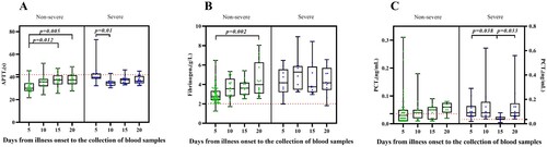
To observe the dynamic changes in coagulation function indicators after the onset of COVID-19 in patients, we regrouped the two groups of patients based on the time from onset to the collection of blood samples and classified them into groups every five days ((A)). The reference values considered for each coagulation index were as follows: APTT 29–42 s, D-dimer 0.5 mg/L, and fibrinogen 2 g/L.
Figure 1. Dynamic changes of coagulation indices and inflammation indices in severe and non-severe COVID-19 patients. Timeline charts illustrate the changes of coagulation and inflammation parameters in 172 peripheral blood samples, among which 119 were samples of non-severe patients and 53 were of severe patients. The symbols and error bars show medians, 25% and 75% percentiles.
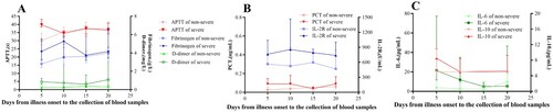
As time progressed, the D-dimer levels of non-severe COVID-19 patients showed a trend of an initial increase followed by a decrease, reaching the highest value (0.385 mg/L), which was within the normal baseline range, on day 15. In contrast, an opposite trend was observed in the D-dimer levels of patients with severe COVID-19; their D-dimer level reached the lowest value (0.595 mg/L), which was higher than the normal range, on day 15.
Fibrinogen levels in non-severe COVID-19 patients increased with time, the lowest value (2.82 g/L) on day 5 was higher than the normal baseline range, and the data obtained on day 20 was significantly higher compared to day 5 (P = .002, (B)). On the other hand, fibrinogen levels in severe COVID-19 patients showed unstable changes, the lowest value (3.76 g/L) was on day 15, which was higher than the normal baseline value.
Figure 2. Dynamic changes of APTT, fibrinogen and PCT in severe and non-severe COVID-19 patients. Timeline charts illustrate the changes of APTT, fibrinogen and PCT in the 172 peripheral blood samples. Red dotted lines represent the upper limit of the reference value of the three indicators.
Comparison of initial indices of inflammation between severe and non-severe COVID-19 patients
The results revealed that IL-2R, IL-6, IL-10, and PCT in the severe group were significantly higher than those in non-severe group (767.5 pg/mL vs. 565.5 pg/mL (P = .024), 11.435 pg/mL vs. 3.195 pg/mL (P = .005), 5 pg/mL vs. 5 pg/mL (P = .009), 0.07 ng/mL vs. 0.037 ng/mL (P = .000), respectively) ().
Dynamic changes of inflammation indices in severe and non-severe COVID-19 patients
The groups for observing the dynamic changes of inflammatory factors in COVID-19 patients were the same as the groups made for observing the dynamic changes of coagulation function ((B and C)). The reference values of each inflammatory factors under consideration were as follows: IL-2R <200 U/mL, IL-6 ≤ 7 pg/mL, PCT 0.030–0.036 ng/mL.
The level of IL-2R in non-severe COVID-19 patients showed unstable changes, and the lowest value was recorded on day 20 (468 U/mL), which was above the normal range. While in severe COVID-19 patients, IL-2R showed an initial increase followed by a decrease, recording their lowest value on day 20 (738 U/mL); however, this value was higher than the upper limit of the normal range. In patients with non-severe COVID-19, IL-6 initially decreased which was followed by an increase, reaching the normal range before day 15, and exceeding the upper limit of the normal range on day 20. On the contrary, IL-6 in severe COVID-19 patients was higher than the upper limit of normal before day 15, after which it dropped to the normal range and remained stable. The value of PCT in non-severe COVID-19 patients was in a continuously increasing trend, reaching a level higher than the upper limit of normal after day 5. While in severe COVID-19 patients, the value of PCT remained consistent, with the lowest value (0.04 ng/mL) recorded on day 15; however, this value was higher than the upper limit of the normal range and was significantly lower than that on day 5 and day 20 (P < .05, (C)).
Comparison of correlations of inflammatory factors and coagulation indices between severe and non-severe COVID-19 patients at different blood sample collection times
The results of the analyses indicated that fibrinogen in severe COVID-19 patients was strongly correlated with IL-2R, IL-6, IL-10, and PCT on day 10 (r = 0.912, P = .004; r = 0.949, P = .001; r = 0.846, P = .016; r = 0.905, P = .013, respectively) (). In addition, D-dimer of severe COVID-19 patients was strongly correlated with IL-2R on day 5 (r = 0.895, P = .003), and strongly correlated with IL-2R, IL-6, and PCT on day 10 (r = 0.734, P = .016; r = 0.768, P = .009; r = 0.746, P = .033, respectively); it was also strongly correlated with PCT on day 20 (r = 0.873, P = .002)(). However, no correlation between fibrinogen and inflammatory factors was observed in non-severe COVID-19 patients at any time point.
Table 2. Correlations of fibrinogen and inflammatory factors in patients with severe COVID-19 at different blood sample collection times.
Table 3. Correlations of D-dimer and inflammatory factors in patients with severe COVID-19 at different blood sample collection times.
Discussion
Previous study regarding coagulation function of COVID-19 patients revealed that patients with severe disease had higher levels of fibrinogen and D-dimer during the early stage, becoming more significant with disease progression [Citation7]. In our study, we observed that values of APTT, D-dimer, and fibrinogen were significantly different between non-severe and severe COVID-19 patients. Levels of D-dimer and fibrinogen in severe COVID-19 patients were abnormally increased at an early stage and remained above the upper limit of normal, which matches the data reported by previous studies [Citation8, Citation9]. Although, measuring D-dimer levels lacks internationally certified calibrators, quality controls, and possesses inter-laboratory and inter-individual variability [Citation10]. However, it is confirmed that high levels of D-dimer seem to be a major clinical risk factor for patients diagnosed with venous thromboembolic events (VTE) [Citation11]. In patients with non-severe COVID-19, fibrinogen alone was abnormally increased, and the change was not significant. The elevated coagulation indices indicated that patients with COVID-19 are at risk for hypercoagulability [Citation12]; however, this risk of hypercoagulability is more evident in patients with severe COVID-19.
Increased levels of D-dimer and fibrinogen indicate that coagulopathy results from the formation of a large amount of fibrin. Patients with COVID-19 tend to lose the balance between coagulation and fibrinolysis and thus present with significant hypercoagulability [Citation13]. It is hypothesized that the release of tissue factors, lymphocyte cell death, hypoxia, and endothelial damage also contribute to the hypercoagulable state of patients with COVID-19 [Citation14].
The concentration of inflammatory factors was higher in the serum of severe COVID-19 patients than in the serum of non-severe COVID-19 patients, suggesting that cytokine storm is related to disease severity [Citation15]. In critically ill patients, the release of inflammatory mediators possibly leads to the increase in blood viscosity, which further contributes to hypercoagulability [Citation12]. Here, notable differences were present in inflammatory factors such as IL-2R, IL-6, IL-10, and PCT between the two groups. During the early stage of the disease, the inflammatory factors including IL-2R, IL-6, and PCT of severe COVID-19 patients were higher than the normal range and reached the peak value, which indicated that the inflammatory reaction of severe COVID-19 patients was remarkable. Additionally, their D-dimer level was higher than normal during the early stage of the disease, with a strong correlation between D-dimer and the abnormal inflammatory factors, suggesting that the change in D-dimer levels in patients with severe COVID-19 may be related to the inflammatory reaction in the early stage. Fibrinogen in severe COVID-19 patients also showed an abnormal peak at an early stage; however, it was strongly correlated with inflammatory factors including IL-2R, IL-6, IL-10, and PCT only on day 10. Therefore, other factors besides inflammation should be considered for early abnormal fibrinogen levels in severe COVID-19 patients. In patients with non-severe COVID-19, their fibrinogen level increased abnormally but had no correlation with inflammatory factors, which indicated that abnormal fibrinogen in non-severe COVID-19 patients was not caused by systemic inflammatory reaction alone.
Related studies have confirmed that damage to the endothelium leads to abnormal regulation of fibrinolysis by plasminogen activator inhibitor from the endothelium, and the imbalance of fibrinolysis leads to a hypercoagulable state, eventually causing coagulation dysfunction [Citation16–18]. Arterial endothelial cells can express human angiotensin-converting enzyme 2 (ACE2) [Citation19, Citation20], ACE2 interacts with spike (S) protein on the SARS-CoV-2, which ultimately facilitates the entry of the virus into cells [Citation21, Citation22]. Furthermore, histopathological studies on COVID-19 patients have also showed that SARS-CoV-2 thrives in endothelial cells, directly participating in the host inflammatory reaction, thereby promoting endothelial inflammation in organs, which lead to diffuse endothelial inflammation and thrombosis of micro-vessels and macro-vessels in the venous and arterial circulation [Citation23]. Here, fibrinogen in non-severe COVID-19 patients had no correlation with inflammatory factors at any time point, and therefore, it can be speculated that the abnormal increase of fibrinogen in these patients may be the result of endothelial inflammation caused by ACE2-mediated SARS-CoV-2 entering the endothelial cells. Endothelial cells not only cause abnormal coagulation leading to a state of hypercoagulability, but they also participate in immune cell infiltration and cytokine production during a viral infection, consequently playing an important regulatory role in the formation of a cytokine storm [Citation24]. After the invasion of SARS-CoV-2, endothelial injury, coagulation dysfunction, and inflammatory reaction are closely associated. The body’s immune response and inflammatory factors activate the coagulation mechanism, which, in turn, leads to excessive inflammatory reaction [Citation25], which eventually lead to aggravation of the disease. Furthermore, it was found that the number of circulating endothelial cells was substantially higher in patients with severe COVID-19 [Citation26]. The influence of excessive inflammatory reaction on coagulation function in severe COVID-19 patients may mask the abnormal coagulation caused by damaged endothelial cells, while the influence of endothelial cells on coagulation can be better reflected in patients with mild inflammatory reaction. Most importantly, these findings indicate that during the early stage of disease progression, when fibrinogen levels seem to be high and D-dimer levels are lower, patients need to receive treatment immediately. Failing which, progressing to fibrinogen depletion as D-dimer levels increase, would subsequently cause a cytokine storm that could potentially lead to a poor prognosis [Citation27]. Therefore, even if the inflammatory reaction is not serious in COVID-19 patients, the changes of coagulation function should be paid utmost attention to, especially for certain high-risk groups with thrombosis. As part of their treatment, it may be possible to improve the patient’s condition by early anticoagulation therapy and maintaining the normal physiological state of endothelial cells.
Our research has some limitations. The major limitation is that we have merely shown the changes in laboratory indices and analyzed possible explanations. Further experiments are needed to explore whether SARS-CoV-2 directly attacks vascular endothelial cells and causes abnormal coagulation functions, to verify our hypothesis.
Conclusion
We conclude that there are differences in coagulation function and inflammatory factors between patients with non-severe and severe COVID-19. The hypercoagulability risk of patients with severe COVID-19 was more evident which was related to abnormal inflammatory factors, suggesting a relationship between inflammatory factors and abnormal coagulation. Although there was no apparent abnormality in most inflammatory factors in non-severe COVID-19 patients, abnormality was still observed in coagulation function, and had no relationship with any inflammatory factors, which indicated that inflammatory factors alone were not the cause of abnormal coagulation function in COVID-19 patients. During clinical treatment, it is crucial to consider early anticoagulation therapy with monitoring and maintaining endothelial cell stability in patients with no apparent severe inflammatory reaction.
Disclosure statement
No potential conflict of interest was reported by the author(s).
Additional information
Funding
References
- van der Poll T, Herwald H. The coagulation system and its function in early immune defense. Thromb Haemost. 2014;112(4):640–648.
- Guan WJ, Ni ZY, Hu Y, et al. Clinical characteristics of coronavirus disease 2019 in China. N Engl J Med. 2020;382(18):1708–1720.
- Tang N, Li D, Wang X, et al. Abnormal coagulation parameters are associated with poor prognosis in patients with novel coronavirus pneumonia. J Thromb Haemostasis. 2020;18(4):844–847.
- Castelli R, Gidaro A. Abnormal hemostatic parameters and risk of thromboembolism among patients with COVID-19 infection. Journal of Hematology. 2020;9(1-2):1–4.
- Wang D, Hu B, Hu C, et al. Clinical characteristics of 138 hospitalized patients with 2019 novel coronavirus-infected pneumonia in Wuhan, China. JAMA. 2020;323(11):1061–1069.
- National Health Commission & National Administration of Traditional Chinese Medicine 2020. Diagnosis and Treatment Protocol for Novel Coronavirus Pneumonia (Trial Version 7). Chin Med J 2020;133(9):1087–1095.
- Bi X, Su Z, Yan H, et al. Prediction of severe illness due to COVID-19 based on an analysis of initial fibrinogen to albumin ratio and platelet count. Platelets. 2020;31(5):674–679.
- Hardy M, Michaux I, Lessire S, et al. Prothrombotic hemostasis disturbances in patients with severe COVID-19: individual daily data. Data Brief. 2020;33:106519.
- Hardy M, Michaux I, Lessire S, et al. Prothrombotic disturbances of hemostasis of patients with severe COVID-19: A prospective longitudinal observational study. Thromb Res. 2021;197:20–23.
- Hardy M, Lecompte T, Douxfils J, et al. Management of the thrombotic risk associated with COVID-19: guidance for the hemostasis laboratory. Thromb J. 2020;18:17.
- Kampouri E, Filippidis P, Viala B, et al. Predicting venous thromboembolic events in patients with coronavirus disease 2019 requiring hospitalization: an observational retrospective study by the COVIDIC initiative in a Swiss University hospital. Biomed Res Int. 2020;2020: 9126148.
- Terpos E, Ntanasis-Stathopoulos I, Elalamy I, et al. Hematological findings and complications of COVID-19. Am J Hematol. 2020;95(7):834–847.
- Nougier C, Benoit R, Simon M, et al. Hypofibrinolytic state and high thrombin generation may play a major role in SARS-COV2 associated thrombosis. J Thromb Haemost. 2020;18(9):2215–2219.
- Iba T, Levy JH, Levi M, et al. Coagulopathy in COVID-19. J Thromb Haemost. 2020;18(9):2103–2109.
- Liu J, Li S, Liu J, et al. Longitudinal characteristics of lymphocyte responses and cytokine profiles in the peripheral blood of SARS-CoV-2 infected patients. EBioMedicine. 2020;55:102763.
- Vallet B, Wiel E. Endothelial cell dysfunction and coagulation. Crit Care Med. 2001;29(7 Suppl):S36–S41.
- Witkowski M, Landmesser U, Rauch U. Tissue factor as a link between inflammation and coagulation. Trends Cardiovasc Med. 2016;26(4):297–303.
- Keller TT, Mairuhu ATA, de Kruif MD, et al. Infections and endothelial cells. Cardiovasc Res. 2003;60(1):40–48.
- Kowalczuk S, Bröer A, Tietze N, et al. A protein complex in the brush-border membrane explains a hartnup disorder allele. FASEB J. 2008;22(8):2880–2887.
- Silver MA, Bohnert M, Beck AT, et al. Relation of depression of attempted suicide and seriousness of intent. Arch Gen Psychiatry. 1971;25(6):573–576.
- Kuba K, Imai Y, Rao S, et al. A crucial role of angiotensin converting enzyme 2 (ACE2) in SARS coronavirus-induced lung injury. Nat Med. 2005;11(8):875–879.
- Li W, Moore MJ, Vasilieva N, et al. Angiotensin-converting enzyme 2 is a functional receptor for the SARS coronavirus. Nature. 2003;426(6965):450–454.
- Varga Z, Flammer AJ, Steiger P, et al. Endothelial cell infection and endotheliitis in COVID-19. Lancet. 2020;395(10234):1417–1418.
- Teijaro JR, Walsh KB, Cahalan S, et al. Endothelial cells are central orchestrators of cytokine amplification during influenza virus infection. Cell. 2011;146(6):980–991.
- Wang J, Saguner AM, An J, et al. Dysfunctional coagulation in COVID-19: from cell to bedside. Adv Ther. 2020;37(7):3033–3039.
- Guervilly C, Burtey S, Sabatier F, et al. Circulating endothelial cells as a marker of endothelial injury in severe COVID-19. J Infect Dis. 2020;222(11):1789–1793.
- Grobler C, Maphumulo SC, Grobbelaar LM, et al. Covid-19: The rollercoaster of fibrin(ogen), D-dimer, Von willebrand factor, P-selectin and their interactions with endothelial cells, platelets and erythrocytes. Int J Mol Sci. 2020;21(14):5168.