

ABSTRACT
Background
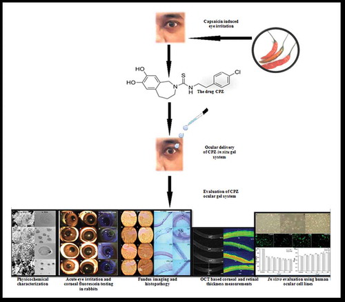
Defense personnel utilize capsaicin-based ocular sprays as non-lethal agents for law implementation during instances of mob violence. This study involves the capsaicin antagonist Capsazepine and the investigation of whether Capsazepine’s antagonistic approach can be favorably utilized for defense utilization to block capsaicin-initiated pain and inflammation via the ocular pathway.
Research design and methods
Ocular capsazepine in situ gels were prepared with polymers Pluronic F-127 and Chitosan; optimized formulation was quantified in ocular tissues chromatographically and by in vivo live ocular imaging; anti-inflammatory efficacy was determined by eye irritation testing, corneal and retinal imaging, ocular prostaglandin estimation, and by viability and proliferation testing using human ocular cell lines, etc.
Results
A physicochemically stable Capsazepine in situ gel was formulated which showed little ocular irritation, considerable transcorneal permeation; was precisely quantified in ocular tissues by gas chromatography and in vivo live ocular imaging; showed anti-inflammatory properties against capsaicin by eye imaging experiments, prostaglandin declination and showed acceptable cytocompatibility when studied using human ocular cell lines.
Conclusions
The fabricated in situ Capsazepine gel system might be promising for ocular delivery as it appears a pharmacologically potent and safe development, suitable for utilization in the ocular clinical therapy, provided there is additional research to substantiate it.
Graphical Abstract
Acknowledgments
Harshita Krishnatreyya is thankful and extends heartfelt gratitude to Dr. Jayanta Barooah from the MSN Hospital, Nagaon, Assam, India, for providing the facility of the OCT and Fundus camera for experimental eye imaging. The author extends gratitude to Dr. Angarag Bhagawati, Tezpur Eye Hospital, Assam, India for providing the fundus imaging facility. The author is thankful to the Defence Research Laboratory (DRL), Defence Research and Development Organisation (DRDO), Ministry of Defence, Govt. of India, for providing necessary research fellowship, instrumental and chemical facilities for this work. The author is also grateful to the Pharmaceutical and Fine Chemical Technology, Department of Chemical Technology, University of Calcutta, Kolkata, India for providing necessary administrative support in carrying out this part of the Ph.D. work. Authors are thankful and gratefully acknowledge all the anonymous reviewers for their valuable suggestions that helped us improve this manuscript.
Author contributions
H Krishnatreyya, A Saha, and P Chattopadhyay designed the study and critically reviewed it. HK and H Hazarika performed all experiments and statistical analyses and wrote the manuscript; S Mandal helped in the fluorescent observation of the HCEpiC and HRMEC cells; NS Bora provided advice during cell culture experiments; S Kishor performed the GC-MS analysis of the samples; YD Bhutia, D Goyary, and S Karmakar helped in the procurement of experimental animals, chemicals, and reagents, etc., necessary for all experimental procedures.
Declaration of interest
The authors have no relevant affiliations or financial involvement with any organization or entity with a financial interest in or financial conflict with the subject matter or materials discussed in the manuscript. This includes employment, consultancies, honoraria, stock ownership or options, expert testimony, grants or patents received or pending, or royalties.
Reviewer disclosures
Peer reviewers on this manuscript have no relevant financial or other relationships to disclose.