Every year, students on the MSc in Medical Art at the University of Dundee present their work as part of the Duncan of Jordanstone College of Art & Design ‘Master’s Show’. This year, due to the ongoing COVID-19 situation, the Showcase is online (https://www.dundee.ac.uk/masters-showcase/2021/medical-art). Following are some highlights from the 2021 exhibition ().
Figure 1. Konstantinos Alexandrou – Image of the adult form of the parasitic helminth H. polygyrus invading the intestinal wall, from the animation ‘The immune modulating activities of Heligmosomoides polygyrus’.
Figure 2. Konstantinos Alexandrou – The mechanism of action of one of the molecules the parasitic helminth releases, from the animation ‘The immune modulating activities of Heligmosomoides polygyrus’.
Figure 3. Samuel Dapaah – still from pupillary light reflex pathway animation – eyeball with oculomotor nerve (CN III).
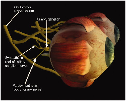
Figure 4. Samuel Dapaah – still from pupillary light reflex pathway animation – schematic representation of pathway.
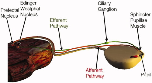
Figure 5. Marra Evans – Still image of the sagittal view of the brain highlighting the anterior olfactory cortex, from ‘Sniffing Out a Diagnosis for Parkinson’s Disease, An Animation’.
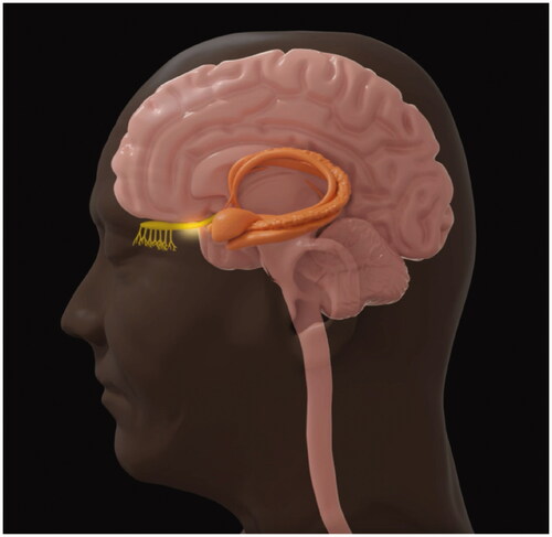
Figure 6. Marra Evans – Still image of a closeup of the olfactory nerve and olfactory bulb, from ‘Sniffing Out a Diagnosis for Parkinson’s Disease, An Animation’.
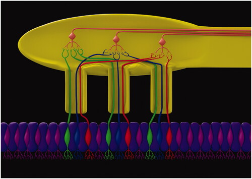
Figure 7. Sophia Lappe – Illustration of week one of development shows the implantation of the blastocyst into the uterine wall.
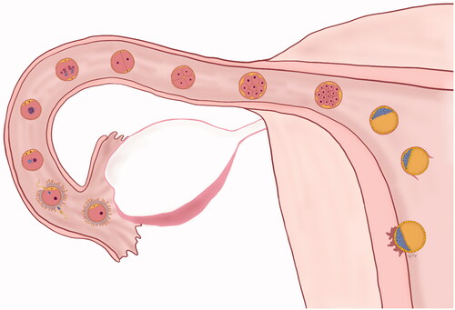
Figure 8. Sophia Lappe – Models of the developing human embryo from days 22 (left) to 28 (right).
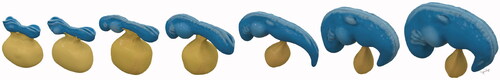
Figure 9. Alphy Pullely – The above visuals depict 2D images of 3D models of four congenital kidney anomalies (a) Ectopic Kidney, (b) Pancake Kidney, (c) Horseshoe Kidney, (d) Supernumerary Kidney. These variations were modelled using Zbrush 3D software and high quality 2D images were rendered using Keyshot 8 software.
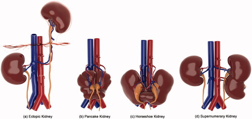
Figure 10. Reba (Rebekah) Sadok – Down Syndrome facial muscles 3D model.
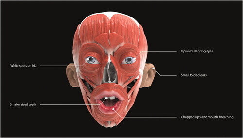
Figure 11. Reba (Rebekah) Sadok – Down Syndrome tongue 3D model.
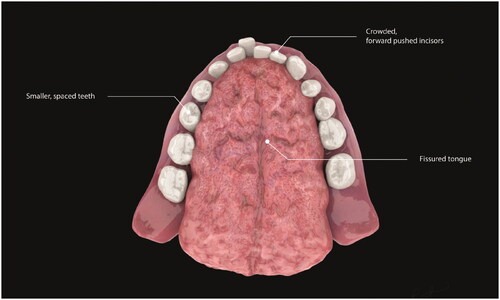
Figure 12. Emily Wallace – Emily Wallace – Open foetal surgery – This illustration shows an incision made on a pregnant person’s abdomen to access a foetus who has spina bifida. The defect on the lower back can now be closed to prevent further damage to the spinal nerves.
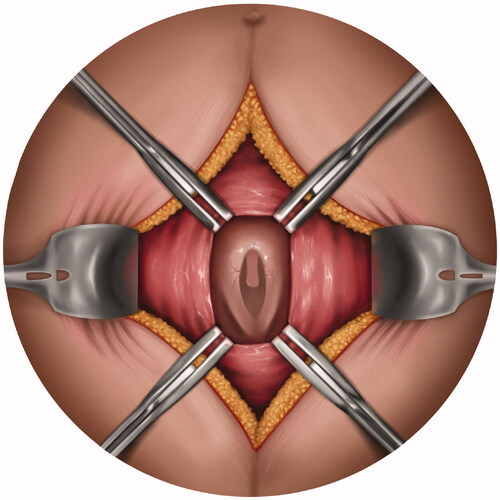
Figure 13. Emily Wallace – Types of Spina bifida – This set of illustrations show the anatomical variations of spina bifida occulta, meningocele and myelomeningocele.
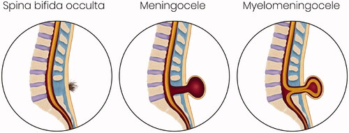
Figure 14. Emily Wallace – Types of Spina bifida – This set of illustrations show the anatomical variations of spina bifida occulta, meningocele and myelomeningocele.
Figure 15. Abigail De Rancourt De Mimérand – 2D illustration of the base of the skull showing the cranial foramina. Taken from a website on the Anatomy of the Nervous System.
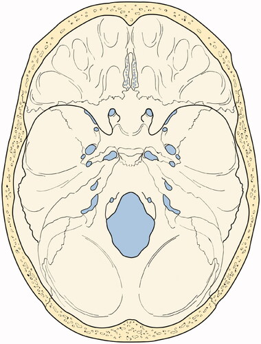
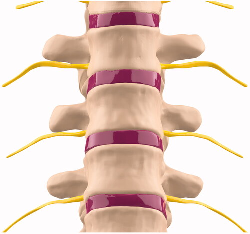
Figure 16. Abigail De Rancourt De Mimérand – Close-up still image of a 3D model of the thoracic vertebral column highlighting the intervertebral discs.