Abstract
Background We have previously shown that patients with instability of the anterior syndesmosis benefit from an anatomical reconstruction. It is not known whether this is because of restored kinematics.
Methods In a prospective study of 5 patients, we assessed clinical findings and tibiofibular kinematics, evaluated by radiostereometry, before and after reconstruction of a chronic syndesmotic injury.
Results We found no statistically significant differences in tibiofibular kinematics before and after reconstruction. The kinematics of the fibula relative to the tibia during external rotation stress differed from that known in asymptomatic volunteers, but the differences were not typical enough to differentiate between patients and healthy subjects. Clinical examination and ankle scores, however, showed that all patients benefited from reconstruction of the anterior syndesmosis.
Interpretation Radiostereometry is not an adequate technique to diagnose chronic syndesmotic instability or to demonstrate restoration of the kinematics of the ankle as a cause of the beneficial effect of anatomical reconstruction of the syndesmosis.
Several studies have shown that a rupture of the anterior tibiofibular and/or deltoid ligament results in instability (Close Citation1956, Mullins and Sallis Citation1958, Rasmussen et al. Citation1982, Xenos et al. Citation1995, Beumer et al. Citation2003a).
Patients with instability of the anterior syndesmosis of the ankle benefit from an anatomical reconstruction of the anterior tibiofibular syndesmosis by a surgical technique which showed promising results in a retrospective study (Beumer et al. Citation2000). In this study, we prospectively assessed the clinical result of reconstructive surgery of the anterior syndesmosis. A second aim was to determine whether abnormal tibiofibular motion could be demonstrated with radiostereometry (RSA) in patients with syndesmotic instability.
Patients and methods
We studied 5 patients (2 women) with unilateral chronic syndesmotic instability. Their mean age was 32 (22–36) years ().
Table 1. Patient data
The suspicion of chronic instability of the anterior syndesmosis of the ankle was based on medical history, physical examination, radiographs (plain and stress, anteroposterior and lateral) and MRI. The patients underwent arthroscopy of the ankle and if syndesmotic instability was found, they participated in the study. The study was approved by the medical ethics committee of the Erasmus Medical Centre, Rotterdam, the Netherlands. All patients gave written informed consent.
The patients had 1.5 (1–3) years of complaints of the ankle after they had sustained a sprain. All had a sensation of giving way and problems with walking on uneven ground, without frank giving way. In addition, stiffness, limited dorsifiexion, and a recurrent swelling at the level of the syndesmosis were present. All patients indicated pain at the anterior syndesmosis and 3 out of 5 had tenderness around the medial and lateral malleolus also. The squeeze (Hopkinson et al. Citation1990), external rotation (Boytim et al. Citation1991), and fibula translation test (Ogilvie-Harris et al. Citation1994) were positive (i.e. painful) in all patients, while no patient had a positive (i.e. unilaterally increased mobile) anterior drawer test. No patient showed signs of hypermobility according to Beighton et al. (Citation1983). The complaints had not disappeared with nonoperative treatment which consisted of anti-infiammatory medication and plaster immobilization, followed by exercises to improve proprioception and strength.
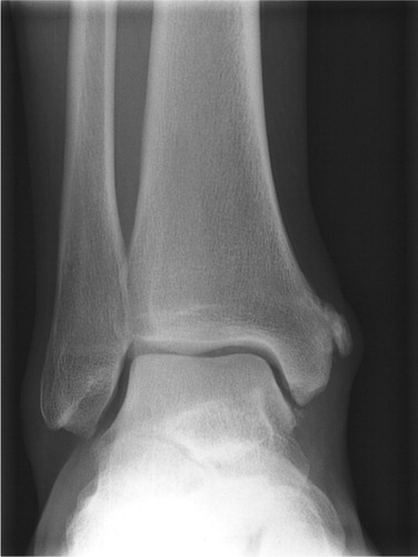
In 2 patients, plain ankle radiography showed a periosteal reaction at the medial malleolus above the insertion of the deltoid ligament, that might have resulted from a tear of the periosteum together with the attachment of the deltoid ligament (). No patient showed increased talar tilt or anterior translation on stress radiographs. MRI findings were not conclusive with regard to syndesmotic injury, as the anterior tibiofibular ligament cannot be visualized along its entire length in the orthogonal planes of conventional MRI—and no reports had been presented at that time on the MRI findings of chronic syndesmotic injuries. The periosteum at the medial malleolus and the deltoid ligament of the 2 patients with calcification at the medial malleolus showed thickening and loss of fascicular detail with MR imaging, indicating old injury.
Figure 1. Avulsion of the periosteum above the insertion of the deltoid ligament.
After arthroscopic confirmation of the diagnosis of syndesmotic instability by demonstrating increased movement of the fibula, easy access of the test probe into the syndesmosis, and increased tibiofibular width by easy turning of the transverse end of the test probe (Ogilvie-Harris et al. Citation1994, Beumer et al. Citation2000), 5 tantalum markers (0.8 mm) were placed in the involved distal tibia and fibula each, controlled by fluoroscopy during arthroscopy to provide optimal marker distribution.
Before arthroscopy, and 6 months after reconstruction, we assessed the function of the ankle with the squeeze test, the external rotation test, and the fibula translation test, as well as the Tegner-activity level score (Tegner and Lysholm Citation1985), the Karlsson (Citation1991) score, the Sefton et al. (Citation1979) score, and the Ankle-Hindfoot scale (Kitaoka et al. Citation1994). About 6 weeks after the arthroscopy, reconstruction of the anterior syndesmosis was performed by medialization and cranialization of the tibial insertion of the slack anterior tibiofibular ligament, after a syndesmotic set screw was placed during compression of the mortise (Beumer et al. Citation2000). Postoperative treatment consisted of 6 weeks of non-weight bearing in a below-knee plaster, followed by removal of the screw under local anesthesia. Thereafter, weight bearing and unlimited exercise were allowed.
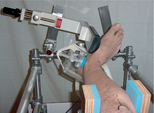
RSA examinations were performed in one or more of the following ways: A. Standing non-weight bearing, B. Standing weight bearing, C. Supine neutral, D. Supine with the application of a 7.5-Nm external rotation stress at the base of the first metatarsal (; Beumer et al. Citation2003a). These examinations were performed on the day before reconstruction (A, B, C, D), the day after reconstruction (C), 6 weeks after reconstruction (just after the syndesmotic setscrew had been removed; C), and 3 months after reconstruction (A, B, C, D).
Figure 2. The RSA external rotation stress examination
Weight-bearing examinations were performed while the patients stood on a pedestal. External rotation stress and also the “standard neutral” examinations were done in the supine position. Displacements of the fibula relative to the tibia during neutral weight bearing versus neutral non-weight bearing (“neutral weight bearing”), during external rotation stress versus neutral supine (“7.5-Nm external rotation stress”), and also in the standard neutral position after reconstruction versus before reconstruction (“standard neutral post-reconstruction”) were assessed, and expressed as 3 translation parameters and 3 rotation parameters of the fibula relative to the tibia. Positive directions for translations along the coordinate axes were lateromedial (transverse axis), caudocranial (longitudinal axis), and posterioranterior (sagittal axis). Positive directions for rotations about the coordinate axes were plantar flexion (transverse axis), internal rotation (longitudinal axis), and adduction (sagittal axis).
We scanned the radiographs with a Vidar VXR-12 scanner (Vidar, Lund, Sweden), at 150 dots per inch resolution and 8-bit gray scale resolution. The measurement of marker coordinates in the digitized radiographs, the three-dimensional reconstruction of the marker positions, and the micromotion analysis were done with RSA-CMS (MEDIS, Leiden, the Netherlands), a software package that performs the RSA procedure automatically in digitized or digital radiographs (Valstar et al. Citation2000).
Reliability of RSA depends on nonlinear marker distribution. The condition number provides a geometrical interpretation of the distribution of the markers in 3-D space (Söderkvist and Wedin Citation1993). We used the same upper limits for the condition number as described in the literature, i.e. a condition number lower than 80–90 representing a reliable distribution (Börlin et al. Citation2002). Double exposures of all 4 examinations were made in 2 patients in order to assess the reproducibility of the measurements.
Statistics
We assessed normality of the data with the Shapiro-Francia W’test. Differences between pre- and postoperative values were performed using paired t-tests. 95% confidence intervals for differences in pre- and postoperative values were calculated using the t-distribution. All tests were two-sided and had 5% significance level.
The pre- and postoperative displacements of the fibula relative to the tibia during neutral weight bearing and external rotation stress were compared with data assessed from asymptomatic volunteers (Beumer et al. Citation2003b) (see appendix). Differences between the patients in this study and those asymptomatic volunteers were analyzed with Wilcoxon Sum Rank Test (Mann-Whitney U-test), with the significance level set at p < 0.05.
Results
6 months after reconstruction, the ankles of all 5 patients had improved, as shown by the various ankle scores (). No patient complained of instability or pain anymore. After the operation, all the syndesmotic tests had turned from positive to negative.
The condition number for the tibia was 23 (14–32), and that for the fibula was 17 (12–24). No segment was excluded from analysis. These condition numbers lay well below the limits we set, so that reliable translations and rotations could be calculated. We assessed the reproducibility of the different examinations by repeating 8 of them. The standard deviations for the rotations about the transverse, longitudinal and sagittal axes were 0.4, 0.7, and 0.3 degrees, and the standard deviations for the translations were 0.2, 0.2, and 0.3 mm ().
Table 2. Reproducibility of the examination assessed by double exposures of 2 patients
Neutral weight bearing
Pre- and postoperative displacement of the fibula showed a large interindividual variation (). There were no statistically significant differences between the weight-bearing kinematics of the preoperative and postoperative examinations, nor between the kinematics of the patients in this study and the healthy volunteers during weight bearing.
Table 3. Neutral weight bearing compared to neutral non-weight bearing. Fibular displacements relative to the tibia for 5 patients
Standard neutral post-reconstruction
Directly after reconstruction, the fibula had translated (p = 0.04) in a medial direction by on average 1.1 (0.21–2.34) mm, thus decreasing tibiofibular width (). After 6 weeks (removal of the syndesmotic setscrew) and 3 months, it was seen that tibiofibular width gradually returned to its approximate preoperative value. When the direct postoperative data were compared to the preoperative situation, reduced translations of the fibula in a cranial (p = 0.04) and posterior (p = 0.04) direction were found during the neutral supine investigations.
Table 4. Standard neutral post-reconstruction (supine postoperative versus to preoperative). Fibular displacementsrelative to the tibia for 5 patients
We found no statistically significant differences during the neutral supine investigations between the preoperative findings and those recorded 6 weeks and 3 months after reconstruction.
7.5-Nm external rotation stress
Preoperative displacement of the fibula relative to the tibia showed a mean posterior translation of 0.97 (0.08–2.45) mm and 2.32 (0.91–3.49) degrees external rotation when compared to the neutral situation. Postoperative (3 months) versus preoperative displacement of the fibula relative to the tibia showed smaller posterior translations (mean 0.78 mm, range −0.02–1.32; p = 0.35) and external rotation (mean 1.74°, range 0.06–3.94; p = 0.14) ().
Table 5. External rotation stress (7.5 Nm) compared to neutral (supine). Fibular displacements relative to the tibia for 5 patients
In comparison with the asymptomatic volunteers, the patients displayed smaller displacements during the external rotation stress investigation before reconstruction, for medial (p = 0.01) and posterior (p = 0.04) translation, as well as for external rotation (p = 0.04). In comparison with the control ankles, smaller displacements were found 3 months after reconstruction for medial (p = 0.005) and posterior (p = 0.003) translation, and external rotation (p = 0.04), as well as larger cranial displacements (p = 0.05).
Discussion
We used RSA to assess displacements at the tibiofibular syndesmosis before and after reconstruction of the anterior syndesmosis, as conventional radiography is unreliable and not sensitive when repeated radiographs are necessary (McDade Citation1975, Beumer et al. Citation2004). The data acquired were compared with the kinematics of healthy volunteers who took part in an RSA study that was performed to obtain “normal” values (Beumer et al. Citation2003b).
Post-reconstruction differences in tibiofibular kinematics relative to preoperative findings were not statistically significant. In contrast to a previous cadaveric study (Beumer et al. Citation2003a), we were unable to demonstrate syndesmotic instability as a statistically significant increased posterior translation of the fibula during the external rotation stress examination in these patients. This is probably because we did not have an individual “intact” situation.
Furthermore, the result of the above examination in cadavers may be different from the chronic situation in patients because of healing of the ligament in the chronic situation. In all the syndesmotic reconstructions we performed, a continuous but slack anterior tibiofibular ligament was found. The cadaveric study, however, is best compared with an acute ligament rupture. For ethical reasons, we chose not to perform the RSA external rotation stress examination in the contralateral ankle.
Preoperative external rotation stress examinations resulted in an external rotation of the fibula of between 0.91 and 3.49 degrees. 2 patients exhibited radiological features that might indicate old deltoid injury. One of these patients showed external rotation smaller than average; the other had the largest external rotation found preoperatively. It has been described that deltoid ligament rupture results in increased talar external rotation which increases further after additional sectioning of the syndesmosis (Rasmussen 1985).
In view of the functional instability (Freeman Citation1965), an unexpected observation was that our patients showed smaller displacements of the fibula relative to the tibia during the external rotation stress investigation (for medial and posterior translation, as well as external rotation) than the asymptomatic volunteers. Apart from the wide range of values found in healthy volunteers, and combined with the small number of patients in our study, several reasons might be given for this finding. Firstly, with respect to positioning of the ankle, plantarflexion and dorsiflexion are known to change the tibiofibular relationship. The ankles of the patients and the healthy volunteers were placed in the testing device by 2 investigators (AB, BAS) according to the zero starting position for the foot (American Academy of Orthopedic Surgeons Citation1965). It is therefore unlikely that a positioning error would be the reason for this observation. Secondly, one might speculate that the fibulas of the volunteers could show increased displacement if they suffered from generalized joint laxity, since they underwent reconstruction of the lateral collateral ligaments of the contralateral ankle in the past. This, however, was not the case—as none showed signs of hypermobility and none had complaints of instability, or a positive anterior drawer or syndesmotic stress test (Beumer et al. Citation2003b). Also, it was shown previously with RSA that the volunteers had tibiofibular kinematics that were different to those of patients with bilateral chronic lateral instability of the ankle (Löfvenberg et al. Citation1990).
Another reason for the smaller displacements of the fibula in these patients compared to the volunteers could be the effect of posttraumatic changes around the syndesmosis, as have been described by Ogilvie-Harris et al. (Citation1994). During arthroscopy, however, the triad described by these authors (disruption of the interosseous and posterior inferior tibiofibular ligament and chondral fracture of the posterolateral portion of the tibial plafond) was not found. Differences in pain sensation between the patients and the volunteers during the stress examination and reactive (in)voluntary muscle tensioning can be the reason for smaller displacements during the stress examination.
The most likely reason for the small displacements (especially external rotation) of the fibula in these patients during the stress examination is an altered position of the fibula due to rupture of the anterior tibiofibular ligament. A cadaveric study showed that the fibula rotates on average 1.5 (0.1–3.4) degrees externally after transection of the anterior tibiofibular ligament (Beumer et al. in press). This is in accordance with findings from studies not using RSA (Close Citation1956, Rasmussen et al. Citation1982, Xenos et al. Citation1995). One might assume that the fibula remains in this position when no reduction is performed and secured in the acute situation.
The combination of the fibula being in this externally rotated position and changes in viscoelastic behavior of the ligament due to injury (Frank and Shrive Citation1994) could account for the smaller displacements of the fibula found during the external rotation stress examination in these patients, when compared to the healthy volunteers.
Directly after reconstruction of the syndesmosis, a decrease in tibiofibular width of 1 mm was found due to application of the syndesmotic setscrew. This had returned to the preoperative situation 3 months after reconstruction, indicating that the screw had temporarily fixated the syndesmosis. Furthermore, this finding suggests that overtightening of the syndesmosis will not easily be achieved in the ligamentous repair we perform.
We could not reveal the syndesmotic instability by the RSA external rotation stress examination. As the displacements measured may be largely infiuenced by voluntary muscle tensioning and movement, and since RSA is an invasive procedure—not suitable for diagnostic imaging in daily practice—we conclude that the RSA external rotation stress examination is not the proper tool to assess chronic syndesmotic instability in a patient who has not been anesthetized.
No competing interests declared.
Related Research Data
- American Academy of Orthopaedic Surgeons. Joint motion. Method of measuring and recording. Churchill Livingstone, Edinburgh 1965
- Beighton P H, Solomom L, Soskolne C L. Articular mobility in an African population. Ann Rheum Dis 1983; 32: 413–8
- Beumer A, Heijboer M P, Fontijne W P J, Swierstra B A. Late reconstruction of the anterior distal tibiofibular syndesmosis. Good outcome in 9 patients. Acta Orthop Scand 2000; 71: 519–21
- Beumer A, Valstar E R, Garling E H, Van Leeuwen W J, Sikma W, Niesing R, Ranstam J A, Swierstra B A. External rotation stress imaging in syndesmotic injuries of the ankle. Comparison of lateral radiography and radiostereometry in a cadaveric model. Acta Orthop Scand 2003a; 74(2)201–5
- Beumer A, Valstar E R, Garling E H, Niesing R, Ranstam J, Löfvenberg R, Swierstra B A. Kinematics of the distal tibiofibular syndesmosis. Radiostereometry in 11 normal ankles. Acta Orthop Scand 2003b; 74(3)337–43
- Beumer A, Van Hemert W L, Niesing R, Entius C A, Ginai A Z, Mulder P G, Swierstra B A. Radiographic measurement of the distal tibiofibular syndesmosis has limited use. Clin Orthop 2004, 423: 227–34
- Beumer A, Valstar E R, Garling E H, Niesing R, Ginai A Z, Ranstam J, Swierstra B A. Effects of ligament sectioning on the kinematics of the distal tibiofibular syndesmosis. A radiostereometric study of 10 cadaveric specimens based on presumed trauma mechanisms and suggestions for treatment. Acta Orthop., In press
- Börlin N, Thien T, Kärrholm J. The precision of radiostereometric measurements. Manual vs. digital measurements. J Biomechanics 2002; 35: 69–79
- Boytim M J, Fischer D A, Neumann L. Syndesmotic ankle sprains. Am J Sports Med 1991; 19: 294–8
- Close R. Some applications of the functional anatomy of the ankle joint. J Bone Joint Surg (Am) 1956; 38(4)761–82
- Frank C B, Shrive N G. The Ligament. Biomechanics of the musculo-skeletal system, B M Nigg, W Herzog. John Wiley and Sons, Chisester 1994; 106–32
- Freeman M A. Instability of the foot after injuries to the lateral ligament of the ankle. J Bone Joint Surg (Br) 1965; 47(4)669–77
- Hopkinson W J, St. Pierre P, Ryan J B, Wheeler J H. Syndesmosis sprains of the ankle. Foot Ankle 1990; 10: 325–30
- Karlsson J. Evaluation of the ankle joint function: the use of a scoring scale. The Foot 1991; 1: 15–9
- Kitaoka H B, Alexander I J, Adelaar R S, Nunley J A, Myerson M S, Sanders M. Clinical rating systems for the anklehindfoot, midfoot, hallux, and lesser toes. Foot Ankle Int 1994; 15: 349–53
- Löfvenberg R, Kärrholm J, Selvik G. Fibular mobility in chronic lateral instability of the ankle. Foot Ankle 1990; 11: 22–9
- McDade W C. Treatment of ankle fractures. Instr Course Lect 1975; 24: 251–93
- Mullins J F P, Sallis J G. Recurrent sprain of the ankle joint with diastasis. J Bone Joint Surg (Br) 1958; 40: 270–3
- Ogilvie-Harris D J, Reed S C. Disruption of the ankle syndesmosis. Diagnosis and treatment by arthrosopic surgery. Arthroscopy 1994; 10: 561–8
- Rasnyssen O, Tovborg-Jensen I, Boe S. Distal tibiofibular ligaments. Analysis of function. Acta Orthop Scand 1982; 53: 681–6
- Sefton G K, George J, Fitton J M, McMullen H. Reconstruction of the anterior talofibular ligament for the treatment of the unstable ankle. J Bone Joint Surg (Br) 1979; 61: 352–4
- Söderkvist I, Wedin P. Determining the movements of the skeleton using well-configured markers. J Biomechanics 1993; 26(12)1473–7
- Tegner Y, Lysholm J. Rating systems in the evaluation of knee ligament injuries. Clin Orthop 1985, 198: 43–9
- Valstar E R, Vrooman H A, Toksvig-Larsen S, Ryd L, Nelissen R G H H. Digital automated RSA compared to manually operated RSA. J Biomechanics 2000; 33: 1593–99
- Xenos J S, Hopkinson W J, Mulligan M E, Olson E J, Popovic N A. The tibiofibular syndesmosis. Evaluation of the ligamentous structures, methods of fixation, and radiographic assessment. J Bone Joint Surg (Am) 1995; 77: 847–56
Appendix. Fibular displacements relative to the tibia for 11 normal ankles. Values are mean (range). From Beumer et al. 2003b