In February 2000, a 59-year-old woman with bilateral congenital hip dislocation underwent cement-less total hip arthroplasty with alumina-on-alumina bearings on the right side. The implants used were a ceramic sandwich cup, a titanium alloy stem, and a 28-mm alumina ceramic head (Kyocera, Kyoto, Japan). The sandwich cup consisted of an alumina ceramic liner housed in an ultra-high molecular weight polyethylene shell that was held in a titanium alloy metal shell.
The patient had good clinical results until July 2001, when she noticed a crepitus deep in the right hip during motion. Radiographs revealed a comminuted fracture of the ceramic liner. The patient underwent revision surgery in August 2001. 9 large pieces of the ceramic liner and many small fragments were found along with black-stained peri-prosthetic tissues. The cause of the ceramic liner fracture may be the concentration of stress at the rim of the thin ceramic liner, and the steep cup placement may have enhanced the risk of liner fracture (Hasegawa et al. Citation2003). The ceramic fragments were removed, and synovectomy that was as complete as possible was performed. When we removed the polyethylene liner and the screws of the metal shell, we observed that the shell was not loose, and therefore it was not removed. The head was removed. The femoral stem was not loose, and the taper was undamaged macroscopically. After the joint was lavaged with a pulsatile lavage system (Zimmer, Warsaw, IN), a modular polyethylene liner without a ceramic liner (inner diameter, 26 mm) was implanted. The ceramic head was replaced with a 26-mm cobalt-chromium head (Kyocera).
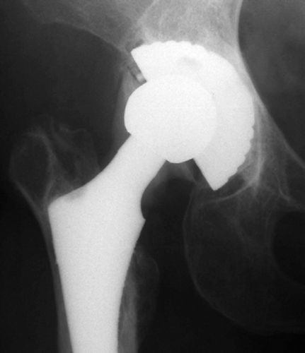
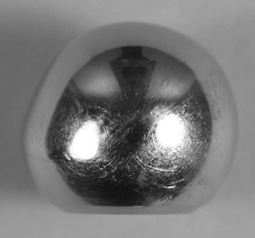
The patient returned to full activity without discomfort in the hip. About 2 years later, in September 2003, a radiograph taken at another institution revealed a radio-dense shadow around the stem neck (). The patient underwent a second revision at our institution in May 2004. A black-colored effusion filled the pseudocapsule. We found considerable black staining in the peri-prosthetic tissues. The black-stained granulation was excised. The cobalt-chromium femoral head was severely worn (). Alumina particles, ranging in size from 1 to 2 mm, were found loose within the pseudocapsule. The polyethylene liner was embedded with small particles. We removed the acetabular metal shell, although well-fixed, and replaced it with a new cementless metal shell. The well-fixed femoral stem was revised because it was not compatible with a ceramic component, and given the circumstances, another cobalt-chromium head would be likely to fail. A polyethylene liner was inserted into the metal shell and a new alumina head was placed on the new taper.
Figure 1. Anteroposterior radiograph taken 25 months after revision because of ceramic liner fracture, demonstrating metallosis around the stem neck. Note the loss of the superior part of the cobalt-chromium head.

Figure 2. The removed cobalt-chromium head showing loss of the superior part of the head because of severe wear.

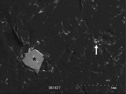
Histological examination showed intensive metallosis corresponding to black-stained fibrosis containing many macrophages filled with metallic particles, as well as extracellular metallic particles. Wear of the retrieved femoral head and polyethylene liner was measured with a three-dimensional coordinate measuring apparatus (Mitsutoyo Corp., Tokyo, Japan). Volumetric head wear was 455 mm3(165 mm3/year), and the maximum linear wear of the head and the polyethylene liner was 1.16 mm (0.42 mm/year) and 0.30 mm (0.11 mm/ year), respectively. Scanning electron microscopy (S-3000N; Hitachi, Tokyo, Japan) of the worn femoral head revealed a badly scratched surface. Small particles embedded in the polyethylene liner were retrieved. Excised black discolored synovium was digested and centrifuged. The particles were collected from the sediment and filtered. These particles from the polyethylene and synovium were examined using scanning electron microscopy and energy-dispersive radiographic analysis (EDAX HIT S-3200N; Hitachi, Tokyo, Japan). The analysis revealed sharp-edged particles that were composed of aluminum and oxygen, that ranged in size from 0.1 to 0.7 mm, and which originated from the fractured aluminum oxide ceramic liner. Irregular-shaped small particles were also found, and these particles were analyzed for the presence of cobalt, chromium, and molybdenum; they originated from the cobalt-chromium femoral head ().
Figure 3. Scanning electron micrograph of the retrieved polyethylene liner showing a sharp-edged alumina particle (asterisk) and irregular-shaped metal particle (arrow) embedded in the polyethylene. The elements contained in the material were identified using energy-dispersive radiographic analysis.

The patient had an uneventful recovery, and had neither hip pain nor radiographic abnormality at the 1-year follow-up.
Discussion
Revision hip arthroplasty after ceramic fracture can be problematic. Allain et al. (Citation2003) reported the results of a short-term (average 3.5 years) multicenter study of 105 patients who underwent revision surgery to treat fracture of a ceramic head. Macroscopic metallosis was found in 20 of the 33 repeat revisions. Particles of alumina were found in 13 hips. 20 of the 26 stainless-steel femoral heads were distorted, indicating metallic wear. They concluded that the cup should be removed at the time of revision, even if it appears normal macroscopically, because microscopic ceramic particles may be embedded in it. In their study, the new ceramic femoral heads were at least as durable as the original ceramic femoral head implants, and prevented wear of the femoral head, but cobalt-chromium heads also provided satisfactory results. In contrast, the authors advised against the use of a stain-less-steel femoral head because of the abnormal wear that can occur. Kempf and Semlitsch (Citation1990) reported a case of metallosis in a patient with a stainless-steel femoral head. In that case, the surgeon merely replaced the fractured ceramic head with a new stainless-steel femoral head, and the cemented all-polyethylene cup remained in situ.
Metal heads made of cobalt-chromium alloy are believed to be more resistant to abrasive wear than heads made of stainless steel (Kempf and Semlitsch Citation1990, Allain et al. Citation2003). However, to our knowledge, our report is the first to indicate that the severe wear of the cobalt-chromium femoral head can be caused by alumina particles. Cobalt-chro-mium alloy has Vickers hardness 240–450. This is not much different from stainless-steel alloy (170– 350). In contrast, the hardness of aluminium oxide is approximately 2,000 (Dearnley Citation1999). Thus, a similar phenomenon was seen when cobalt-chro-mium was used instead of stainless steel. Thorough debridement of the ceramic particles must be performed, although elimination of all particles seems to be impossible. To remove all fragments of a fractured ceramic implant, the resection borders should be designed in healthy tissue at a sufficiently safe distance from the macroscopically visible focal margins. Because it is impossible to require such resection borders in revision surgery, surgeons have to perform synovectomy as extensively as possible to remove as much of the ceramic particles as possible. One solution is to use adapters that—like an extension piece—are fitted onto the deformed cone to create a new cone. Because these adapters are made of metal, there is no jump in the elasticity module on the incongruent—now cone-adapter boundary surface—as occurs when a ceramic head is used directly. The adapter boundary surface facing the hip head possesses mechanical qualities that are comparable to those of the cone of a new stem (Matziolis et al. Citation2003).
Our report provides relatively early information about the difficulties with ceramic particles, and it is still not quite clear how these cases should be treated.
The authors thank Masaru Ueno, Ph.D., of Japan Medical Materials, Osaka, Japan, for technical assistance during the analysis of the retrieved prosthesis.
- Allain J, Roudot-Thoraval F, Delecrin J, Anract P, Migaud H, Gartallier D. Revision total hip arthroplasty performed after fracture of a ceramic femoral head. A multicenter survivorship study. J Bone Joint Surg (Am) 2003; 85: 825–30
- Dearnley P A. A review of metallic, ceramic and surface-treated metals used for bearing surfaces in human joint replacements. Proc Inst Mech Eng [H] 1999; 213: 107–35
- Hasegawa M, Sudo A, Hirata H, Uchida A. Ceramic acetabular liner fracture in total hip arthroplasty with a ceramic sandwich cup. J Arthroplasty 2003; 18: 658–61
- Kempf I, Semlitsch M. Massive wear of a steel ball head by ceramic fragments in the polyethylene acetabular cup after revision of a total hip prosthesis with fractured ceramic ball. Arch Orthop Trauma Surg 1990; 109: 284–7
- Matziolis G, Perka C, Disch A. Massive metallosis after revision of a fractured ceramic head onto a metal head. Arch Orthop Trauma Surg 2003; 123: 48–50