Abstract
Background The aims of the present study were to assess the development of hip dysplasia in children with bilateral spastic cerebral palsy and to evaluate the factors that influence the progression.
Patients and methods 76 children, 42 with spastic quadriplegia and 34 with diplegia, were included in the study. Their mean age at the first radiographic examination was 3.5 (1–11) years. The patients were followed up until operative treatment (54 subjects) or until the most recent radiograph in those who did not undergo hip surgery. The mean length of follow-up was 4.8 (1–13) years. On the initial and most recent radiographs, the migration percentage (MP) was measured, which is the percentage of the femoral head lateral to the acetabular rim.
Results The mean MP of the side with the largest displacement was 25% (-18–66) at the initial radiographic examination and 51% (9–100) at the last follow-up. The mean increase in MP was 7% (-2–33) per year. Linear multiple regression revealed that gait function and age were the most important variables that influenced the rate of MP progression. Children who could not walk had significantly greater MP progression per year (12%) than those who walked with or without support (2%). In the quadriplegics, the maximal yearly increase in MP was 13% under 5 years of age and 7% in older children. This difference was statistically significant, whereas no significant difference in relation to patient age was seen in the diplegics.
Interpretation There is a pronounced trend towards displacement of the hips in quadriplegic CP patients who are under 5 years of age and cannot walk. Because hip dislocation may lead to severe problems, close follow-up is important in finding the appropriate time for hip surgery in order to avoid progression towards dislocation. The risk of severe hip dysplasia is considerably less in spastic diplegia. ▪
There is increased risk of hip dysplasia and dislocation in cerebral palsy (CP) with bilateral involvement. The risk of hip dislocation varies with the degree of disability (Howard et al. Citation1985, Vidal et al. Citation1985). Dislocation may lead to pain and severe problems with ambulation, sitting balance, perineal nursing care, and decubitus ulceration (Samilson et al. Citation1972, Letts et al. Citation1984). Although the natural history of hip development has been evaluated in some reports (Reimers Citation1980, Vidal et al. Citation1985, Miller and Bagg Citation1995), the risk factors have not been sufficiently evaluated and the rate of deterioration of the hips has not been clearly established. This is important, because the effectiveness of prophylactic treatment such as intrathecal baclofen or adductor tenotomy cannot be properly assessed unless the development of the hip joints in unoperated children has been well characterized.
The aims of the present study were to assess the rate of progression of hip dysplasia in unoperated children with bilateral spastic CP and to evaluate the factors that influence the development of the hips.
Patients and methods
Children with bilateral spastic CP were recruited among patients who were referred to our departmentfor orthopedic evaluation. To be included in the study, the children had to be under 12 years of age at the time of the first pelvic radiograph and they should not have undergone previous orthopedic surgery in the hip region. The patients had been treated with physiotherapy of various kinds and to different extents, but none had undergone specific treatment such as botulinum toxin in the hip muscles or intrathecal baclofen to reduce the degree of spasticity. Because the aim of the study was to assess the progression of the lateralization of the hips, only patients with femoral head coverage above 30% in the worst hip at the initial radiograph were included.
The material comprised 76 subjects (39 boys) with spastic CP, with a mean age of 3.5 (1–11) years at the time of the primary radiographs. 42 patients had quadriplegia (QP) and 34 had diplegia (DP). Their gait function was assessed according to 3 categories: walking without support, walking with support by sticks, crutches or a walking frame on wheels, and no walking capacity. The number of patients in each category was 19, 15, and 42, respectively. There was a strong correlation between the type of CP and gait function, as 40 of the 42 quadriplegics could not walk (2 could walk with support), whereas all except 2 of the diplegics had gait function (13 with support and 19 without support).
The study was based on anteroposterior radio-graphs of the pelvis and hip joints with the child in the supine position. Care was taken to position the child correctly with the legs parallel and—as far as possible—to avoid rotation of the pelvis and legs. The film-focus distance was 115 cm. The following parameters were measured: migration percentage (Reimers Citation1980), acetabular index (Hilgenreiner Citation1925), center-edge (CE) angle (Wiberg Citation1939), and pelvic obliquity (). Pelvic obliquity is the angle between the horizontal line and the line between the lowest points of the pelvic bones on the right and left side.
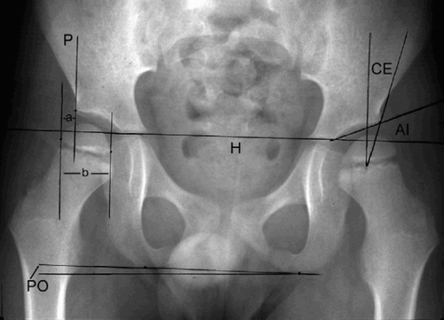
Figure 1. A 6-year-old boy with diplegia, showing the radiographic measurements.The migration percentage (MP) is the lateral displacement of the femoral head (a/b × 100).The other radiographic parameters are indicated (AI:acetabular index;CE:center-edge angle;PO:pelvic obliquity).H is Hilgenreiner's line and P is Perkins'line.AI is the slope of the acetabular roof, which is the angle between the acetabular roof and Hilgenreiner's line.The CE angle is the angle between the perpendicular line through the center of the femoral head and the line between this and the lateral acetabular rim.
The migration percentage (MP) is the percentage of the femoral head lateral to the acetabulum (lateral to Perkins'line), measured parallel to Hilgenreiner's line. When the lateral margin of the femoral head was medial to Perkins'line, the MP was given a negative value. When the whole femoral head was lateral to Perkins'line, the MP was registered as 100%. The relationship between MP and femoral head coverage is that their sum is 100%. The hips were classified as normal (MP under 20%), “at risk” (MP 20–32%), subluxation (MP 33–89%), and dislocation (MP 90% or higher). Hips with MP less than 33% were called “located” in order to distinguish them from subluxation. Based on the initial and the last radiograph, the mean MP progression per year was calculated. The worst hips (the side with the largest MP) in each patient at the initial and follow-up radiographs were used for this calculation.
When assessing the development of the hips with time, the classification of Kalen and Bleck (Citation1985) was used. A change in MP of less than 10% is termed “unchanged”, whereas a decrease of 10% or more is called “improved” and an increase of 10% or more is termed “worse”. For a normal hip to become worse, the MP had to reach a value over 20% at follow-up.
The patients were followed up until operative treatment (54 patients), or until the most recent radiograph in those who had not undergone hip surgery (22 patients). The mean length of follow-up was 4.8 (1–13) years. The mean age of the patients at the most recent radiograph was 8.3 (2–19) years.
Statistics
The statistical differences between groups were assessed by t-tests and chi-square test when 2 groups were compared, and analysis of variance (ANOVA) when more than 2 groups were compared. Correlation between parameters was evaluated by Pearson's correlation coefficient (r). The risk factors for MP progression were evaluated with the use of linear multiple regression. The underlying assumptions were checked using Mallow's Cp, Cook's d and the deleted residuals (Kleinbaum et al. Citation1987). The significance level was set at 0.05.
Results
The mean initial MP was 25% (-18–66) and the MP at the last radiographic examination was 51% (9–100). There was a significant correlation between the MP of the right and left side, with a correlation coefficient of 0.65 at the primary radiographs and 0.30 at follow-up.
The mean progression in MP was 7.1% (-2–33) per year. There was a clear association between type of CP and deterioration of the hips, as the MP progression was 11% per year in QP and 2.0% in DP (, ). The difference was highly significant (p < 0.001). The lateral displacement increased with decreasing functional level, as the MP progression was 12% per year in subjects with-out gait function, 2.7% in those with gait function with support, and 0.9% in subjects who could walk without support (). The difference between patients who could not walk and those who could walk was significant (p < 0.001). In the diplegic group, the yearly increase in MP tended to be less in those who could walk independently compared to those who had to use support when walking (p = 0.06).
Figure 2. A boy with spastic quadriplegia. A. The hips are normal at 2 years of age. B. The right hip is severely subluxated at 10 years of age (MP increase 8.5% per year in the right hip and 4.2% inthe left).
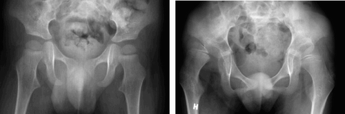
Table 1. Migration percentage (mean, SD) in relation to type of CP and gait function a
There was a significant association between increase in MP and age (), as children under 5 years of age had greater MP progression per year than older children (p = 0.02). The differencebetween the age groups was significant only for the quadriplegics, whose MP progression per year was 13% in the youngest group and 7.3% in those over 5.0 years of age (p = 0.02). The corresponding results in the diplegics were 2.0% under 5 years and 1.8% above 5.0 years (p = 0.9). When the children above and below 8 years of age at the initial radiograph were compared, the youngest group had greater MP progression per year than the older group (p = 0.02).
Table 2. Migration percentage (mean, SD) related to patient age at the initial radiograph, and type of CP a
There was no significant correlation between initial MP and yearly increase in MP. The MP progression was greater (p = 0.02) when there was initial subluxation (10% per year) than when both hips were initially located (5.6%). 7 patients had initial MP of 50% or more (6 unilaterally and 1 bilaterally). These hips deteriorated, and went on to dislocation in 4 cases and increased subluxation in 3.
Linear multiple regression analysis was performed to evaluate the variables that had the greatest influence on the rate of MP progression. Gait function and age were the only variables with significant influence. Gait function (with or without support) explained 32% of the MP progression per year, and age explained 5%. The multiple regression equation was: MP progression per year = 23.0 – (0.57 × age) – (9.6 × gait function). In this equation, age is patient age in years at the initial radiograph; no gait function is 1 and walking ability with or without support is 2.
Initially, both hips were located in 51 patients (27 normal and 24 “at risk”) and 25 patients had subluxation in one or both hips. When assessing the development of the hips with time, using the classification of Kalen and Bleck (Citation1985), 30 patients had unchanged hips, 45 patients developed worse hips, and the hips of 1 diplegic improved during the follow-up period. Again, better prognosis was seen in DP than in QP, as 12 patients with DP (35%) and 33 with QP (78%) got worse hips. Total dislocation occurred in 14 quadriplegic patients. The mean age at dislocation was 6.5 (4–15) years. No patients in the diplegic group had dislocation.
Unilateral improvement (decrease in MP of 10% or more) occurred in 8 patients (3 with DP and 5 with QP). The opposite hip deteriorated in all 5 quadriplegics. There was alteration from located hips to subluxation in 1 child, from subluxation to more pronounced subluxation in 2, and from subluxation to dislocation in 2 cases. Thus, unilateral improvement was of no benefit to the patients.
The radiographic parameters acetabular index (AI), CE angle, and pelvic obliquity are shown in . The mean CE angle was smaller at follow-up than at the initial examination (p = 0.01) and the mean AI was larger (p < 0.001), whereas there was no significant difference in PO. There was a high correlation between MP and CE angle (r = –0.93), and between MP and AI (r = 0.55), but there was no correlation between MP and pelvic obliquity.
Table 3. Radiographic measurements (mean, SD) other than migration percentage
Discussion
Because the patients were referred for orthopedic evaluation, the present sample does not represent a true cross section of the CP population. Scrutton et al. (Citation2001) reported that one-third of unselected patients with bilateral CP could walk without support, which is a larger proportion than the one quarter in the present study. The reason for the greater severity in the present material is that there were more quadriplegics than diplegics, whereas diplegia is more common in an unselected material (Nordmark Citation2000). None of the present children with quadriplegia had independent gait function, whereas slightly more than one-half of those with DP could walk without support—which is in accordance with the results of Nordmark Citation(2000). Thus, within each group of DP and QP, the present material should be representative.
Radiographic measurements
The most important single qualification of a normal hip joint is an adequate coverage of the femoral head, which can be assessed by the migration percentage and the CE angle. Reimers (Citation1980) found a high correlation between these parameters, and this has been confirmed in the present study. The CE angle is not so easy to measure, however, especially when the femoral head is not completely spherical and the interobserver agreement is inferior to that of the MP (Wiig et al. Citation2002).
Acetabular dysplasia was assessed by the acetabular index. Vidal et al. (Citation1985) reported that acetabular deformity in CP does not develop until after the age of 30 months, indicating that there may not have been enough time for the acetabulum to become abnormal when the femoral head begins to become displaced laterally. Whereas Cooke et al. (Citation1989) felt that the acetabular index was the most powerful radiographic predictor, Vidal et al. (Citation1985) maintained that AI should not be considered as a prognostic indicator below the age of 5 years.
Because MP is an easy measurement to make, is little influenced by the rotational position of the femur (Reimers Citation1980), and has good interobserver agreement (Dobson et al. Citation2002, Wiig et al. Citation2002), I would recommend MP as the main radiographic parameter, which is in agreement with most previous studies (Reimers Citation1980, Vidal et al. Citation1985, Miller and Bagg Citation1995). The acetabular index is a useful parameter in preoperative planning when there is doubt with regard to indications for soft-tissue release, femoral osteotomy or acetabular osteotomy alone or in combination. Pelvic obliquity is a relevant parameter when there is both hip dysplasia and scoliosis.
Because it is the grade of dysplasia in the worst hip that is of most importance to the patient, it seems adequate to include one hip only for each patient in the statistics, which is what was done in the present study. However, this has not been done in previous reports (Reimers Citation1980, Vidal et al. Citation1985, Miller and Bagg Citation1995). If both hips are included, the real progression in MP will be under-estimated, i.e. progression will be less than if only the hip with greatest displacement is included. In 5 quadriplegics, one hip had reduced MP at follow-up whereas the other side had increased MP. This also occurred in 5 of the 45 patientsin the series of Miller and Bagg (Citation1995). It is, however, of no help to the patient that one hip improves if the other becomes worse.
Risk factors for progression of hip migration
The hips of most individuals with CP are normal up to the age of approximately 18 months (Letts et al. Citation1984). Hip dysplasia develops because of muscular imbalance and spasticity. Of the possible risk factors for lateral displacement, multiple regression showed that gait function and age were the only variables of significant influence. Type of CP was not a significant factor, because almost all children with DP could walk whereas almost all the quadriplegics could not walk. Thus, type of CP was confounded in the multiple regression analysis.
The mean increase in MP was 7% per year. When comparing this with previous studies, one has to distinguish between different walking abilities, which has not been done in some reports (Reimers Citation1980, Miller and Bagg Citation1995). In the present study, patients who could walk had considerably less risk of hip displacement than those who could not walk, which is in agreement with the findings of Vidal et al. (Citation1985). This indicates that walking is beneficial to the development of the hips. It seems that not only the capacity to walk, but also the degree of gait function is important since the MP progression tended to be less in the diplegics who could walk independently than in those who needed support.
Significantly larger MP progression per year occurred in younger children than in older children, no matter whether the delineation between the groups was 5 years or 8 years. This is not consistent with the experience of Miller and Bagg (Citation1995) who had 75% quadriplegics in their series and found no significant difference above and below 8 years of age. In other reports, no distinction between age groups was made (Reimers Citation1980). Vidal et al. (Citation1985) only examined children aged 1–5 years and found a mean increase in MP of 5.5% per year. This is lower than in our material, where the yearly increase in MP was 8.2% under 5 years of age. The reason for this discrepancy is unclear, but it could be due to the fact that only the worst hip was used in the present calculations. Another reason could be selection of patients, but this seems less likely since the proportion of quadriplegics was more than 50% in both series.
Type of CP has not been considered in some previous reports (Reimers Citation1980, Miller and Bagg Citation1995). Thus, the annual MP increase of 10% in spastic CP found by Reimers (Citation1980) is difficult to interpret. Vidal et al. (Citation1985) reported an increase of 7–9% per year in quadriplegic bedridden patients. An even greater MP increase of 11% was found in our quadriplegics. The fact that only the hip with the largest lateralization was used for statistics probably contributed to the larger MP increase than previously reported.
In the diplegic group the mean MP progression per year was 2%, which is somewhat less than reported by Vidal et al. (Citation1985) who found an MP increase of 4% per year in children with a potential for independent walking.
Clinical consequences
It would be an advantage in clinical work if there was a cut-off value for MP, such that percentages above this limit would predict deterioration of the hips. Miller and Bagg (Citation1995) found that all hips with MP in the range 60–90% went on to dislocation. The present results showed that all 7 patients with initial MP above 50% had deterioration to dislocation, or severe subluxation. Thus, in order to avoid dislocation, it seems adequate to recommend operative treatment before the MP reaches 50%. Nevertheless, the decision and timing of hip surgery should be based on a combination of clinical and radiographic findings.
The different prognoses according to gait function and age indicate that the follow-up routines should be different. There was a pronounced trend towards deterioration in subjects without gait function. Thus, close follow-up is necessary—especially in children under 5 years of age, in order to detect hips at risk for dislocation. It seems reasonable to start with an anteroposterior radiograph at 1 year of age and repeat this examination on an annual basis until the natural history has been established. In spastic diplegia, the risk of severe hip dysplasia is considerably less and such frequent follow-up is not necessary. A rational policy would be to recommend the first radiograph at 2–3 years of age in diplegics and the next radiograph 3 years later. More frequent follow-up is indicated should clinical deterioration (reduced and asymmetric abduction) occur.
No competing interests declared.
Related Research Data
- Cooke P H, Cole W G, Carey R P L. Dislocation of the hip in cerebral palsy. J Bone Joint Surg (Br) 1989; 71: 441–6
- Dobson F, Boyd R N, Parrot J, Nattrass G R, Graham H K. Hip surveillance in children with cerebral palsy. J Bone Joint Surg (Br) 2002; 84: 720–6
- Hilgenreiner H. Zur Frühdiagnose und Frühbehandlung der angeborenen Hüftgelenkverrenkung. Med Klin 1925; 21(38)1425–9
- Howard C B, McKibbin B, Williams L A, Mackie I. Fac-tors affecting the incidence of hip dislocation in cerebral palsy. J Bone Joint Surg (Br) 1985; 67: 530–2
- Kalen V, Bleck E E. Prevention of spastic paralytic dislocation of the hip. Dev Med Child Neurol 1985; 27: 17–24
- Kleinbaum D G, Kupper L L, Muller K E. Applied regression analysis and other multivariate methods. 2. ed. PSW-Kent Publishing Boston. 1987
- Letts M, Shapiro L, Mulder K, Klassen O. The windblown hip syndrome in total body cerebral palsy. J Pediatr Orthop 1984; 4: 55–62
- Miller F, Bagg M R. Age and migration percentage as risk factors for progression in spastic hip disease. Dev Med Child Neurol 1995; 37: 449–55
- Nordmark E. Measurements of function in children with cerebral palsy (thesis). Lund University Studentlitteratur, 2000: 1–78
- Reimers J. The stability of the hip in children. Acta Ortop Scand (Suppl 184) 1980; 51: 12–9
- Samilson R L, Tsou P, Aamoth G, Green W. Dislocation and subluxation of the hip in cerebral palsy. J Bone Joint Surg (Am) 1972; 54: 863–73
- Scrutton D, Baird G, Smeeton N. Hip dysplasia in bilateral cerebral palsy: incidence and natural history in children aged 18 months to 5 years. Dev Med Child Neurol. 2001; 43: 586–600
- Vidal J, Deguillaume P, Vidal M. The anatomy of the dys-plastic hip in cerebral palsy related to prognosis and treatment. Internat Orthop (SICOT) 1985; 9: 105–10
- Wiberg G. Studies on dysplastic acetabula and congenital subluxation of the hip joint. Acta Chir Scand (Suppl 58) 1939; 83: 28–38
- Wiig O, Terjesen T, Svenningsen S. Inter-observer reliability of radiographic classifications and measurements in the assessment of Perthes'disease. Acta Orthop Scand 2002; 73: 523–30