Abstract
Background The optimal design of an elbow prosthesis for badly damaged elbows is unkown. We evaluated 23 GSB III semi-constrained (sloppy-hinged) total elbow arthroplasties in 16 consecutive patients with rheumatoid arthritis.
Patients and methods After a mean follow-up period of 5 (2–9) years, we assessed quality of the cementing technique, signs of aseptic loosening, patient satisfaction, range of movement, and determined the Hospital for Special Surgery (HSS) elbow score. 3 patients had died before follow-up; thus, 20 replacements in 16 patients were available for clinical and radiographic study. All patients had endstage rheumatoid arthritis (RA) of the elbow joint.
Results In 2 patients, humeral components were revised due to malorientation. 1 arthroplasty was revised due to aseptic loosening of the humeral component. There were 4 cases of intraoperative fracture which healed uneventfully. The total rate of complications was thus one-third. In 17 of 40 components, the cementing technique was rated as marginal or inadequate. We found no association between cementing technique and loosening. The arc of extension/flexion increased by 19° (0–80), and the range of pronation/supination increased by 31° (0–130). There were no cases of infection or ulnar nerve dysfunction. At the latest follow-up, the HSS elbow score was 84 (40–100) points. 11 of 20 elbows were rated as excellent, 4 elbows were rated as good, 2 elbows were rated as fair, and 3 elbows were rated as poor. 14 of 16 patients were satisfied with the result and the 2 patients who were not satisfied had persistent pain.
Interpretation Despite the inherent problems of cementing in small-calibre medullary cavities, the clinical outcome of the GSB III arthroplasty was encouraging for patients with-end stage RA. The rate of overall complications compared favorably with other studies of semiconstrained elbow arthroplasty for end-stage RA. Most complications of the series were minor and did not necessitate revision. ▪
Rheumatoid arthritis (RA) often involves the elbow, causing joint destruction, instability, pain and reduced functional ability. At advanced stages, replacement arthroplasty may be necessary to relieve pain and improve range of movement (Swedish Council on Technology Citation2000). Due to joint destruction, poor bone stock, high risk of fracture, poor skin quality, and the difficulty of cementing both humerus and ulna properly, the use of replacement arthroplasties in these patients is fraught with complications.
Most studies on elbow joint replacement have been published by authors involved in the design and first clinical trials of the implants (Ewald et al. Citation1980, Gschwend et al. Citation1996, Citation1999, Inglis et al. Citation1997, Baksi Citation1998, Gill and Morrey Citation1998, Kudo Citation1998). Since 1990, the Gschwend-Scheier-Bähler III (GSB III) (Sulzer Medical, Wintherthur, Switzerland) semi-constrained total elbow replacement has been the implant of choice at our department for patients with end-stage RA of the elbow (–). The design allows preservation of the distal humeral condyles and their muscular attachments. The GSB III elbow prosthesis is semi-constrained (sloppy-hinged), its design combining intrinsic stability with sufficient varus and valgus laxity to reduce peak forces at the interfaces.
Figure 1 a. Preoperative lateral view. b. Preoperative anteroposterior view. Pre- and postoperative radiographs of the GSB III elbow arthroplasty in a 58-year-old woman with severe rheumatoid destruction of the elbow joint.
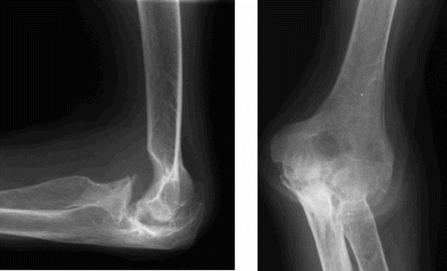
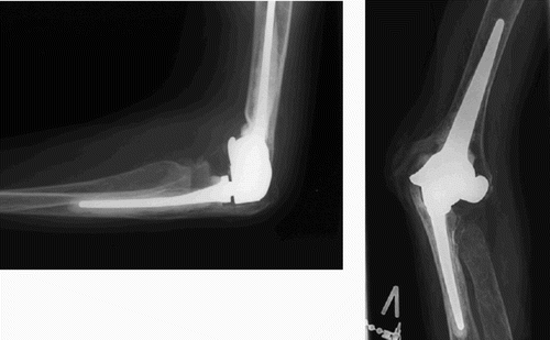
Figure 2 a. Postoperative lateral view. The arrows mark adequate cement mantle around the ulnar stem extending past the tip, and the inadequate cement mantle of the humeral stem, which does not extend past the tip of the component. b. Postoperative anteroposterior view. Note the preservation of the distal humeral condyles with muscular and ligamentous attachments, thereby improving soft tissue stability of the arthroplasty and reduction of peak forces at the interfaces. The radial head has routinely been removed. Pre- and postoperative radiographs of the GSB III elbow arthroplasty in a 58-year-old woman with severe rheumatoid destruction of the elbow joint.
We present a 2–9-year clinical and radiographic evaluation of 23 GSB III elbow replacements in 16 patients.
Patients and methods
Between 1990 and 1997, 23 primary GSB III, semiconstrained total elbow joint replacements were performed in 19 patients (16 women) with RA. The main indications were pain, instability and functional impairment. All patients had radiographic evidence of severe joint destruction due to rheumatoid arthritis. Mean age at surgery was 64 (31–76) years. Prior to replacement surgery, 2 patients had had open synovectomy of the joint and 2 patients had had excision of the radial heads. 12 arthroplasties were performed on the right, dominant side and 8 arthroplasties were performed on the left, non-dominant side. A senior surgeon (CHJ) performed 17 procedures, and another senior surgeon (S S-H) performed the other 6.
Operative technique
A 1-g dose of methicilline was administered preoperatively. The patient was placed in the lateral decubitus position with a tourniquet around the upper arm, and the elbow flexed 90° over a padded support. The joint was exposed through a posterior midline incision. The ulnar nerve was identified and protected, but not routinely transposed anteriorly. The triceps tendon and underlying muscle was split longitudinally and mobilized from the olecranon, medial to lateral with 2–3 mm slivers of bone at the insertion sites. The triceps tendon and slivers of bone were kept in continuity distally with the ulnar periosteum forming a soft tissue/bone sheath. The collateral ligaments were identified and preserved. The radial heads were removed, followed by anterolateral synovectomy. The distal humerus and proximal ulnae were prepared by power saw using the cutting jigs supplied. The distal humeral condyles with muscular attachments were preserved. Following reduction with the proper trial components, the medullary cavities were cleansed by pulse lavage, dried, and the final components were cemented.
After the curing period, the components were assembled. The soft tissue/bone sheath was sutured and anchored through small drill holes. The mean operating time was 120 (110–195) min. Postoperatively, the elbow was immobilized in a plaster-of-Paris slab cast in 30° flexion for 2 weeks, followed by physiotherapy of non-weight-bearing exercises of the wrist, elbow and shoulder. We find it important to protect the cicatrice and skin with this regime, because these patients often have vulnerable skin and soft tissues due to prior medical treatment. The skin sutures were removed after 3 weeks.
For the first 17 replacements we used Palacos cement (Biomet, Warsaw, IN), but we found it difficult to introduce this high-viscosity cement into the medullary cavity of the ulna. Since 1996, we have used low-viscosity Simplex (Stryker/Howmedica, Allendale, NJ) introduced by retrograde filling instead, using a Ch 16 catheter mounted on a 60-mL syringe. The cement is stored at a maximum of 5° to enhance its low viscosity. This method facilitates achievement of a circumferential cement mantle.
Clinical and radiographic evaluation
16 patients with 20 replacements were available for radiographic and clinical follow-up examination, mean 5 (2–9) years after the index operation. 3 patients (3 GSB III) with the replacements in situ had died of unrelated causes, 24, 32, and 33 months postoperatively. In 2 of these patients, the prosthesis had done well according to the patient records. In the third patient, the prosthesis had disassembled 7 days after the operation, necessitating revision.
At the latest follow-up, patients were rated according to the Hospital for Special Surgery (HSS) elbow score (Figgie et al. Citation1989, Inglis et al. Citation1997). A maximum of 40 points was assigned for complete pain relief, a maximum of 20 points was assigned for excellent overall function, a maximum of 12 points was assigned for full strength (lifts 2.5 kg to 90°), and a maximum of 28 points was assigned for a full range of movement (140° flexion, pronation < 60°, supination < 60°); giving a total of 100 points. A score of 90–100 points is excellent, a score of 80–89 points is good, a score of 70–79 points is fair, and a score of less than 70 points is poor. Preoperatively, no consistent clinical scoring was recorded. The range of movement was recorded preoperatively. In addition to the clinical evaluation, the overall satisfaction of the patient was assessed.
We evaluated the quality of the cementing technique according to Schneeberger et al. (Citation2000). This was considered adequate when the mantle extended past the tips of the components, marginal if the mantle reached but did not extend past the tips of the components, and inadequate if the mantle did not reach the tip of the component.
Loosening of the cementbone interface was rated type I if a radiolucent line of 1 mm or more in width involved less than 50% of the total interface, type II if the radiolucency involved more than 50% of the total interface, type III if the radiolucency involved the entire interface, and type IV if there was radiographic evidence of gross loosening.
Statistics
We used Chi-Square tests to assess the relationship between adequacy of cementing and aseptic loosening. Paired-samples t-tests were used to investigate the relationship between aseptic loosening and follow-up period. The relationship between follow-up period and HSS score was investigated by Spearman’s correlation coefficient. Schnee-berger’s classification of adequacy of cementing and grades of aseptic loosening were dichotomized for statistical purposes into adequate/inadequate cementing technique and radiographic loosening/no loosening. All calculations were performed using SPSS statistical software version 12 (SPSS Inc., Chicago, IL). The significance level was set at < 0.05.
Results
Clinical evaluation
At the follow-up, the mean HHS elbow score was 84 (40–100) points. 11 of 20 elbows rated excellent, 4 elbows rated good, 2 elbows rated fair and 3 elbows rated poor. 14 of 16 patients were satisfied with the results of the replacements. 2 patients (with type III and type IV aseptic loosening) were dissatisfied, mainly due to pain. 11 of the 20 replacements caused no pain, 6 replacements caused slight pain, 1 replacement caused moderate pain related to day-to-day activity, 1 replacement caused pain at rest, and 1 replacement caused severe pain at rest and during day-to-day activity. 13 elbows had stage V destruction according to the classification of Larsen et al. (Citation1977), 6 elbows had stage IV destruction, and 1 elbow had stage III destruction.
Preoperatively, extension/flexion motion was 89° (40–140), increasing to 108° (65–140) postoperatively. Motion thus increased by an average of 19° (0–80). The mean preoperative range of pronation/supination was 119° (50–180). Postoperatively, the mean range of pronation/supination increased to 150° (0–180). The mean range of pronation/supination increased by 31° (0–130) (). There was no significant relationship between follow-up period and HSS score (r = 0.04; p = 0.9).
Table 1. Range of movement (ROM), preoperatively and postoperatively
Radiographic evaluation ( and )
17 of 40 components (7 humeral and 10 ulnar) showed marginal cementing technique, and 4 of these components had radiographic loosening (types II–IV) (2 humeral, 2 ulnar). 23 components were adequately cemented, but 2 of these (humeral) had signs of loosening. In the 6 arthroplasties performed with low-viscosity cement, 5 ulnar and all humeral components had adequate cementing technique. We found no relationships between adequacy of cementing and aseptic loosening at follow-up (humeral components: OR 0.53 (95% CI 0.3–0.85), p = 0.08; ulnar components: OR 1.0 (95% CI 0.1–8.9), p = 0.7). There were no relationships between humeral loosening or ulnar loosening, and length of follow-up (humeral: (−25–54); ulnar: p = 0.5, (8–53–32), p = 0.5).
Table 2. Results of the cementing technique
Table 3. Radiological signs of aseptic loosening at the latest follow-up
Complications
Complications occurred in 7 of the 20 arthroplasties. In 1 case of gross aseptic loosening of the ulnar component, the patient rated 40 on the HSS score, and revision surgery was planned. In 2 patients, the humeral components were malpositioned—which led to dislocation in one case and limited flexion in the other. Both components were revised, and the clinical and radiographic results of the revisions were excellent. 3 fractures of the ulnar epicondyles and 1 fracture of the radial epicondyle occurred intraoperatively, 3 of which required temporary fixation with k-wires. The fourth fracture was observed on the routine follow-up radiographs 2 months postoperatively, and was treated by non-weight-bearing exercises. Fractures healed uneventfully without any adverse effect on the clinical outcome. No patients experienced superficial or deep infections or ulnar neuropathy postoperatively.
Discussion
Due to the severe joint destruction of the elbow because of RA, replacement surgery is usually fraught with high complication rates. We have treated end-stage RA of the elbow joint with the semi-constrained (sloppy-hinged) GSB III prosthesis (introduced in 1978). Its design allows preservation of the collateral ligaments and the distal humeral condyles, thereby preserving the extensor and flexorpronator muscle origins. Ideally, these features will contribute to postoperative stability of the implant and reduce peak forces at the interfaces (Herren et al. Citation2001, McKee et al. Citation2003). In our study of 20 GSB III semi-constrained total elbow arthroplasties in 16 patients with RA, the overall complication rate was one-third, which is comparable to other studies using semi-constrained designs (Ramsey et al. Citation1999, Hildebrand et al. Citation2000, Kelly et al. Citation2004). 1 of the 20 replacements was revised due to aseptic loosening, and 5 replacements were reoperated for other reasons. We had no cases of infection and no cases of ulnar neuropathy. In a series of 36 GSB III replacements, and after an average of 14 years follow-up, Gschwend et al. (Citation1996) reported aseptic loosening in 3 cases, and 9 elbows were reoperated due to disassembly. There were 3 cases of deep infection. In a series of 14 GSB III replacements followed up for 6 years, Schneeberger et al. (Citation2000) reported aseptic loosening in 4 of 14 replacements and deep infection in 1. In a recent study of 28 GSB III replacements followed for 8 years, 5 patients were reoperated (Kelly et al. Citation2004). There were 2 cases of persistent ulnar nerve paresthesia, 2 cases of superficial infection, and 1 case of deep infection.
One semi-constrained elbow prosthesis that is commonly used in the United States for end-stage rheumatoid arthritis is the Mayo-Coonrad device. Morrey and Adams (Citation1992) reported the results of 58 such replacements in 54 patients who were followed for 4 years. The cement mantle was satisfactory in all ulnar components, and satisfactory in 54 of the humeral components. Flexion-extension motion increased by an average of 23°, and pronation/supination motion increased by an average of 32°. 53/58 patients reported no pain at the latest follow-up, and 52 of the patients achieved an excellent or good result. However, 8 patients sustained condylar fractures, 4 patients were infected, and 1 patient had ulnar neuropathia. 6 patients were reoperated.
In a comparison of an unconstrained elbow replacement (the Ewald prosthesis) in 12 elbows, and the Mayo-Coonrad semiconstrained replacement in 14 elbows with RA, Wright et al. (Citation2000) reported the results of 3 years of follow-up. There was 1 revision due to aseptic loosening in the semi-constrained group. Apart from this failure, all the patients reported satisfactory results of surgery.
Ramsey et al. (Citation1999) reported 6-year follow-up results of 19 Coonrad-Morrey semiconstrained elbow arthroplasties. There were 4 cases with aseptic loosening and fractures of the humeral condyles. Hildebrand et al. (Citation2000) reported complications in 26/51 Coonrad-Morrey semiconstrained arthroplasties followed for 4 years, including 10 cases of ulnar nerve dysfunction, 9 condylar and olecranon fractures, and 3 cases of infection.
In addition to 2 cases of malposition of the humeral components and 1 case of dislocation, the majority of the complications in our series were minor and manageable without adverse effects on the clinical outcomes. There were no superficial or deep infections, which we attribute to our use of a plaster-of-Paris slab cast for 2 weeks postoperatively. In contrast to comparable series, in which ulnar nerve injury or entrapment has been a common complication of the procedure (Hilde brand et al. Citation2000, Kelly et al. Citation2004), none of our patients had ulnar neuropathy. 17/40 components showed marginal or inadequate cementing technique, similar to the findings of Schneeberger et al. (Citation2000). However, in 6 of our replacements fixated with low-viscosity cement, only 1 ulnar component had a marginal cement mantle and there was no radiolucency in any of the 12 components. We will thus continue the use of low-viscosity cement.
No competing interests declared.
Author contributions
CHJ and SJ: Follow-up examinations, radiography and manuscript. MR: Follow-up examinations. SS-H: Radiography, follow-up examinations.
Related Research Data
References
- Baksi D P. Sloppy hinge prosthetic elbow replacement for post-traumatic ankylosis or instability. J Bone Joint Surg (Br) 1998; 80: 614–9
- Ewald F C, Scheinberg R D, Poss R, Thomas W H, Scott R D, Sledge C B. Capitellocondylar total elbow arthroplasty. J Bone Joint Surg (Am) 1980; 62: 1259–63
- Figgie M P, Inglis A E, Mow C S, Figgie H E, III. Salvage of non-union of supracondylar fractures of the humerus by total elbow arthroplasty. J Bone Joint Surg (Am) 1989; 71: 1058–65
- Gill D R, Morrey B F. The Coonrad-Morrey total elbow arthroplasty in patients who have rheumatoid arthritis. J Bone Joint Surg (Am) 1998; 80: 1327–35
- Gschwend N, Simmen B R, Metajovsky Z. Late complications in elbow arthroplasty. J Shoulder Elbow Surg 1996; 5: 86–96
- Gschwend N, Scheier N H, Baehler A R. Long-term results of the GSB III elbow arthroplasty. J Bone Joint Surg (Br) 1999; 81: 1005–12
- Herren D B, O'Driscoll S W. An K N Role of collateral ligaments in the GSB-linked total elbow prosthesis. J Shoulder Elbow Surg 2001; 10: 260–4
- Hildebrand K A, Patterson S D, Regan W D, MacDermid J C, King G J. Functional outcome of semiconstrained total elbow arthroplasty. J Bone Joint Surg (Am) 2000; 82: 1379–86
- Inglis A E, Inglis A E, Jr, Figgie M M, Asnis L. Total elbow arthroplasty for flail and unstable elbows. J Shoulder Elbow Surg 1997; 6: 29–36
- Kelly E W, Coghlan J, Bell S. Five to thirteen-year follow-up of the GSB III total elbow arthroplasty. J Shoulder Elbow Surg 2004; 13: 434–40
- Kudo H. Non-constrained elbow arthroplasty for mutilans deformity in rheumatoid arthritis. J Bone Joint Surg (Br) 1998; 80: 234–9
- Larsen A, Dale K, Eek M. Radiographic evaluation of rheumatoid arthritis and related conditions by standard reference films. Acta Radiol 1977; 18: 481–91
- McKee M D, Pugh D M, Richards R R, Pedersen E, Jones C, Schemitsch E H. Effect of humeral condylar resection on strength and functional outcome after semiconstrained total elbow arthroplasty. J Bone Joint Surg (Am) 2003; 85: 802–7
- Morrey B F, Adams R A. Semiconstrained arthroplasty for the treatment of rheumatoid arthritis of the elbow. J Bone Joint Surg (Am) 1992; 74: 479–90
- Ramsey M L, Adams R A, Morrey B F. Instability of the elbow treated with semiconstrained total elbow arthroplasty. J Bone Joint Surg (Am) 1999; 81: 38–47
- Rozing P. Souter-Strathclyde total elbow arthroplasty. J Bone Joint Surg (Br) 2000; 82;: 1129–34
- The Swedish Council on Technology Assessment in health care Rheumatic diseases-surgical treatment. Acta Orthop Scand (Suppl 294) 2000:; 28–33
- Schneeberger A G, Hertel R, Gerber G. Total elbow replacement with the GSB III prothesis. J Shoulder Elbow Surg 2000; 9: 135–8
- Wright T W, Wong A M, Jaffe R. Functional outcome comparison of semiconstrained and unconstrained total elbow arthroplasty. J Shoulder Elbow Surg 2000; 9: 524–31