Abstract
Background Bleeding in the calf or ankle joint may lead to ankle equinus deformity, particularly in childhood and during adolescence. We assessed the long-term functional and radiographic results after Achilles tendon lengthening for ankle equinus deformity in hemophiliacs.
Patients and methods Between 1975 and 1986, 30 hemophilic patients with pes equinus were surgically managed by Achilles tendon lengthening. Of these, 23 were followed up prospectively twice a year for an average of 13 (1–24) years. The mean age at operation was 29 (12–46) years. The clinical results were documented according to the score of the Advisory Committee of the World Federation of Hemophilia (WFH), while radio-graphs were evaluated using the Pettersson score. On average, preoperative ankle equinus deformity was 21 (5–55) degrees. Mean range of motion was 21 (5–42) degrees prior to surgery.
Results At the first postoperative examination 1 year after surgery, 21/23 cases were improved, and 9/21 reached dorsiflexion to at least neutral position. At the last follow-up, ankle equinus deformity was 10 (4–20) degrees on average. 20/23 patients still showed significant improvement compared to their condition before surgery. 7 patients still had complete correction of the equinus deformity, while mean range of motion decreased constantly over the observation period. The clinical score was significantly improved 1 year after surgery and diminished only slightly afterwards. Radio-graphic outcome deteriorated, with scores rising from 4.3 (1–10) points preoperatively to 7.3 (3–12) points at last follow-up.
Interpretation Most patients treated for hemophilic pes equinus by Achilles tendon lengthening experienced long-term benefit concerning the equinus deformity, but gradually lost overall movement of the ankle joint. Progression of the ankle arthropathy cannot be hindered. ▪
Bleeding in the juvenile and adolescent hemophiliac with no or inadequate substitution therapy often occurs in the calf muscles and the ankle joint (Ahlberg 1965, Gamble et al. Citation1991). Fibrous atrophy after recurrent bleeding may cause an ankle equinus deformity, which functionally, may be complicated by an extension deficit of the knee (Ribbans and Philipps Citation1996).
In cases for which non-surgical management options remain ineffective, moderately simple operative techniques such as Achilles tendon lengthening and simultaneous dorsal capsulotomy have been suggested (Stagnara et al. Citation1965, Niemann Citation1971, Ribbans and Philipps Citation1996). We assessed long-term clinical and radiographic results after Achilles tendon lengthening for hemophilic equines deformity.
Patients and methods
Between 1975 and 1986, 30 hemophiliacs with secondary equinus deformity of the ankle due to recurrent bleeding episodes underwent Achilles tendon lengthening. Of these, 23 patients—all with severe hemophilia A (factor VIII less than 1%)—could be followed up. 12 were operated on the right side, and 11 on the left side. The mean age at the time of operation was 29 (12–46) years. Average follow-up was 13 (1–24) years. Clinical outcome was based on the Advisory Committee of the World Federation of Hemophilia (WFH) clinical score (Pettersson and Gilbert Citation1985), while radio-graphic results were evaluated using the Pettersson score (Pettersson et al. Citation1980). Pain (0–3 points), bleeding (0–3 points) and physical examination (0–12 points) make up the clinical score (WFH, 0 points normal). The physical examination score is based on joint swelling (0 or 2), muscle atrophy (0–1), axial deformity (0–2), crepitus on motion (0–1), range of motion (0–2), flexion contracture (0 or 2) and instability (0–2) points. The radiographic score ranges from 0 points for a healthy joint to 13 points for massive radiographic destruction. We assessed the clinical score at least twice a year, and the radiographic score every second year.
Range of motion (ROM) of the ankle was determined as follows. When knee arthropathy did not inhibit movement, the patient's knee was bent 90 degrees. For clinical practicability, we measured the angle between the lateral foot margin and the ventral tibia. As suggested by AO, the neutral position was defined as a 90-degree angle between tibia and foot. ROM was the sum of passive movement in the direction of maximum plantar and dorsal flexion.
Factor replacement
FactorVIII to achieve an activity of 30–40% was performed preoperatively. Directly before surgery, factor activity was raised to 80–100% by giving approximately 30–40 units offactorVIIIper kg body weight. A coagulation test was performed postoperatively and, if required,factorVIII was again given. FactorVIIIactivity was kept at 60% up to the fourth postoperative day and subsequently maintained at 50% until day 14 postoperatively. In the following period, factorVIIIactivity was held at 20%. After rehabilitation, a return to usual concentrations was allowed.
Operative technique
The indication for surgical management was a persistent or a progressively deteriorating equinus position of the ankle. Lengthening was performed by an open Z-incision of the tendon and, if considered necessary, a simultaneous posterior capsule release of the ankle joint. In cases where there was a tendency of hindfoot valgus, the distal lateral limb of the split tendon was detached; in patients with a hindfoot varus, the distal medial limb of the incised tendon was detached. The Achilles tendon was lengthened to such an extent that the ankle joint was in a neutral position (0 degrees). Slackness of the tendon was avoided. Postoperatively, the lower leg and foot were fixed with a circular cast in a neutral position over a period of 6 weeks. After this, intensive physiotherapy was performed and patients were fully permitted to bear weight.
Statistics
Postoperative improvement of maximum dorsiflexion after Achilles tendon lengthening and also changes of clinical and radiographic scores were statistically assessed by paired t-test, at a significance level of 0.05. The courses of the severity of equinus deformity and the total range of motion (pre- and postoperatively) are graphically pre-sented with mean values and the corresponding 95% confidence intervals. Analysis was done using SPSS version 11.0 for Windows.
Results
The WFH clinical score improved from 4.2 (1–8) to 2.1 (0–6) points 1 year after surgery (p < 0.001) and increased slightly to 2.7 (1–4) points at the last follow-up (p = 0.001). At the last examination, 12 ankles showed clinical improvement, 1 ankle showed deterioration postoperatively, and 10 other ankles were unchanged. The 6 patients who were assessed for up to 10 years after surgery showed an average improvement of 3 points, whereas the 17 patients who were assessed for more than 10 years after surgery improved clinically by an average of 1 point. 6 patients had reduced pain on a continuous basis while 17 patients remained unchanged at the last follow-up.
The radiographic score increased from mean 4.4 (1–10) points preoperatively to 7.3 (3–12) points at last follow-up 1–24 (13) years (p < 0.001). The radiographs of 18 patients showed postoperative deterioration, 4 remained unaltered and 1 patient showed improvement. In the 6 patients assessed 10 years or less after surgery, the score diminished by 2 points on average, while it diminished by an average of 4 points in the other patients.
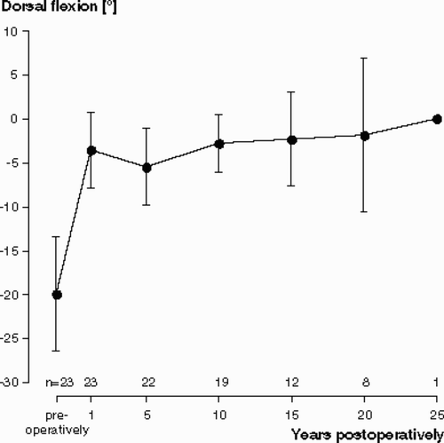
The average preoperative equinus position was 21 (5–42) degrees. Postoperatively, 9/23 patients reached dorsiflexion to at least neutral position, while the average postoperative equinus deformity in the other patients was 5 (2–20) degrees (p < 0.001). 21/23 patients showed significant improvement compared to their status before surgery. At last follow-up, the mean ankle equinus contracture was 10 (4–20) degrees. At first follow-up 1 year after surgery, the 20 patients with a measurable improvement in the equinus contracture had an average postoperative improvement from –23 degrees to –7 degrees dorsiflexion. Overall, these remained unchanged over the subsequent observation period. Even after 15 and 20 years, the improvement in equinus deformity was about the same as after initial surgery (). 7 patients still had complete persistent correction of the initial deformity.
Figure 1. Average change in the severity of equinus deformity pre- and postoperatively (with 95% confidence interval;negative values mean equinus deformity and positive values mean dorsiflexion beyond neutral position). Deformity showed an average improvement of 16° after tendon lengthening, and remained almost unchanged over subsequent years (n = number of patients).
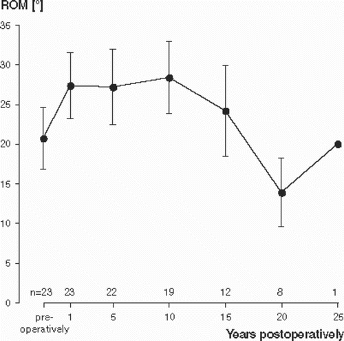
The preoperative ROM was mean 21 (5–42) degrees and increased 1 year after surgery to mean 28 (10–50) degrees (p = 0.003). At last follow-up, ROM was improved (10 degrees on average) in only 8 patients and had deteriorated (9 degrees on average) in 15 patients, suggesting that later on, an improvement of the dorsiflexion was frequently associated with a loss of plantar flexion. ROM was more or less unchanged up to 10 years postoperatively, but later on tended to deteriorate ()—possibly due to an increase in the hemophilic arthropathy of the ankle.
Figure 2. Mean values of total range of motion (ROM) in the ankle joint preoperatively and postoperatively (with 95% confidence interval) after Achilles tendon lengthening for hemophilic equinus deformity (n = number of patients).
Except for delayed wound healing in 1 patient, there were no complications.
Discussion
Achilles tendon lengthening may be used in the treatment of hemophilic pes equines for contractures less than 30°. Larger contractures may require corrective wedge-osteotomy, or an Ilizarov technique (Ribbans and Ress Citation1999) should be considered. Interestingly, we found no suture insufficiencies in our patients, despite the fact that in some cases a marked lengthening of the Achilles tendon was performed. Ribbans and Rees (Citation1999) recommended a subcutaneous technique but admitted that open surgery allows more extensive correction. They cited numerous potential complications of lengthening procedures such as wound break down, sural nerve injury, postoperative hematoma and adhesions. We did not observe these complications in our patients.
Although no surgery of the ankle joint was performed, tendon lengthening had a positive effect on the postoperative clinical ankle score of the patients. Tendinous correction and the simultaneously achieved indirect slackening of the periarticular soft-tissue structures seem to normalize the motion pattern of the ankle joint.
By Achilles tendon lengthening, the ankle's overall range of motion is only slightly improved, as a gain of dorsal flexion is typically associated with loss of plantar flexion. Johnson and Babbitt (Citation1985) were able to show a correlation between the loss of ROM and successive deterioration of articular cartilage in the hemophiliac. One can therefore hypothesize that the documented loss of ROM in our patients preoperatively is not solely a result of the ankle equinus deformity, but is probably multifactorial. Thus, Achilles tendon lengthening addresses only part of the problem.
For activities of daily living, the ability to dorsiflect the foot is of importance. While normal gait with an equinus deformity of 30° is not possible, a patient with a pes equinus of only 10° shows almost normal gait when treated with an in-shoe heel wedge (Perry Citation1992). Mobility and elasticity of the contract capsule of a hemophilic joint is obviously not improved by lengthening of the Achilles tendon, yet by altering the possible ROM to a functionally better region, overall, gait is enhanced. In our patients associated loss of plantar flexion was of no functional concern.
We found that Achilles tendon lengthening is an acceptable method for treatment of hemophilic ankle quinus deformity. However, in the long run there was a tendency of overall loss of ankle ROM. This may be due to the associated arthropathy of the ankle that is almost always present. Most of our patients had some degree of arthropathy of the ankle (detected radiographically) before surgery. Whether loss of motion is due to a progressive arthropathy cannot be answered by our study.
The radiographic assessment of our patients showed that, despite surgical management, joint damage progressed. This finding is not new. In previous studies, we have found that osteotomy of hemophilic knee and hip joints did not necessarily prevent further destruction (Wallny et al. Citation2002, Citation2003). Furthermore, untreated or only non-operatively managed equinus deformities also show progressive arthropathy (Rodriguez-Merchan Citation1999). It therefore seems that with or without surgical management, deterioration of the ankle joint is inevitable—and is most likely not a result of Achilles tendon lengthening.
No competing interests declared.
Author contributions
TW patient assessment, writing the manuscript. HBL patient assessment, hematological supervision. CN statistics. CK evaluation of data, translation/writing manuscript. PP evaluation of data.
- Ahlberg A. Haemophilia in Sweden. Acta Orthop Scand (Suppl) 1965; 77: 20–45
- Gamble J G, Bellah J, Rinsky L A, Glader B. Arthropathy of the ankle in haemophilia. J Bone Joint Surg (Am) 1991; 73: 1008–15
- Johnson R P, Babbitt D P. Five stages of joint disintegration compared with range of motion in haemophilia. Clin Orthop 1985; 201: 36–42
- Niemann K M. Surgical correction of flexion deformities in hemophilia. Am Surg 1971; 37: 685–90
- Pettersson H, Gilbert M S. Classification of the hemophilic arthropathy. Diagnositc imaging in hemophilia, H Pettersson, M S Gilbert. Springer, New York 1985; 56–68
- Pettersson H, Ahlberg A, Nilsson I M. A radiologic classification of hemophilic arthropathy. Clin Orthop 1980, 149: 153–9
- Perry J. Ankle and foot gait deviations. Gait analysis. Slack Inc, Thorofare, NJ 1992; 193–5
- Ribbans W J, Phillips A M. Hemophilic ankle arthropathy. Clin Orthop 1996, 328: 39–45
- Ribbans W, Rees J L. Management of equines contractures of the ankle in haemophilia. Haemophilia 1999;; 5: 46–52, (Suppl 1)
- Rodriguez-Merchan E C. Therapeutic options in the management of articular contractures in haemophiliacs. Haemophilia 1999; 5: 5–9, (Suppl)
- Stagnara P, Fauchet R, Thouverez J P, Belleville J. Ténotomie du tendon d'Achille pour équinisme du pied chez un hémophile B. Hemostase 1965; 5: 191–3
- Wallny T, Brackmann H H, Hess L, Seuser A, Hofmann P, Kraft C N. Long-term follow after varus osteotomy for haemophilic arthropathy of the hip. Haemophilia 2002; 8: 149–52
- Wallny T, Saker A, Hofmann P, Brackmann H H, Nicolay C, Kraft C N. Long term follow-up after osteotomy for haemophilic arthropathy of the knee. Haemophilia 2003; 9: 69–75