Abstract
Background There is no concensus on the optimal treatment time for unstable hips in the newborn. We analyzed the efficiency of a treatment program that has been used for 10 years at our hospital, in which all unstable hips (subluxatable, Barlow-positive and Ortolani-positive) are treated with the von Rosen splint for 6 weeks.
Patients and methods Between 1988 and 1997, 32,171 children were born alive at the hospital. During this period 247 children had a clinically unstable hip diagnosed. 223 of the 247 children underwent a radiographic follow-up after 5–15 years.
Results 1 patient with bilateral instability and treated with a splint for 6 weeks showed a dislocated left hip at the radiographic examination at 8 months, which is part of the screening program, and needed operative treatment. 1 patient did not follow the treatment program and showed a dislocated hip at the age of 3. Another 4 patients required more treatment than the 6 weeks with the splint.
We found no dysplastic hips at the radiographic follow-up. There was no late dysplasia and there were no late dislocations in children born in Lund between 1988 and 1997 who were diagnosed at other Swedish centers that treat developmental dysplasia of the hip (DDH).
Interpretation We conclude that the present screening and 6-week treatment in a von Rosen splint prevent almost all cases of late dysplasia and late dislocation of the hip.
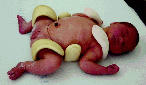
It is generally accepted that early treatment of Neonatal Instability of the Hip (NIH) prevents dislocation (Palmen Citation1984). There are, however, variations in the screening procedure, the splinting technique, the duration of splinting, and the follow-up routines (Emneus Citation1966, Fredensborg Citation1976, Palmen Citation1984, Sahlstrand et al. Citation1985, Krikler and Dwyer Citation1992). We analyzed the efficiency of a treatment program that has been used for 10 years in Lund, Sweden, in which subluxatable (unstable, but not dislocatable) and Barlow-positive and/or Ortolani-positive hips are treated with the von Rosen splint () for 6 weeks. With this program, we reduced the treatment time in the von Rosen splint from 12 to 6 weeks and sonography was introduced as a diagnostic tool for NIH.
Figure 1. The von Rosen splint.
Patients and methods
During the 10-year period 1988–1997, 32,171 children were born alive at the University Hospital in Lund. The pediatricians routinely examined all hips during the first days of life, and those hips that were judged to be unstable were referred for a second examination in the orthopedic department by two senior orthopedic surgeons. During this period, 247 children (68 boys and 179 girls) had an unstable hip diagnosed. In doubtful cases sonography was performed, but in cases with clear clinical instability, treatment was started with a von Rosen splint. The NIH was left-sided in 137 children, right-sided in 41, and bilateral in 68. In 1 child the side of instability could not be determined from the patient's medical record. 4 children with teratological dislocations were excluded from this analysis. All 247 children were treated with the von Rosen splint for 6 weeks, after which the hips were re-examined clinically and with sonography using an anterior dynamic technique (Harcke et al. Citation1984, Dahlström et al. Citation1986, Harcke and Grissom Citation1990, Harcke Citation1992). If the displacement of the femoral head exceeded one quarter of its diameter, measured by sonography, the hip was diagnosed as being unstable. If the hips were stable, treatment was discontinued. If the hips were unstable, treatment was extended for 3 weeks more. 2 weeks after completion of the treatment, a repeat examination with sonography was performed. At 8 months, a final clinical and radiographic examination was done. Radiographs were taken in the anteroposterior direction with the legs straight. The radiographs were analyzed with regard to the presence of dislocation, acetabular dysplasia, and development of the femoral head (). Furthermore, 223 children (90%) completed a radiographic follow-up after 5–15 years at which time acetabular index (Hilgenreiner Citation1925), CE-angle (Wiberg Citation1939) and the spherical index (Fredensborg Citation1976) were determined. The percentage migration (Reimers Citation1980) was calculated in cases with borderline acetabular index or borderline CE-angles. An acetabular index of < 20° was considered normal (Tönnis Citation1976) and a spherical index of > 35 was considered to be normal (Fredensborg Citation1976). We used the Severin (Citation1941) definition concerning the CE-angles; for patients aged 6–13 years, >19° is normal and 15–19° is uncertain. We measured CE-angles and spherical index in 275 primary unstable hips (241 patients with 306 unstable hips minus 24 patients with 31 unstable hips) and in 159 normal hips. Acetabular index was measured in 206 hips (and the results from patients who received additional treatment and hips with ossification of the triradiate cartilage have been excluded).
Table 1. Treatment program for neonatal hip instability
All treatment failures were also evaluated clinically. All other centers in Sweden that treat DDH were contacted and asked whether any late dislocation or dysplastic hip had been diagnosed in patients born in Lund between 1988 and 1997.
Statistics
Differences in paired measurements were tested using paired t-test. If the data distribution was asymmetric, the p-value from the t-test was compared with the p-value obtained from the corresponding non-parametric test: Wilcoxon signed-rank test.
Results
Of the 247 children with NIH, 244 had stable hips after both 6 and 8 weeks, as judged clinically and by sonography. 1 patient with unstable hip at 6 weeks had extended treatment for 3 weeks in a von Rosen splint. 1 hip had not stabilized with splinting and was operated on with adductor-psoas tenotomy and hip spica for 8 weeks, followed by an abduction splint for another 3 months. The hip developed normally. 1 girl with left-sided unstable hip did not show up for examination at 8 weeks and 8 months, and the parents had terminated the treatment with the von Rosen splint themselves. She was referred to our orthopedic clinic at the age of 3 years with leg length discrepancy. Radiographs revealed a dislocated hip. She was operated on with open reduction and Chiari osteotomy. After operation, the hip developed satisfactorily and at the latest examination at the age of 8, the patient had no leg length discrepancy and walked without any limp.
Of the 244 children with stable hips, 241 showed no dysplasia at the radiographic examination at 8 months. 2 hips showed minor dysplasia with acetabular indexes of 34° at 8 months and were treated with an abduction splint for 3 months. The radiographic follow-up examination showed normal hips. 1 girl with bilateral instability, and treated with von Rosen for 6 weeks, revealed a dislocated left hip at the radiographic examination at 8 months. She was operated on with adductor-psoas tenotomy and closed reduction, and the hip developed normally (). Thus, altogether 6 children required additional treatment ().
Figure 2. Patient no. 1 at 8 months. Left hip dislocated. Patient no. 1 at 9 years of age. Left hip normal.
Table 2. Treatment failures
217 children (275 originally unstable hips) had no dysplasia at the radiographic follow-up at 5–15 years. Of the remaining 24 children (31 unstable hips), 19 would not participate at the late radiographic follow-up and 5 patients were living abroad. These 24 patients all had normal radiographs at the 8-month radiographic control.
At the 5–15 year radiographic follow-up, the mean CE-angle was 30° (15–44) in the 275 primary unstable hips and 29° (19–45) in the 159 normal hips. The mean acetabular index was 14° (5–27) in 206 unstable hips and 14° (4–23) in 111 normal hips. The mean spherical index was 48% (37–52) for the 275 unstable hips and 48% (36–50) for the normal hips (). We found 9 hips with borderline acetabular index and 9 hips with border-line CE-angles (). The radiographs were reevaluated. All had normal Reimers indexes below 20% and were not regarded as dysplastic. We found no hips with spherical index below 35.
Table 3. CE-angles, acetabular indices and spherical indices. P-values for comparison of unstable and stable hips in unilateral cases
Table 4. The 17 hips (16 children) with borderline angles at the 5–15 year follow-up
At the other centres treating DDH in Sweden, no late CDH had been diagnosed in children born in Lund 1988–1997. In unilateral cases (n = 159), we compared unstable hips with the stable side and found significant differences for CE-angles (p = 0.007) and for spherical indexes (p = 0.04), but not for acetabular indexes (p = 0.4) ().
Discussion
The frequency of children treated for NIH in Lund was 7.7 in 1,000. The mean frequency of treatment in Sweden has been estimated to be 12 in 1,000 by Palmen Citation(1984). The incidence of congenital dislocation of the hip before the introduction of screening in Sweden was 0.8 in 1000 (Palmen Citation1984). The expected number of patients with dislocated hips in our material, without screening and treatment, would thus have been about 25. Our treatment program with 6 weeks of splint immobilization was not sufficient in 6 cases (). The dislocated hip (no. 1) was discovered at the radio-graphic examination, which is part of the screening program at 8 months of age. Whether extended treatment with the von Rosen splint would have prevented the dislocation or not is impossible to know. It is, however, possible that additional use of static ultrasound would have revealed a dysplasia even though the hip was stable at 6 weeks (Graf Citation1992). Static ultrasound investigation may also have contributed to early diagnosis of the 2 hips with minor dysplasia at 8 months (numbers 2 and 3). 1 hip (no. 4) did not become stable even with the treatment extended to 12 weeks, and had probably not become stable without operation1 hip (no. 5) stabilized after 3 weeks of extended treatment. The last patient (no. 6) did not follow instructions for treatment or screening, and these types of failures are impossible to avoid in any screening or treatment program.
Our study was not undertaken to reduce the number of patients undergoing treatment, but rather to reduce the treatment time. 8 weeks of treatment has been shown to be sufficient (Sahlstrand et al. Citation1985). Some authors have recommended 6 weeks of treatment for unstable hips and 3 months for dislocatable hips (Düppe and Danielsson Citation2002). In order to show that 6 weeks of treatment is sufficient for both unstable and dislocatable hips, a late radiographic investigation was necessary to prove that no late dysplasia had occurred.
The second examination with sonography did not contribute to the diagnosis of instability, and as a consequence we now perform sonography only at 6 weeks. It may be of value to adopt static and dynamic approaches at the same time when using sonography after 6 weeks (Graf Citation1992, Poul et al. Citation1998). The distribution of CE angles and spherical indexes at the 5–15-year radiographic follow-up are similar to those presented by Fredensborg (Citation1976). The small but significant difference in the CE-angle and the sperical index between the normal and the affected hip in unilateral cases () is not considered to have any clinical relevance.
We found no late dysplasia at the radiographic follow-up among the 241 patients with standard treatment, although some of the measurements were borderline values. No late dislocation or dysplasia in children born 1988–1997 in Lund has been diagnosed at Swedish centers treating DDH. It is unlikely that a dislocated hip would not be recognized in Sweden. However, it is possible that a dysplastic hip without symptoms might not be diagnosed. If we discount patients born in Lund between 1988 and 1997 who have either moved abroad with a dislocated hip or have an unsymptomatic dysplastic hip which has not been recognized, we can conclude that the screening and treatment program presented here prevents almost all cases of congenital dislocations and late dysplasia of the hip.
We thank Jonas Björk Ph.D. for his help with statistical analysis.
No competing interests declared.
Author contributions
JG measuring radiographs. GH study design, data collection, analyzing results. HL-P study design, data collection, analyzing results, manuscript.
- Dahlström H, Ohlberg L, Friberg S. Sonography in congenital dislocation of the hip. Acta Orthop Scand 1986; 57: 402–6
- Döppe H, Danielsson L G. Screening of neonatal instability and of developmental dislocation of the hip. A survey of 132,601 living newborn infants between 1956 and 1999. J Bone Joint Surg (Br) 2002; 84(6)878–85
- Emneus H. Some new aspects of the treatment of congenital dislocation of the hip (CDH) according to Palmén-von Rosen. Acta Orthop Scand 1966; 37: 311–6
- Fredensborg N. The results of early treatment of typical congenital dislocation of the hip in Malmo. J Bone Joint Surg (Br) 1976; 58: 272–8
- Graf R. Hip sonography-how reliable? Sector scanning versus linear scanning? Dynamic versus static examination. Clin Orthop 1992, 281: 18–21
- Harcke H T. Imaging in congenital dislocation and dysplasia of the hip. Clin Orthop 1992, 281: 22–8
- Harcke H T, Grissom L E. Performing dynamic sonography of the infant hip. Am J Roentgenol 1990; 155: 837–44
- Harcke H T, Clarke N M, Lee M S, Borns P F, MacEwen G D. Examination of the infant hip with real-time ultrasonography. J Ultrasound Med 1984; 3: 131–7
- Hilgenreiner H. Zur Fröhdiagnose und Fröhbehandlung der angeborenen Höftgelenksverrenkung. Med Klein 1925; 21: 1385
- Krikler S J, Dwyer N P. Comparison of results of two approaches to hip screening in infants. J Bone Joint Surg (Br) 1992; 7: 701–3
- Palmen K. Prevention of congenital dislocation of the hip. The Swedish experience of neonatal treatment of hip joint instability. Acta Orthop Scand, 1984(Suppl 208)1–107
- Poul J, Garvie D, Grahame R, Saunders A J. Ultrasound examination of neonate's hip joints. J Pediatr Orthop 1998; 7: 59–61
- Reimers J. The stability of the hip in children: a radiological study of the results of muscle surgery in cerebral palsy. Acta Orthop Scand 1980, Suppl 184
- Sahlstrand T, Malmgren N, Ahlgren S A, Helgason H, Nilsson J. Management of neonatal hip instability: an analysis of the efficiency in a consistent treatment program. J Pediatr Orthop 1985; 5: 540–5
- Severin E. Contribution to knowledge of congenital dislocation of hip joint: late results of closed reduction and arthrographic studies of recent cases. Acta Chir Scand 1941; 84(Suppl 63)1–142
- Tönnis D. Normal values of the hip joint for the evaluation of X-rays in children and adults. Clin Orthop 1976, 119: 39–47
- Wiberg G. Studies on dysplastic acetabula and congenital subluxation of the hip joint: with special reference to the complication of osteoarthritis. Acta Chir Scand 1939, Suppl 58: 83