Abstract
Background The indication for acquiring angiographic embolization in the initial treatment of severe pelvic fractures is controversial. We describe the characteristics and outcome of 31 patients with traumatic pelvic bleeding who underwent percutaneous angiography with embolization according to a standardized protocol.
Patients and methods During an 8.5-year period, 1,260 patients were treated for pelvic trauma. We performed a prospective registration of the 46 patients who underwent angiography, and report the 31 patients who had signs of significant arterial injury on angiography, necessitating embolization.
Results The rate of significant arterial injury after pelvic trauma was 2.5%. All patients had been subjected to high-energy injuries and all were severely injured as measured by the Injury Severity Score: 41 (17–66). Pelvic arterial injury was observed with all types of pelvic trauma, including isolated acetabular (4/31) and sacral fractures (3/31). The internal iliac artery or its branches was injured in 28 of 31 patients. Survival rate after embolization was 84%, and correlated inversely with increasing patient age. None of the patients died of bleeding.
Interpretation Our findings show that significant pelvic arterial injuries occur in a minority of patients after pelvic trauma, and predominantly affect patients with multiple high-energy injuries regardless of fracture type. The effect of angiographic embolization was good.
Major pelvic arterial injuries are rare but potentially lethal, and necessitate further interventional resuscitative treatments when bleeding is profuse (Ben-Menachem et al. Citation1991). In order to improve survival, several different algorithms for the initial treatment have been proposed (Agnew Citation1994, Gruen et al. Citation1994, Pohlemann et al. Citation1996, Ertel et al. Citation2001). Angiography is widely used as part of these protocols, and is regarded as a safe and effective method of achieving arterial control. However, few studies have described the indications for angiography or characterized the patient population who present with significant arterial bleeding after pelvic trauma.
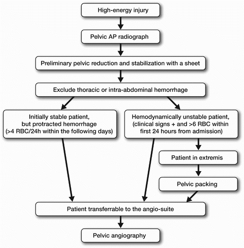
At our regional trauma center, a protocol for the initial treatment of patients with pelvic trauma has been employed since 1994 (Tötterman and Røise Citation2002) (). In this standardized protocol the indication for percutaneous pelvic angiography is tied to predetermined transfusion criteria. In this investigation, we studied the characteristics of 31 patients with pelvic trauma in need of angiographic embolization who were treated according to the protocol, and determined the effects of embolization on transfusion requirements and patient survival.
Patients and methods
Patients with pelvic ring fractures necessitating angiography were registered prospectively from February 1995 to July 2003. During this time, 1,260 patients with high-energy and low-energy pelvic ring fractures were treated at our hospital. 31 poly-traumatized patients with pelvic injury were dead on arrival. 46 patients fulfilled the transfusion criteria for requiring angiography. This showed arterial injury in 31 patients, who all underwent angiographic embolization (AE) and constituted the study population.
Demographic data, surgical interventions, angiographic procedure, and complications were registered prospectively. Injuries were coded according to the Abbrevated Injury Scale, 1990 (AAAM Citation1990), for calculation of Injury Severity Score (ISS) according to Baker et al. (Citation1974). Pelvic fractures were classified according to A.O.-O.T.A. (OTA 1996). The angiographies were reviewed retrospectively for detailed arterial anatomy. The number of transfusions was recorded, including the number of units of packed red blood cells (RBC) given perioperatively if surgery was performed within 24 h of embolization (n = 16). Angiography time was registered as procedure time, including transferals to and from the angiography suite.
Mean age at injury was 40 (9– 82) years. All patients had been involved in high-energy traumas. Traffic accidents accounted for 23 of the cases, falls from heights accounted for 7 and a skiing injury for 1. All patients were severely injured, as measured by ISS: mean 41 (17– 66). Additional injuries were observed in 30 patients (), 26 of whom fulfilled the criteria for polytrauma—defined as injury to two or more organ systems with ISS exceeding 15. No sex-related differences in injury severity were seen.
Table 1. Additional injuries in the 31 patients
Pelvic ring fractures constituted 24 of the 31 fractures; 8 patients had simultaneous ace-tabular fractures. All types of pelvic fracture were observed, with a predominance of type B (13: B1 = 0; B2 = 7; B3 = 6), followed by type C (10: C1 = 6; C2 = 1; C3 = 3), and type A (1). A substantial number of patients with isolated acetabular (4) and sacral fractures (3) was observed. The fracture was open in 5 patients.
18 patients were admitted directly from the site of injury, and the remaining patients via a referring hospital. The time from injury to admission was 6 h (10 min to 2 days). Transferring the patient via a referring hospital increased the time from injury to admission by 2 (2–48) hours. Patients admitted directly from the site of injury had higher ISS (mean 47) compared to patients admitted via a local hospital (mean 41), but the difference was not significant (p = 0.2).
Indications for angiography
According to the protocol, angiography was indicated in two groups of patients ():
1) Hemodynamically unstable patients who, in addition to crystalloid volume replacement, required ≥ 6 units of packed RBCs (1 unit = 300 mL) within 24 h of the accident. Hemodynamic instability was defined by the presence of a minimumof 3 of the following clinical signs: tachycardia, delayed capillary refill < 2 sec, hypotension < 90 mmHg, reduced level of consciousness, or reduced pulse pressure.
2) Patients who lacked clinical signs of hemodynamic instability, but demonstrated signs of protracted bleeding, as measured by hemoglobin and base excess, necessitating 4 units of RBCs daily in the days that followed. In these patients,blood transfusions were initiated with hemoglobin count of less than 8 g/100 mL in younger patients, and < 9 g/100 mL in patients older than 65 years.
AE was considered effective when: (a) no radiological signs of extravasation were seen on angiography after AE, (b) when no further interventions in the form of repeat embolization or pelvic packing were needed, and (c) when patients survived the first 24 h after AE.
In patients with unstable pelvic fractures, the pelvic injury was stabilized in the emergency department using noninvasive pelvic sheeting placed around the trochanteric regions, as described by Routt et al. (Citation2002) and Bottlang et al. (Citation2002). The pelvic volume is reduced by traction and internal rotation of the hips. The ends of the sheet are crossed, pulled taut and secured with clamps. External fixation was reserved for: (1) patients who underwent noninvasive stabilization and a successful embolization, but continued to demonstrate clinical signs of pelvic bleeding, indicating venous bleeding (1 patient), and (2) patients who developed venous stasis of the lower extremities due to the pelvic sheeting (2 patients). Traction was applied in dislocated acetabular fractures.
In initially hemodynamically unstable patients (20/31), supraumbilical diagnostic peritoneal lavage (DPL) was performed in 14 cases to exclude intra-abdominal hemorrhage. 14 patients who were in the process of exanguinating underwent extra-peritoneal pelvic packing as a salvage procedure (first described by Pohlemann et al. (Citation1995)), supplemented by emergency thoracotomy with aortic clamping when needed. 14 patients underwent surgery for associated injuries. Internal fracture fixation was performed in 14 patients.
Statistics
Means were compared using the paired-samples t-test, except for variables with a markedly skewed distribution, where a Mann-Whitney U-test was used. An independent samples t-test was used to compare means between survivors and non-survi-vors. A 5% significance level was used. Statistical analysis was carried out using SPSS version 12.0 software.
Unless stated otherwise, the results are given as mean values (range).
Results
Profuse initial pelvic bleeding was the indication for angiography in 20 of the 31 patients. In the remaining 11 patients, angiography was performed due to protracted bleeding. AE was performed within 14 h (2 h to 2 days) from injury. Only 10 patients underwent AE within 6 h of injury. Once in the emergency department, AE was performed within 9 h (40 min to 2 days). In the 20 patients who had signs of profuse bleeding at admission, AE was performed within 3 (0.7–6) h. The angiographic procedure time was 2 h 10 min (75 min to 4 h), including transferals between the ICU and the angiography suite.
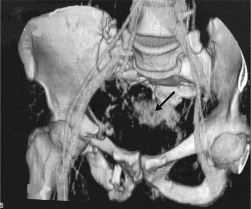
Most patients (28/31) had sustained injuries to the internal iliac artery (IIA) (). Only 3 patients had injury to the main posterior trunk, while the others had injuries involving the branches of the IIA. Multiple arterial injuries were observed in 6 patients. Injuries involving only the anterior or the posterior branches were most common, and occurred in 23 patients: 16 had injury only to the anterior IIA-branches and 7 only to the posterior branches ().
Figure 2. CT-reconstruction of an unstable pelvic fracture with injury to the lateral sacral artery (arrow).

Table 2. Distribution of arterial injuries as seen on angiography
According to the predefined criteria for evaluation of the effect of AE, AE was considered to be effective in 29 of the 31 patients.
5 patients died during hospitalization, none as a consequence of constant pelvic bleeding. The causes of death were head injury in 2, multiple organ failure in 2, and thoracic injury in 1. Those who died and 21/26 of the survivors were polytraumatized. Survivors were younger (36 years) than non-survivors (63 years; p = 0.004) (). The average ISS was 52 in non-survivors and 42 in survivors (p = 0.1). The procedure time for AE was similar for survivors and for non-survivors.
Table 3. Characteristics of survivors and non–survivors. Values are median (range) if not otherwise stated
Patients received a total of 36 (6–156) RBCs during hospitalization.When transfusion requirements were adjusted with respect to time, 27 patients showed a reduction in requirements within 24 h of AE, 3 patients had increased transfusion requirements, and 1 patient had unaltered transfusion requirements related to the procedure. The need for transfusions decreased from 0.032 units RBCs/min prior to AE, to 0.005 units RBCs/min within 24 h of AE (p < 0.005). Protracted bleeding after angiography necessitated repeat AE in 3 patients, which was successful in all cases.
No complications secondary to the AE were registered. However, 3 patients developed gluteal muscle necrosis following extensive degloving injures to the gluteal areas and they underwent several revisions. In these cases the initial trauma was interpreted as being the primary cause of muscle ischemia, but pelvic AE may have contributed to its development. No ischemic ruptures of the bladder or rectum were registered.
Discussion
In multiply injured patients with pelvic ring injuries, exsanguinating bleeding is a major cause of death within the first 24 h of injury (Pohlemann et al. Citation1996, Heckbert et al. Citation1998, Eastridge et al. Citation2002). Thus, treatment decisions during this time interval have a significant influuence on patient survival and later function (Kregor and Routt Citation1999).
In order to improve the prognosis of patients with severe pelvic trauma, standardized protocols for the initial treatment have been presented (Burgess et al. Citation1990, Pohlemann et al. Citation1994, Wong et al. Citation2000). Angiographic embolization is considered a safe and effective method to control arterial bleeding and is being increasingly used in the initial treatment of the hemodynamically unstable patients (Velmahos et al. Citation2000). Of all patients with pelvic trauma, 0.01–2.3% undergo AE (Poole et al. Citation1991, Agolini et al. Citation1997, Demetriades et al. Citation2002). The corresponding rate for patients with unstable pelvic injury is 9–80% (Demetriades et al. Citation2002, Miller et al. Citation2003), depending on the patient population, the facilities and the indications for angiography. With our protocol, 4% of all patients with pelvic trauma fulfilled the criteria for angiography and 2.5% of all patients admitted with pelvic trauma during the study period underwent AE. Significant arterial injury was observed predominantly in patients with multiple high-energy injuries.
Several studies have reported good radiological results of AE in control of arterial bleeding (Agolini et al. Citation1997, Perez et al. Citation1998, Wong et al. Citation2000, Cook et al. Citation2002, Fangio et al. Citation2005). This was confirmed in our study, where arterial occlusion was accomplished in all patients and a significant reduction in transfusion requirements was observed in most of them.
Despite successful embolization, mortality in patients who undergo AE after pelvic trauma remains high, at 14–47% (Agolini et al. Citation1997, Velmahos et al. Citation2000, Citation2002, Wong et al. Citation2000, Fangio et al. Citation2005). In our material the mortality rate was 16%. However, none of the patients died of exanguination. Nevertheless, as the indication for angiography varies, comparison of mortality between different trauma centers is difficult. Generally, both mortality and ISS seem to be higher in patients who require angiography than in patients with pelvic fracture who do not (Agnew Citation1994, Agolini et al. Citation1997). Furthermore, it is generally recognized that time to control of bleeding is important. Agolini et al. (Citation1997) described a significant increase in mortality with a delay of over 3 h to achieve embolization in 15 patients who underwent AE. In our study, almost half of the patients were admitted from another hospital and only one-third of the patients had AE performed within 6 h of injury. However, we found no significant difference in time to AE between survivors and non-survivors. Nevertheless, the delay in AE may have contributed to death in the 2 patients who later died of multiple organ failure.
In our study the angiographic procedure time was over 2 h, longer than previous reports of 1.5 h (Agolini et al. Citation1997, Fangio et al. Citation2005). The difference may be partly explained by the fact that we included transportation to and from the angiography suite. We found it important to register this time since the ability to monitor or intervene on a patient during transferals is reduced. The long procedure time emphasizes the need for supplementary procedures to control bleeding in the severely unstable patients.
Predominance of injuries to the internal iliac artery or its branches, as previously reported by O’Neill et al. (Citation1996), was verified in our study. Thus, one should pay special attention to the branches of this artery. Furthermore, we found multiple bleeding sites to be common, as also reported by other authors (O’Neill et al. Citation1996, Fangio et al. Citation2005).
In order to recognize patients predisposed to severe pelvic bleeding, several authors have have investigated the correlation between different fracture characteristics and pelvic hemorrhage (Meyers et al. Citation2000, Grainger and Porter Citation2003, Pérez and Alocover Citation2004, Sarin et al. Citation2005). The results are controversial, and most studies have concentrated on unstable pelvic fractures (Agolini et al. Citation1997, Kregor and Routt Citation1999, Cook et al. Citation2002). However, major pelvic bleeding has also been observed in other types of pelvic ring fractures (Cryer et al. Citation1988, Patel et al. Citation1996, Meyers et al. Citation2000, Ruotolo et al. Citation2001, Grainger and Porter Citation2003, Pérez and Alcover Citation2004). In our study major arterial hemorrhage was seen in all types of pelvic trauma, but only 34% of the patients had unstable pelvic fractures. Due to the small number of patients, however, no specific pattern of injury could be observed.
We found, as have previous authors, that AE is a safe procedure with few complications (Obaro and Sniderman Citation1955, Hietala Citation1978, Hare and Holland Citation1983, Takahira et al. Citation2001, Ramirez et al. Citation2004). However, to detect more subtle complications after AE, such as erectile dysfunction or ischemic nerve injuries, a different study design is required.
The limitations of our study are the small number of patients and the heterogeneous population regarding type of fracture, patient age, associated injuries and trauma mechanisms. The strength of the study is the clearly defined set of indications for requiring angiography. By using strict indications for angiography, unnecessary delay in achieving bleeding control may be avoided. Further studies are needed to evaluate whether late mortality may be reduced further by changing the indication for angiography or by reducing the time to AE.
No competing interests declared.
Contributions of authors
AT did the prospective registration, analyzed the data and wrote the article. JBD reviewed angiographic results, discussed results. JEM discussed material, results. NEK reviewed angiographic results, and planning of the study. OR planned the study, discussed results and registration. NOS wrote anesthetic details.
- AAAM Association for the Advancement of Automotive Medicine. Abbreviated injury scale - 1990 revision 1990, Des Plaines IL
- Agnew S G. Hemodynamically unstable pelvic fractures. Ortop Clin North Am 1994; 25: 717–21
- Agolini S F, Shah K, Jaffe J, Newcomb J, Rhodes M, Reed J F, 3rd. Arterial embolization is a rapid and effective technique for controlling pelvic fracture hemorrhage. J Trauma 1997; 43(3)395–9
- Baker S P, O'Neill B, Haddon W, Jr, Long W B, Jr. The Injury Severity Score: a method for describing patients with multiple injuries and evaluating emergency care. J Trauma 1974; 14(3)187–96
- Ben-Menachem Y, Coldwell D M, Young J W. R, Burgess A R. Hemorrhage associated with pelvic fracures; causes, diagnosis, and emergent management. Am J Roentg 1991; 157: 1005–14
- Bottlang M, Simpson T, Sigg J, Krieg J C, Madey S M, Long W B. Noninvasive reduction of open-book pelvic fractures by circumferential compression. J Orthop Trauma 2002; 16(6)367–73
- Burgess A R, Eastridge B J, Young J W, Ellison T S, Ellison P S, Jr, Poka A, Bathon G H, Brumback R J. Pelvic ring disruptions: effective classification system and treatment protocols. J Trauma 1990; 30(7)848–56
- Cook R E, Keating J F, Gillespie I. The role of angiography in the management of haemorrhage from major fractures of the pelvis. J Bone Join Surg (Br) 2002; 84(2)178–82
- Cryer H M, Miller F B, Evers F, Rouben L R, Seligson D L. Pelvic fracture classification: correlation with hemorrhage. J Trauma 1988; 28(7)974–80
- Demetriades D, Karaiskakis M, Toutouzas K, Alo K, Velma-hos G, Chan L. Pelvic fractures: epidemiology and predictors of associated abdominal injuries and outcomes. J Am Coll Surg 2002; 195(1)1–10
- Eastridge B J, Starr A, Minei J P, O'Keefe G E, Scalea T M. The importance of fracture pattern in guiding therapeutic decision-making in patients with hemorrhagic shock and pelvic ring disruptions. J Trauma 2002; 53(3)446–50, discussion 450–1
- Ertel W, Keel M, Eid K, Platz A, Trentz O. Control of severe hemorrhage using C-clamp and pelvic packing in multiply injured patients with pelvic ring disruption. J Orthop Trauma 2001; 15(7)468–74
- Fangio P, Asehnoune K, Edouard A, Smail N, Benhamou D. Early embolization and vasopressor administration for management of life-threatening hemorrhage from pelvic fracture. J Trauma 2005; 58(5)978–84
- Grainger M F, Porter K M. Life threatening haemorrhage from obturator vessel tear as a result of pubic ramus fraccture: a case report. Injury 2003; 34(7)543–4
- Gruen G S, Leit M E, Gruen R J, Peitzman A B. The acute management of hemodynamically unstable multiple trauma patients with pelvic ring fractures. J Trauma 1994; 36: 706–11
- Hare W S, Holland C J. Paresis following internal iliac artery embolization. Radiology 1983; 146(1)47–51
- Heckbert S R, Vedder N B, Hoffman W, Winn R K, Hudson L D, Jurkovich G J, Copass M K, Harlan J M, Rice C L, Maier R V. Outcome after hemorrhagic shock in trauma patients. J Trauma 1998; 45(3)545–9
- Hietala S O. Urinary bladder necrosis following selective embolization of the internal iliac artery. Acta Radiol Diagn 1978, 19: 316–20
- Kregor P J, Routt M L, Jr. Unstable pelvic ring disruptions in unstable patients. Injury (Suppl 2) 1999; 30 B: 19–28
- Meyers T J, Smith W R, Ferrari J D, Morgan S J, Fancoise R J, Echeverri J A. Avulsion of the pubic branch of the inferior epigastric artery: a cause of hemodynamic instability in minimally displaced fractures of the pubic rami. J Trauma 2000; 49: 750–3
- Miller P R, Moore P S, Mansell E, Meredith J W, Chang M C. External fixation or arteriogram in bleeding pelvic fracture: initial therapy guided by markers of arterial hemorrhage. J Trauma 2003; 54(3)437–43
- Obaro R O, Sniderman K W. Avascular necrosis of the femoral head as a complication of complex embolization for severe pelvic haemorhage: a case report. Br J Radiol 1955; 68: 920–2
- O'Neill P A, Riina J, Sclafani S, Tornetta III P. Angiographic findings in pelvic fractures. Clin Orthop 1996, 329: 60–7
- OTA. Orthopaedic Trauma Association Committee for Coding and Classification: Fracture and dislocation compendium. J Orthop Trauma, O T A, 1996; 10: 1–154
- Patel N H, Matsuo R T, Routt M L, Jr. An acetabular fracture with superior gluteal artery disruption. Am J Roentgenol 1996; 166: 1074
- Perez J V, Hughes T M, Bowers K. Angiographic embolisation in pelvic fracture. Injury 1998; 29(3)187–91
- Pérez M U, Alocover H A. Hypovolemic shock due to a fracture of the superior pubic ramus in a young man. Case report. Injury 2004; 35(1)80–2
- Pohlemann T, Bosch U, Gänsslen A, Tscherne H. The Hannover experience in management of pelvic fractures. Clin Orthop 1994, 305: 69–80
- Pohlemann T, Gänsslen A, Bosch U, Tcherne H. The technique of packing for control of hemorrhage in complex pelvic fractures. Tech Orthop 1995; 9(4)267–70
- Pohlemann T, Gänsslen A, Schellwald O, Culemann U, Tscherne H. Outcome evaluation after unstable injuries of the pelvic ring. Article in German. Unfallchirurg 1996; 99(4)249–59
- Poole G, Ward E, Muakkassa F, Hsu H S. H, Griswold J A, Rhodes R S. Pelvic fracture from major blunt trauma outcome is determined by associated injuries. Ann Surg 1991; 213: 532–8
- Ramirez J I, Velmahos G C, Best C R, Chan L S, Demetriades D. Male sexual function after bilateral internal iliacartery embolization for pelvic fracture. J Trauma 2004; 56(4)734–9, discussion 739–41
- Routt M L, Jr, Falicov A, Woodhouse E, Schildhauer T A. Circumferential pelvic antishock sheeting: A temporary resuscitation aid. J Orthop Trauma 2002; 16(1)45–8
- Ruotolo C, Savarese E, Khan A, Ryan M, Kottmeier S, Meinhard B P. Acetabular fractures with associated vascular injury: a report of two cases. J Trauma 2001; 51(2)382–6
- Sarin E L, Moore J B, Moore E E, Shannon M R, Ray C E, Morgan S J, Smith W R. Pelvic fracture pattern does not always predict the need for urgent embolization. J Trauma 2005; 58(5)973–7
- Takahira N, Shindo M, Tanaka K, Nishimaki H, Ohwada T, Itoman M. Gluteal muscle necrosis following transcatheter angiographic embolisation for retroperitoneal haemorrhage associated with pelvic fracture. Injury 2001; 32: 27–32
- Tötterman A, Røise O. Initial management of patients with severe pelvic injuries. Presentation of a protocol applied at Ulleval University Hospital, Oslo, Norway. Article in Norwegian. Scand J Trauma Emerg Med 2002; 10(1)22–6
- Velmahos G C, Chahwan S, Falabella A, Hanks S E. Angiographic embolization for intraperitoneal and retroperitoneal injuries. World J Surg 2000; 24(5)539–45
- Velmahos G C, Toutouzas K G, Vassiliu P, Sarkisyan G, Chan L S, Hanks S H, Berne T V, Demetriades D. A prospective study on the safety and efficacy of angiographic embolization for pelvic and visceral injuries. J Trauma 2002; 53(2)303–8
- Wong Y-C, Wang L-J, Ng C-J, Tseng I-C, See L-C. Mortality after successful transcatheter arterial embolisation in patients with unstable pelvic fractures: rate of blood transfusion as a predictive factor. J Trauma 2000; 49(1)71–5

