Abstract
Background In uncemented total hip arthroplasty with hydroxyapatite coating, early weight bearing is frequently practiced but there is still not much evidence to support this recommendation.
Method In a prospective randomized study we evaluated the effect of partial and full weight bearing after cementless total hip arthroplasty (ABG; Stryker-Howmedica) using radiostereometric analysis (RSA). Between February 1996 and February 2000, 43 consecutive patients (mean age 53 (41–63) years, 23 women) with hip osteoarthrosis received an uncemented and hydroxyapatite-coated prosthesis with an anteverted stem. All patients were operated in a standardized way by three experienced surgeons and they were randomized to partial (P) or full (F) weight bearing during the first 6 weeks after surgery. The patients in the partial weight bearing group were equipped with a pressuresensitive insole signaling when their load exceeded the prescribed weight limit.
Results At 3-month follow-up, the mean proximal (+)/ distal (-) migration of the stem was -0.14 mm (-1.93– 0.11) in group P and -0.31 mm (-4.30–0.16) in group F (p = 0.6). At 1-year follow-up, the mean migration was –0.17 mm (-2.18–0.21) and -0.28 mm (-4.31–0.11), respectively (p = 0.9). There was no significant difference in stem rotations either (p < 0.2). The cup translations, rotations, and femoral head penetration were similar in the two groups (p < 0.1). There were no re-operations during the first year.
Interpretation We did not find any adverse effect of full weight bearing immediately after operation, which justifies use of this regimen after uncemented total hip arthroplasty of the ABG type.
Despite the fact that there has been widespread use of uncemented total hip arthroplasty (THA) with hydroxyapatite coating for more than a decade, there is still not much evidence to recommend full weight bearing immediately after surgery. Functional recovery may be promoted by immediate weight bearing (Rao et al. Citation1998, Kishida et al. Citation2001) and periprosthetic demineralization may be reduced, but primary stability and ingrowth may be jeopardized by premature loading of the implant.
It has been shown that osseous ingrowth is inhibited by excessive micromotion at the interface between implant and bone (Pilliar et al. Citation1986). However, if rigid initial stability is acquired at surgery, ingrowth of bone onto uncemented implants will occur (Jasty et al. Citation1997, Pilliar et al. Citation1986). Recently, satisfactory clinical results have been obtained using weight bearing immediately after uncemented total hip arthroplasty (Chan et al. Citation2003, Boden et al. Citation2004). Few studies have used a randomized design, however, and we have not been able to find any study in which early weight bearing has been studied with high-precision radiostereometry.
In order to evaluate the effects of partial and full weight bearing after cementless total hip arthroplasty, we designed a prospective randomized study. Our hypothesis was that immediate weight bearing after uncemented total hip arthroplasty would lead to enhanced motion during the first 3 months. We used RSA (Selvik Citation1989) to measure three primary outcome parameters: migration of the stem and cup and the penetration of the femoral head.
Patients were examined after 3 months and at 1 year.
Patients and methods
43 consecutive patients entered the study between February 1996 and February 2000, after giving informed consent. Inclusion criteria were noninflammatory arthrosis of the hip, age between 45 and 65, and having given informed consent. Premenopausal women and patients who suffered from a bone disease or who used or had used corticosteroids were excluded. The study was approved by the Ethics Committee of Gothenburg University (no. 175–96) and it was conducted according to the Declaration of Helsinki, 1975.
Before the operation, the patients were stratified on the basis of sex and weight (< 75 kg or ≥ 75 kg) and randomized (using envelopes) to partial (P) or full (F) weight bearing for the first 6 weeks postoperatively. There were 20 men and 23 women with a mean age of 53 (41–63) years, mean weight of 79 (59–140) kg, and mean preoperative Harris hip score of 46 (23–57) with no significant difference between the two groups (). 35 patients had primary OA, 3 had secondary OA due to dysplasia, and 4 were operated for other reasons. All patients received an uncemented hip prosthesis (ABG I; Stryker-Howmedica) with an anteverted stem. The hemispherical titanium acetabular cup of this prosthesis is coated with pure hydroxyapatite (HA) to a thickness of 60 µm (chemical purity of 99.99%, crystallographic composition of 98–99%) and fitted with a polyethylene insert. Initial mechanical stability is achieved by press fit. Our implants were supplied with small spikes, which were fixed to the metallic shell. The titanium femoral stem is tapered proximally but straight distally. It is designed to provide anchoring only at the metaphyseal portion of the stem, which is coated with HA.
Table 1. Patient data; mean and (range)
A modular 28-mm cobalt-chrome head was used in all cases. The operations were performed by 3 experienced surgeons in a standardized way, without osteotomy of the trochanter. During surgery tantalum markers (0.8–1.0 mm in diameter) were inserted into the periacetabular bone, the cup, and the proximal femur. Markers had been inserted into the stem by the manufacturer. 2 markers were inserted into the prosthetic shoulder and 1 at the tip. The rotational stability of the femoral component intraoperatively was determined by use of a torque wrench (Harris et al. Citation1991). This device reproduces the out-of-plane forces seen at the hip joint in vivo. When a torque (maximum 23 Nm) is applied to the torque wrench, rotational micromotion of the prosthesis relative to the femoral cortex can be measured. The patients were mobilized on the day after surgery.
The patients in the F group (with full or unrestricted weight bearing) were immediately instructed to walk with 1 crutch alone or without external support whenever possible. These instructions were reinforced by the physiotherapist during the hospital stay and the patients also received a home exercise program to carry out full weight bearing on the operated leg. Regular contact with a physiotherapist was established to encourage as much weight bearing as possible.
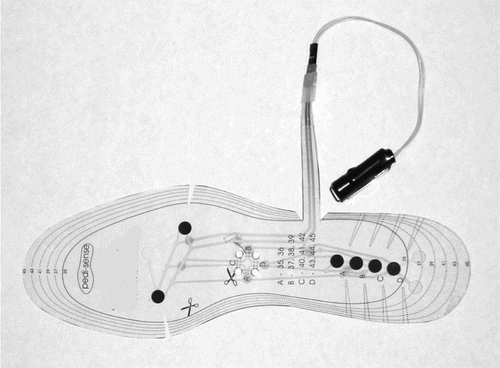
The patients in the P group (protected weight bearing) were equipped with a battery-operated pressure-sensitive auditory device incorporated in the sole of the shoe (pedi-sense; Aggero Produkt & Affärsutveckling AB, Göteborg, Sweden) (). This auditory device has 2 pressure-activated sensors in the forefoot and 1 in the heel. It gives an auditory signal when the load exceeds the prescribed weight limit. The load was calibrated to a maximum of 30 kg. About a week before the operation, the patients were given instruction on how to perform protected weight bearing using 2 crutches and using the auditory device for feedback. They were seen regularly by a physiotherapist after the operation and given a home exercise program involving protected weight bearing.
Figure 1. The battery-operated pressure-sensitive auditory device (Pedi-sense; Aggero AB, Göteborg, Sweden).

Radiostereometry
Radiostereometric examinations with the patient in supine position were done postoperatively (after 5– 7 days), after 3 months, and after 1 year. Uniplanar technique was used with the calibration cage positioned under the examination table (Selvik, Citation1989, Kärrholm Citation1989, Kärrholm et al. Citation1997, Valstar et al. Citation2005). The rotations of the stem were measured in relation to the 3 cardinal axes. Subsidence of the gravitational center of the stem markers and the center of the femoral head was calculated. Migration of the femoral stem in relation to the bone was only studied in terms of subsidence. The migration of the cup was measured as rotations around the three axes and translations of the center of the cup. The translations of the center of the femoral head using the cup markers as fixed reference segment were evaluated to measure the femoral head penetration, hereafter referred to as wear. For all measurements and calculations, we used the UmRSA system (RSA Biomedical, Umeå, Sweden). In order to maintain the precision of the measurements, the RSA was only done if at least 3 welldefined markers could be identified with a scatter corresponding to a condition number of < 120 and stability corresponding to 0.3 mm (mean error of rigid body fitting (Selvik Citation1989).
Patients who were lost to follow-up
In one patient, a 60-year-old man who was randomized to full weight bearing, a fracture in the femoral cortex occurred peroperatively. The postoperative regimen was therefore changed and the patient had to be excluded from the study, but he continued to participate in the clinical and radiological follow-up. In another patient, a 56-year-old woman, a fracture was first suspected at the 3-month follow-up. This patient was randomized to partial weight bearing and continued her mobilization according to the protocol. No examinations were missing, but not all radiostereometric calculations could be done—mainly due to insufficient visualization of the tantalum markers in the periacetabular bone on the postoperative radiographs (see number of calculations in and ). In 4 patients (all in group F) insufficient numbers of tantalum markers in the proximal femur were found immediately after surgery or at the 1-year follow-up. Thus, 38 femoral stems (19 in group P and 19 in group F) could be evaluated using RSA. There were no reoperations during the first year and none of the patients were planned for revision at the 1-year follow-up; nor were there any other patients lost to follow-up for reasons other than those mentioned above.
Table 2A. Stem migration and rotation at the 3-month follow-up (all hips)
Table 2B. Stem migration and rotation at the 3-month follow-up (all hips)
Table 3. Penetration (wear) at 1-year follow-up (signed values, mm)
Statistics
The study was designed to detect a difference in stem subsidence of 0.2 mm after 2 months, based on the assumption of an SD of 0.2 between the two groups (< 80% probability). To include the randomization stratification criteria with sex and weight as covariates, binary logistic regression was used. The measurements of the primary parameters were not normally distributed at 3 months (Shapiro-Wilk W = 0.3–0.99) and therefore statistical evaluation was performed with non-parametric tests (Mann-Whitney). Any difference was considered to be statistically significant when p < 0.05. We used SPSS for Windows for all statistical evaluations.
Results
Migration of center of stem ()
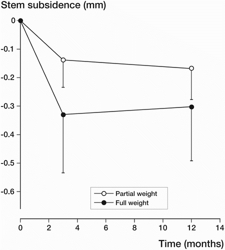
At the 3-month follow-up, the mean proximal (+) or distal (–) migration of the stem in the two groups (P and F) was –0.14 mm (–1.93 to 0.11) and –0.31 mm (–4.30 to 0.16) respectively (p = 0.6, Mann Whitney U-test) (). At the 1-year follow up, the mean migration was –0.17 mm (–2.18 to 0.21) and –0.28 mm (–4.31 to 0.11) respectively (p = 0.9, Mann Whitney U-test) (). The rotations of the stem were small and not significantly different (p < 0.2, Mann Whitney-U test).
In the patient who was excluded from the study because of proximal femoral fracture at the time of surgery (first detected on postoperative radio-graphs), distal migration of the center of the stem was –0.99 mm at three months and –1.09 mm at 1 year. In the patient in the P group who had a suspected fracture of the femoral cortex at the 3month follow-up, distal migration values were 0.11 and 0.08 mm, respectively.
Penetration of the femoral head—wear
At 1 year, the mean proximal penetration of the femoral head in the proximal direction (wear) was high (0.22 mm in the P group and 0.17 mm in the F group), but the difference was not statistically significant (). The mean values for medial/ lateral wear and anterior/posterior wear were less than 0.15 mm and were unaffected by the two types of postoperative mobilization (p < 0.6). The mean total or 3-dimensional wear reached 0.37 mm in the P group and 0.30 mm in the F group (p = 0.7).
Cup migration
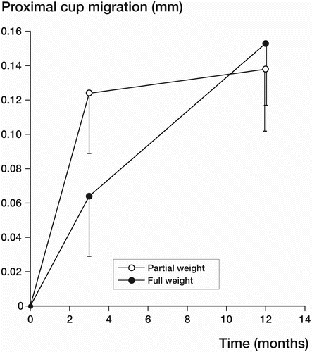
The translations of the center of the cup and cup rotations were not statistically significantly different on either of the follow-up occasions (p < 0.2) ( and ).
Figure 2b. Mean proximal migration of the center of the cup (with SE) at the 3-month and 1-year follow-up.

Table 4A. Cup migration at the 3-month follow-up (all hips)
Table 4B. Cup migration at the 12-month follow-up (all hips)
Discussion
The optimum form of postoperative management after uncemented THA is unclear; partial weight bearing for 6–12 weeks has often been the policy (Kim et al. Citation1999, Aldinger et al. Citation2003, Jacobsen et al. Citation2003), mostly based on the results of animal studies (Pilliar et al. Citation1986) These cannot simply be extrapolated to humans, however. To date, it has not been confirmed in clinical studies that optimal clinical results and bone ingrowth depend on restricted weight bearing.
By partial weight bearing, functional recovery may be inhibited and muscle atrophy and loss of bone mineral density increased. In addition, load on the contralateral hip and upper extremities is significantly higher when less weight is put on the operated lower limb (Rao et al. Citation1998). Immediate weight bearing after uncemented THA shortens the length of stay in hospital, reduces the risk of deep venous thrombosis (Leali et al. Citation2002), and promotes the functional recovery with less usage of ambulatory devices (Kishida et al. Citation2001).
The effectiveness of prescribing limited weight bearing has been debated. Tveit et al. (Citation2001) measured weight bearing in 15 patients who had been operated with an uncemented or hybrid hip arthroplasty and who had been instructed by a physiotherapist to use crutches in order to support 30% of their body weight. None of the patients managed to comply with the directive. It was concluded that the compliance to prescribed limited weight bearing is low.
We have not found any published study in which the effect of weight bearing on early implant migration was evaluated using RSA. The previous studies were not randomized and did measure migration on conventional radiographs, or did not focus on the fixation of the components (Ritter et al. Citation1995, Rao et al. Citation1998, Andersson et al. Citation2001, Chan et al. Citation2003).
Ritter et al. (Citation1995) found that ingrowth of bone in single-stage, bilateral uncemented THA was not adversely affected by weight bearing if initial stability of both the metaphyseal and diaphyseal portions of the femur had been achieved. This conclusion may not have been valid, however, because of the lack of a protected weight bearing control group.
Some authors have studied other effects of the postoperative treatment after insertion of uncemented implants. Andersson et al. (Citation2001) did not find significant differences in hip extension, muscle strength, gait velocity, pain, and walking pattern 6 months after surgery between 10 patients who practiced late weight bearing and 11 patients who practiced immediate weight bearing.
Rao et al. (Citation1998) studied femoral subsidence on conventional radiographs after bilateral uncemented THA and used patients who had undergone unilateral uncemented THA as a control group. Although increased initial subsidence of the femoral component after immediate weight bearing was seen in the bilateral group, this did not prevent the prosthesis from becoming stable and from achieving bone ingrowth. In our study, the initial subsidence of the femoral stem was similar in both study groups. This may indicate that the design of the prosthesis used in our study is more rigid and stable initially, probably due to its hydroxyapatite coating. Chan et al. (Citation2003) found that full weight bearing after uncemented THA is compatible with good clinical results and stability according to conventional radiography.
It has been shown that migration of the femoral component of uncemented and hydroxyapa-tite-coated femoral components is minimal during the first 2 years (Kärrholm et al. Citation1994, Önsten et al.Citation1996). In our study, 9 femoral stems showed subsidence exceeding 0.20 mm during the first 3 months. However, these stems showed almost no further subsidence at the 1-year follow-up, thus indicating that they had stabilized—as did the femoral stem of the patient in this study with femoral fracture. These findings are consistent with other observations (Grant et al. Citation2005). Subsidence seems to be tolerated in some uncemented hydroxyapa-tite-coated stem designs up until 1 year. This may be explained by the unique quality of hydroxyapatite as a biological adjuvant, which enhances bone ingrowth even across a gap (Overgaard et al. Citation1997).
We are aware of only one previous report of an auditory device to facilitate effective partial weight bearing after uncemented THA. Boden et al. (Citation2004) used the same device as we did, in a prospective study of 20 patients who were operated with a hydroxyapatite-coated total hip arthroplasty.
They found that immediate weight bearing had a positive effect on the bone mineral density around the stem of the prosthesis, and especially its distal part. Stem migration was also evaluated, but with conventional radiography.
In our study, we found no adverse effect of immediate weight bearing on femoral and acetabular stability—both initially and up to 1 year after the operation. The acetabular component showed continued migration in both groups, from 3 months up to 1 year postoperatively. The same migration pattern in the first year has been found by other authors in studies on various uncemented acetabular components (Thanner et al. Citation2000, Digas et al. Citation2004). We believe that the migration pattern of the ABG sockets studied by us corresponds to those previously observed for other designs. A longer follow-up period in the present study would have helped to clarify this issue, but is beyond the topic of our study.
We found no difference in migration between full and restricted weight bearing during the first 6 weeks postoperatively, at 3 months, and at the one-year follow up; nor was there any difference in wear in the present study. The wear rate measured (around 0.2 mm) was, however, high—which is consistent with the comparatively high frequency of radiographically observed wear and acetabular osteolysis reported for the ABG I implant with longer follow-up (Rogers et al. Citation2003, Blacha Citation2004). Because of these problems, the ABG I has undergone several modifications in design. The first-generation ABG stems do, however, have a high survival rate (Tonino et al. Citation2000).
According to the present study, full weight bearing immediately after surgery—as much as can be tolerated—is justified for this implant. It seems probable that this observation would be applicable to many similar stems with an HA coating, but this hypothesis requires further investigation.
Contributions of authors
Planning of the study: JK and LA. Operation: LA, ME, and CS. Follow-up: LA, ME, CS, and TT. Collection and analysis of data: TT. Statistical analysis: TT and JK. Manuscript: TT and JK.
- Aldinger P R, Thomsen M, Mau H, Ewerbeck V, Breusch S J. Cementless Spotorno tapered titanium stems: excellent 10–15–year survival in 141 young patients. Acta Orthop Scand 2003; 74(3)253–8
- Andersson L, Wesslau A, Boden H, Dalen N. Immediate or late weight bearing after uncemented total hip arthroplasty: a study of functional recovery. J Arthroplasty 2001; 16(8)1063–5
- Blacha J. High osteolysis and revision rate with the hydroxy-apatite-coated ABG hip prostheses: 65 hips in 56 young patients followed for 5–9 years. Acta Orthop Scand 2004; 75(3)276–82
- Boden H, Adolphson P. No adverse effects of early weight bearing after uncemented total hip arthroplasty: a randomized study of 20 patients. Acta Orthop Scand 2004; 75(1)21–9
- Chan Y K, Chiu K Y, Yip D K, Ng T P, Tang W M. Full weight bearing after non-cemented total hip replacement is compatible with satisfactory results. Int Orthop 2003; 27(2)94–7
- Digas G, Thanner J, Anderberg C, Kärrholm J. Bioactive cement or ceramic/porous coating vs. conventional cement to obtain early stability of the acetabular cup. Randomised study of 96 hips followed with Radiostereometry. J Orthop Res 2004; 22(5)1035–43
- Grant P, Aamodt A, Falch J A, Nordsletten L. Differences in stability and bone remodeling between a customized uncemented hydroxyapatite coated and a standard cemented femoral stem A randomized study with use of radiostereometry and bone densitometry. J Orthop Res 2005; 23(6)1280–5
- Harris W H, Mulroy R D, Jr, Maloney W J, Burke D W, Chandler H P, Zalenski E B. Intraoperative measurement of rotational stability of femoral components of total hip arthroplasty. Clin Orthop. 1991, 266: 119–26
- Jacobsen S, Jensen FK, Poulsen K, Sturup J, Retpen J B. Good performance of a titanium femoral component in cementless hip arthroplasty in younger patients: 97 arthroplasties followed for 5–11 years. Acta Orthop Scand 2003; 74(4)375–9
- Jasty M, Bragdon C R, Zalenski E, O'Connor D, Page A, Harris W H. Enhanced stability of uncemented canine femoral components by bone ingrowth into the porous coatings. J Arthroplasty 1997; 12(1)106–13
- Kärrholm J. Roentgen stereophotogrammetry. Review of orthopedic applications. Acta Orthop Scand 1989; 60(4)491–503
- Kärrholm J, Malchau H, Snorrason F, Herberts P. Micromotion of femoral stems in total hip arthroplasty. A randomized study of cemented, hydroxyapatite-coated, and porous-coated stems with roentgen stereophotogrammetric analysis. J Bone Joint Surg (Am) 1994; 76(11)1692–705
- Kärrholm J, Herberts P, Hultmark P, Malchau H, Nivbrant B, Thanner J. Radiostereometry of hip prostheses. Review of methodology and clinical results. Clin Orthop 1997, 344: 94–110
- Kim Y H, Kim J S, Cho S H. Primary total hip arthroplasty with a cementless porous-coated anatomic total hip prosthesis: 10- to 12-year results of prospective and consecutive series. J Arthroplasty 1999; 14(5)538–48
- Kishida Y, Sugano N, Sakai T, Nishii T, Haraguchi K, Ohzono K, et al. Full weight-bearing after cementless total hip arthroplasty. Int Orthop 2001; 25(1)25–8
- Leali A, Fetto J, Moroz A. Prevention of thromboembolic disease after non-cemented hip arthroplasty. A multimodal approach. Acta Orthop Belg 2002; 68(2)128–34
- önsten I, Carlsson A S, Sanzen L, Besjakov J. Migration and wear of a hydroxyapatite-coated hip prosthesis. A controlled roentgen stereophotogrammetric study. J Bone Joint Surg (Br) 1996; 78(1)85–91
- Overgaard S, Lind M, Rahbek O, Bunger C, Søballe K. Improved fixation of porous-coated versus grit-blasted surface texture of hydroxyapatite-coated implants in dogs. Acta Orthop Scand 1997; 68(4)337–43
- Pilliar R M, Lee J M, Maniatopoulos C. Observations on the effect of movement on bone ingrowth into porous-sur-faced implants. Clin Orthop 1986, 208: 108–13
- Rao R R, Sharkey P F, Hozack W J, Eng K, Rothman R H. Immediate weightbearing after uncemented total hip arthroplasty. Clin Orthop 1998, 349: 156–62
- Ritter M A, Albohm M J, Keating E M, Faris P M, Meding J B. Comparative outcomes of total joint arthroplasty. J Arthroplasty 1995; 10(6)737–41
- Rogers A, Kulkarni R, Downes E M. The ABG hydroxyapa-tite-coated hip prosthesis: one hundred consecutive operations with average 6-year follow-up. J Arthroplasty 2003; 18(5)619–25
- Selvik G. Roentgen stereophotogrammetry. A method for the study of the kinematics of the skeletal system. Acta Orthop Scand. 1989, Suppl 232: 1–51
- Thanner J, Kärrholm J, Herberts P, Malchau H. Hydroxyapatite and tricalcium phosphate- coated cups with and without screw fixation: a randomized study of 64 hips. J Arthroplasty 2000; 15(4)405–12
- Tonino A J, Rahmy A I. The hydroxyapatite-ABG hip system: 5– to 7–year results from an international multicentre study. The International ABG Study Group. J Arthroplasty 2000; 15(3)274–82
- Tveit M, Kärrholm J. Low effectiveness of prescribed partial weight bearing. Continuous recording of vertical loads using a new pressure-sensitive insole. J Rehabil Med 2001; 33(1)42–6
- Valstar E R, Gill R, Ryd L, Flivik G, Börlin N, Kärrholm J. Guidelines for standardization of radiostereometry (RSA) of implants. Acta Orthop 2005; 76(4)563–72
