Abstract
Purpose We assessed the reliability and practicality of two radiographic measurements—cast index and padding index—of the quality of plaster cast in clinical decision making in forearm fractures in children.
Methods 40 orthopedic surgeons (20 consultants and 20 registrars) were asked to predict the risk of re‐displacement in 5 randomly selected cases. Subsequently, these surgeons were taught these indices and they were then requested to use them to predict re-displacement.
Results Every one of the 40 surgeons showed an increase in the number of correct responses. For consultants, the accuracy improved from 33% to 72% and for the registrars it improved from 28% to 81%. This improvement was statistically significant.
Interpretation We suggest that cast and padding indices are simple, reliable, and reproducible radio‐graphic tools that can be used in clinics and wards to predict re‐displacement of forearm fractures in children following initial reduction.
Midshaft and distal radius‐ulnar fractures account for more than half of all fractures in children aged 18 years and younger (Jones et al. Citation1999, Boyer et al. Citation2002). Good anatomical reduction (Proctor et al. Citation1993, Haddad and Williams Citation1995) followed by application of a properly molded cast is necessary for successful closed treatment (Voto et al. Citation1990, Dicke and Nunley Citation1993, Younger et al. Citation1997). It has been suggested that if the cast is properly molded, one could even treat displaced fractures in the distal third of the forearm using a short‐arm plaster cast, with similar results to those obtained with long‐arm plaster casts (Webb et al. Citation2006). A well‐molded cast, as explained by Charnley (Citation1968), is the one that maintains a 3‐point fixation by exerting pressure at certain precisely determined points on the skeleton and none at others. In this form of immobilization by the cast, the soft tissue hinge at the fracture site is over‐corrected and placed under tension by molding the cast, thus introducing the neutralizing force against the couple being produced by the forces on the proximal and distal segments of the fracture.
Apart from the work by Chess et al. (Citation1994), no other study has attempted to define the quality of the plaster cast in objective, measurable, and reproducible terms.
We assessed whether previously described cast and padding indices (Bhatia and Housden Citation2006) could be validated for clinical decision making when predicting re‐displacement of wrist and forearm fractures in children following initial reduction.
Methods
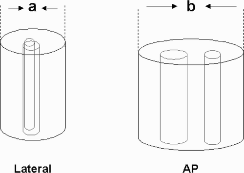
Cast index (Chess et al. Citation1994, Bhatia and Housden Citation2006, Webb et al. Citation2006) is measured as an a/b ratio in which “a” is the cast width in the lateral view and “b” is the cast width in the AP view at the fracture site; as the cast becomes more circular, the value of “a” will approach “b” and the cast index would approach the value of 1 ().
Figure 1. Cast index (a/b) at fracture site. Internal cast width on lateral radiograph (a) and internal cast width on AP radiograph (b).

Padding index (Bhatia and Housden Citation2006), is the ratio of x/y where “x” is the padding thickness under the molded cast in lateral view in the plane of maximum deformity correction and “y” is the maximum interosseous distance in the AP view. In an over‐padded cast, the value of “x” will increase and so the overall value of x/y will tend to increase and there will tend to be a loss of 3‐point fixation of the fracture, as there would be a greater “play” between the plaster cast and the bone, thus rendering the tension on soft tissue at the fracture site ineffective ().
Figure 2. Padding index (x/y). Padding thickness in the plane of deformity correction on lateral radiograph (x) and maximum interosseous space on AP radiograph (y).

The sum total of cast index and padding index has been called the Canterbury index (Bhatia and Housden Citation2006). A reduced fracture in a plaster cast with cast index of more than 0.8, padding index of greater than 0.3, and/or the combined Canterbury index of more than 1.1 should be watched cautiously as it is prone to re‐displacement (Bhatia and Housden Citation2006). Re‐displacement in our study was defined as more than 20 degrees of angulation and/ or more than 50% of translational displacement on check radiographs (Proctor et al. Citation1993, Haddad and Williams Citation1995).
Radiographs (AP and lateral) of 5 randomly selected forearm fractures were presented to 40 orthopedic surgeons working in 5 hospitals in Southern England (20 consultants and 20 registrars) and they were asked to predict re‐displacement (1) by “eyeballing”, and then (2) by using the cast index and padding index (after they had been taught the indices).
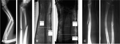
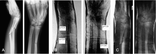
In 2 of these cases, the fractures were widely displaced after trauma () but because of good reduction and well‐molded casts, they had healed without re-displacement. The other 3 cases were moderately displaced (), but even after good reduction they had re‐displaced because of over‐padded or poorly molded casts.
Figure 3. A. Fractures with 100% displacement. B. After reduction, with cast index a/b = 0.69, padding index (x/y) = 0.14, and Canterbury index = 0.69 + 0.14 = 0.83. C. After 3 weeks, showing no re-displacement.

Figure 4. A. Fractures with 50% displacement. B. After reduction, with cast index (a/b) = 0.97, padding index (x/y) = 0.40, and Canterbury index = 0.97 + 0.40 = 1.37. C. After 1 week, showing re-displacement.

As each of the 40 orthopods marked 5 responses, the possible number of responses was 200 (100 for consultants and 100 for registrars). These responses were then marked as being correct or wrong according to the final outcome of the 5 above‐mentioned cases, which was known to the authors. The number that had been assessed correctly was calculated for each of the 20 consultants and each of the 20 registrars both before and after the teaching. The change was tested using Wilcoxon's matched‐pairs signed‐ranks test and a comparison between the responses of consultants and registrars was made using the Mann‐Whitney U test.
We wanted to test these indices in a clinical setting, so most of the cases were presented to the orthopods in busy clinics or on ward rounds; hence, only five cases were selected to keep the exercise of measuring the indices to a manageable time limit.
Results
The accuracy of predicting re‐displacement by “eyeballing” was 33% (i.e. 33 correct responses of 100) for the 20 consultants and 28% for the 20 registrars. After applying the indices, this improved to 72% for the consultants and to 81% for the registrars (p < 0.001). Each of the 40 consultants and registrars showed an increase in the number of correct responses.
In terms of individual improvement of the consultants, (1) 9 consultants scored 1 correct response before indices, but after applying indices, out of these 9, 5 scored 3 correct responses and 4 scored 4 correct responses; (2) 9 consultants scored 2 correct responses before indices, but after applying indices, 3 of them improved their responses to 3, and 6 of them improved to 4; (3) 2 consultants scored 3 correct responses previously, but both improved their score to 4 correct responses after application of indices. These findings represent a significant change (p < 0.001).
For the registrars, (1) 12 registrars scored 1 correct response by “eyeballing”, and this improved to 4 correct responses for all of them after application of indices; (2) 8 registrars scored 2 correct responses before indices, but 7 of these improved to 4, and 1 improved to 5 after application of indices. These findings represent a significant change (p < 0.001). The combined findings for both consultants and registrars were also statistically significant (p < 0.001). This increase in the number of correct responses was not related to the amount of initial displacement of the fractures.
We also analyzed the consultants and registrars who had improved responses. For the consultants, (1) 5 consultants increased their number of correct assessments by 1, (2) 11 consultants increased their correct responses by 2, and (3) 4 consultants increased their correct score by 3.
In the case of registrars, (1)7 registrars increased their correct responses by 2, and (2) 13 increased their correct score by 3. These results are summarized in the Table.
The increase in the number of correct assessments for consultants and registrars before and after training. There was no significant difference in the number of correct assessments before training (Mann‐Whitney U test, p = 0.3), but registrars had a greater increase in the number of correct assessments after training (p = 0.003)
When comparing consultants and registrars, there was no statistically significant difference in the number of correct assessments before training (Mann‐Whitney U Test, p = 0.3), but registrars had a greater increase in the number of correct assessments after training (p = 0.003) (Table).
Discussion
In addition to an initial anatomical reduction (Proctor et al. Citation1993, Haddad and Williams Citation1995), a good plaster cast and meticulous follow‐up are essential components of closed treatment of a fracture (Charnley Citation1968, Chess et al. Citation1994, Bhatia and Housden Citation2006, Webb et al. Citation2006). Younger et al. (Citation1997) suggested that loss of position in the cast zis the most important factor affecting the position at union. In their studies on re‐displacement after closed reduction of forearm fractures in children, Voto et al. (Citation1990) and Dicke and Nunley (Citation1993) concluded that the major underlying factor responsible for re‐displacement or re‐angulation was a loose, poorly molded cast giving poor 3‐point fixation.
In our study, all the orthopedic surgeons predicted re‐displacement by “eyeballing” or from their “gut-feeling” but did not use any objective criteria to assess the quality of the plaster cast or of predicting re-displacement. The number of correct responses was independent of the amount of initial displacement. When they had been taught the indices, the improvement in the accuracy of predicting the re‐displacement after applying the indices was statistically significant, thus demonstrating that improvement was possible in their clinical decision making. This increase in the number of correct responses was independent of the amount of initial displacement of the fractures.
Our study shows that making decisions about redisplacement could be improved by applying these simple, easily reproducible and effective indices in a clinical setting, with no need for any sophisticated instrumentation or for much extra time.
In essence, from the results of this validation study it is clear that cast index and padding index are valuable tools for assessment of plaster cast quality. They are simple, easy to teach, and more accurate than “eyeballing” of post‐reduction plaster casts for making relevant decisions in a clinical setting.
Acknowledgements
The authors thank the editorial staff of Injury journal for granting us permission to reproduce the figures and radiographs of cast and padding indices. We would also like to thank Dr Katherine Ordidge for her help with the statistics.
No competing interests declared.
Contributions of authors
SS and MB did the data collection and wrote the manuscript together, under the guidance of PH.
- Bhatia M, Housden P L. Redisplacement of paediatric forearm fractures: Role of plaster moulding and padding. Injury 2006; 37: 259–68
- Boyer B A, Overton B, Schrader W, Riley P, Fleissner P. Position of immobilization for pediatric forearm fractures. J Pediatr Orthop 2002; 22: 185–7
- Charnley J. The closed treatment of common fractures. Baltimore: Williams and Wilkins. 1968
- Chess D G, Hyndman J C, Leahey J L, Brown D CS, Sinclair A M. Short arm plaster cast for distal pediatric forearm fractures. J Pediatr Orthop 1994; 14: 211–3
- Dicke T E, Nunley J A. Forearm fractures in children. Complications and surgical indications. Orthop Clin North Am 1993; 24: 333
- Haddad F S, Williams R L. Forearm fractures in children: avoiding redisplacement. Injury 1995; 26: 691–2
- Jones K, Weiner D S, Dennis S. The management of forearm fractures in children: A plea for conservatism. J Pediatr Orthop 1999; 19: 811–22
- Proctor M T, Moore D J, Paterson J M H. Redisplacement after manipulation of distal radial fractures in children. J Bone Joint Surg (Br) 1993; 75: 453–4
- Voto S J, Weiner D S, Leighley B. Redisplacement after closed reduction of forearm fractures in children. J Pedatr Orthop 1990; 10: 79–84
- Webb G R, Galpin R D, Armstrong D G. Comparison of short and long arm plaster casts for displaced fractures in the distal third of the forearm in children. J Bone Joint Surg (Am) 2006; 88: 9–17
- Younger A SE, Tredwell S J, Mackenzie W G. Factors affecting fracture position at cast removal after pediatric forearm fracture. J Pediatr Orthop 1997; 17: 332–6