Abstract
Background and purpose The Hansson Twin Hook (HTH) is an alternative to the lag screw in the treatment of trochanteric fractures. In osteoporotic bone, mechanical tests have indicated that the HTH has better fixation properties than the lag screw. We evaluated the fixation stability of the HTH in a large series of elderly patients with trochanteric fractures. Many surgeons were involved in assessment of whether the device was user-friendly.
Patients and methods In a prospective bicentric study, 55 surgeons used the HTH and a standard plate in 157 consecutive patients with trochanteric fractures, 83% of which were unstable. The mean age of the patients was 83 (43–98) years. They were followed regularly clinically and radiographically for at least 4 months, with a radiographic file search at 2 years.
Results Technical errors occurred intraoperatively in 7 cases. The reduction of the fracture was inaccurate in these patients; thus, the HTH had not been placed centrally in the femoral head. One of these errors was immediately and easily corrected without interference with the standard plate, and caused no further problems. 2 of the remaining 6 intraoperative errors developed into failures of fixation during the 2-year period.
Interpretation The HTH gives adequate fixation in the bone of elderly patients with trochanteric fractures and has a low failure rate. It is also easy to use.
The sliding hip screw (SHS) is still considered the gold standard for fixation of trochanteric hip fractures (Parker and Handoll Citation2005) but it has been challenged by use of the intramedullary (IM) nail, the Medoff sliding plate (MSP), and other systems (Ceder Citation2000). The rate of fixation failure is still around 10% for the SHS in unstable trochanteric fractures (Olsson Citation2000). The lag screw for fixation in the femoral head has been unchallenged for many years, and is used both with the standard plate and the IM nail. The Hansson Twin Hook (HTH), a relatively new device, is an alternative to the lag screw for fixation in the femoral head (Olsson et al. Citation2000). In a mechanical study, the HTH showed a different fixation pattern than the standard lag screw when subjected to axial and torsional load (Olsson et al. Citation2002). It resists torsional load better and is as good as the lag screw after 2 mm of bone deformation during axial load. Theoretically, this could be of advantage in the clinical situation, especially for patients with a low bone density. The first clinical study on the HTH showed that it provided sufficient fixation stability, comparable to that offered by a standard lag screw (Olsson et al. Citation2000). However, in that study the HTH was combined with the MSP, which may have contributed to the good clinical results. Another observation was that the HTH allowed a less invasive surgical technique. This led to further design modifications of the HTH; it now has a stronger shaft and stronger hooks to increase the purchase in the bone (Swemac Orthopaedics AB, Linköping, Sweden).
We evaluated the fixation stability of the new HTH in combination with a standard plate in a large series of elderly patients with trochanteric fractures. The operations were performed by many surgeons, which permitted assessment of whether the device was user-friendly.
Patients and methods
The Hansson Twin Hook
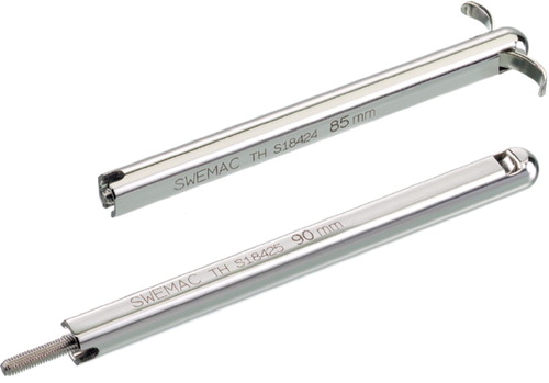
The HTH consists of two parts, an inner sliding component containing the hooks and an outer pin. The outer pin has 2. at sides, which create rotational locking inside the plate barrel. Fixation in the femoral head is achieved by pushing the inner sliding component out through the proximal windows. When the hooks protrude in this way, they bend in arches with their convexity facing the periphery of the femoral head in an anterior and posterior direction. We used the new version of the HTH, in which the diameter of the pin shaft is 8.9 mm—which is 1 mm thicker than the old version (Olsson et al. Citation2002). The hooks have also been made slightly wider and stiffer (). The HTH was combined with a standard 4-hole plate in this study (the Swemac hip plate).
Figure 1. The Hansson Twin Hook (HTH) before and after the protrusion of the hooks.
Surgical technique
After anatomical reduction of the fracture using a traction table and biplane fluoroscopy, a guide pin was inserted centrally into the femoral head at an angle of 135° against the femoral shaft. A channel for the HTH and the plate barrel was reamed. The plate was slid into position and the HTH was inserted through the barrel of the plate into the bone channel. The hooks were then pushed out without rotating or hammering the bone. Finally, the plate was fixed to the femur.
Definition of intraoperative technical errors and failure of fixation
The intended position of the HTH was central in both the anterior-posterior and lateral views of the femoral head and close to the subchondral bone (). An intraoperative technical error was defined as being when one of the hooks was placed through the subchondral bone of the femoral head (). A reoperation with an adjustment or a change of the HTH, leaving the plate in place, can be done percutaneously through a 1-cm long incision under local anesthesia. It was not considered a fixation failure if the fracture underwent uneventful healing without discomfort to the patient. Failure of fixation was classified as cut-out of the HTH through the femoral head, breakage or loosening of the plate, or nonunion. Migration of the HTH in the femoral head without a cut-out or varus angulation of the fracture was not regarded as failure.
Figure 2. The recommended and required position of the HTH is central in both the anterior- posterior and lateral views of the femoral head, and close to the subchondral bone
Figure 3. An intraoperative technical error with one of the hooks placed through the subchondral bone of the femoral head. The cause is usually a non-anatomical reduction of the fracture with sagging, which makes correct placement of the HTH more difficult.
Patients
During 2003, 157 consecutive patients (mean age 83 (43–98) years, 117 women) who had sustained a trochanteric fracture were included in a bicentric study from the Orthopedic Departments of the University Hospital, Malmö, and of the Hospital in Helsingborg. The fractures were graded according to the Jensen-Michaelsen (J-M) classification (Jensen and Michaelsen Citation1975). Patients with a pathological fracture, previous surgery of the proximal femur, or with multiple trauma were excluded. The surgeons in Malmö, 35 of whom performed the operations in this study, had been introduced to the surgical technique as described above in lectures. However, in Helsingborg this technique with the HTH had already been established a few years previously and was a routine procedure for most of the 20 surgeons who performed the operations. Of the surgeons, 36 were certified orthopedic trauma specialists and 19 were registrars.
The same study protocol was used in the two hospitals. Background data were collected when the patients were admitted to hospital. Intraoperative parameters such as type of anesthesia, intraoperative blood loss, and operating time were registered. Blood transfusions, general complications, and the length of the hospital stay were recorded. All patients were allowed full weight bearing as tolerated postoperatively. The patients had a 4-month follow-up with radiographic and clinical examinations. The length of the skin incision, leg length discrepancy, and any discomfort from the osteosynthesis on the lateral aspect of the thigh were recorded at the clinical examination. A radiographic file search was done 2 years postoperatively to find out if any new radiographic examinations of the operated hip had been done after the first 4-month period, and thus to rule out that further fixation failures had occurred.
All patients were given prophylactic antibiotics and anticoagulants according to the standard procedures at the hospitals.
Results
The background parameters were similar in the patient material from Malmö and Helsingborg (Table). 83% of the fractures were unstable (3–4- part fractures). Spinal anesthesia was used in 85% of the patients and general anesthesia was used in 15%. The median operating time (skin to skin) was 45 (15–180) min. The median intraoperative blood loss as estimated by the anesthetic nurse was 0.1 (0–1.7) L and the median units of blood transfusions was 1 (0–10). The average number of operations per surgeon was 3 (1–12). The orthopedic trauma specialists performed 93 operations (59%) and the registrars 64 operations (41%).
3 superficial wound infections were noticed but there was no need for surgical debridement. Furthermore, 1 pulmonary embolus, 5 myocardial infarctions, 2 pressure wounds, and 6 pneumonias were recorded. The median hospital stay was 9 (1–30) days. Of the 109 patients admitted from their own home, 26 (24%) returned directly to their own home at discharge from the hospital, 82 (75%) were discharged to other institutions for rehabilitation or permanent stay, and 1 patient died during the hospital stay.
37 patients (24%) had died by the 4-month follow-up and 5 other patients (3%) had died after 1 year. 17 patients (11%) did not participate in the follow-up: 7 refused further radiographic examinations and 10 gave no reason for not attending. The remaining 103 patients underwent a clinical and a radiographic examination.
The median length of the skin incision was 102 (51–210) mm. The mean leg length shortening was 4.4 (0–20) mm. Discomfort laterally over the hip due to the osteosynthesis was noted in 21 patients (20%).
Background parameters in the 157 patients
Failure of fixation and technical errors
None of the patients showed any breakage or loosening of the plate, and none of them showed radiographic signs of delayed union or nonunion. 2 failures of fixation occurred. The first was in a 93- year-old woman with an intraoperative technical error. The fracture displaced with a subsequent cutout of the HTH through the femoral head within 2 months and a revision with a hip prosthesis was performed. The second case of failure was in an 84-year-old woman with an intraoperative technical error. At the 4-month follow-up, migration of the HTH within the femoral head was discovered. Later, an exchange of the HTH to a shorter one was done and the hooks were positioned slightly more distally in the femoral head. The migration continued, however, and finally a cut-out occurred 22 months after the primary operation. A revision with a hip prosthesis was done.
Technical intraoperative errors (hook penetration) were noted in 5 other patients. All 7 fractures with intraoperative errors, of which 5 were J-M type 5 (4-part fracture), had been inadequately reduced. Thus, the placement of the HTH was not central in the femoral head in the lateral view and consequently one of the hooks had penetrated the subcondral bone (). One of the 5 patients had an adjustment of the HTH during the hospital stay; the hooks were pulled back about 2 mm. All 5 fractures healed with no discomfort for the patient.
Discussion
The later generation of the HTH combined with a standard plate appears to be of value in treating trochanteric fractures. In our study, only 2 failures of fixation (1.3%) occurred. In previous studies on trochanteric fractures treated with the SHS, the rate of fixation failure has varied between 0% and 26% (Sernbo et al. Citation1988, Davis et al. Citation1990, Nungu et al. Citation1991, Barrios et al. Citation1993, Desjardins et al. Citation1993, Radford et al. Citation1993, Stappaerts et al. Citation1995, Leung et al. Citation1996, Buciuto et al. Citation1998, Watson et al. Citation1998, Lunsjö et al. Citation2001b). The most common mode of fixation failure has been a cut-out of the lag screw through the femoral head.
One reason for the low rate of failure with the HTH may be increased rotational stability achieved by the oppositely directed hooks. The lag screw usually gets a firm grip inside the femoral head, when the bone quality is good. In osteoporotic bone, however, there is an increased risk that the lag screw will lose rotational stability once the threads lose their grip (Olsson et al. Citation2002). In contrast, the HTH does not depend on initial fixation strength, since any rotation or compression will impact bone on the supporting area of the hooks, which increases the stability.
Another reason for the low rate of cut-out with the HTH may be that the span of the hooks forces the surgeon to place the HTH more centrally in the femoral head than a lag screw, which is favorable from a mechanical standpoint. Even if one of the hooks has penetrated the subchondral bone, it may be contained within the thick cartilage of the femoral head and would therefore not cause the patient any pain or discomfort. Thus, 5 of the 7 patients with intraoperative errors did not have symptoms that could be ascribed to hook penetration.
It can also be argued that the low rate of fixation failure could be the result of an incomplete followup, i.e. patients lost to follow-up. As expected after a hip fracture (Kanis et al. Citation2003), the mortality rate was relatively high during the first 4 months and some patients may have died before their fixation failure. However, we know from earlier studies (Yoshimine et al. Citation1993, Nakata et al. Citation1994, Lunsjö et al. Citation1995, Citation2001a) that trochanteric fractures operated with sliding screw systems, are in a stable consolidation phase after 3–4 months. Furthermore, 2 years postoperatively we performed a radiographic file search on all patients without finding any further radiographic results of examinations of the operated hips indicating a failure of the fixation. It is unlikely that a patient with symptoms from a fixation failure would not have had such an examination during this time period.
Of the many surgeons involved who had varying degrees of experience with fractures, most had no prior experience of the HTH. Placement of the HTH centrally in the femoral head was adequately done in most cases—but with improper reduction of the fracture, the risk of hook penetration increased. With anatomical reduction of the fracture, this risk should be eliminated. The device must be considered user-friendly considering the low rate of intraoperative errors leading to failures, little intraoperative bleeding, and reasonably short operation time even though the average number of operations with the new method was about 3 per surgeon. An earlier study (Olsson et al. Citation2000) comparing the HTH with a lag screw, both in combination with a 4-hole plate, showed the possibilities of shorter skin incision with the HTH. Also in our study, without aiming for a minimally invasive technique the average length of a skin incision was short. The amount of leg shortening was small in these elderly patients, where most fractures were unstable. It appears that the method would be applicable in routine practice.
At the 4-month follow-up, 1 out of every 5 patients felt discomfort laterally over the hip due to the prominence of the hardware. In significant collapse of the fracture, both the HTH and the lag screw will slide inside the barrel of the plate and protrude into the soft tissue of the lateral thigh. With a lag screw, both it and the plate most be removed to relieve the soft tissue obstacle, since the threads of the lag screw are wider than the barrel. Removal of an HTH, on the other hand, is done with the plate in place and is easily done under local anesthesia. Through a small incision, the hooks are withdrawn to a position inside the outer pin, which can be pulled out without interfering with the barrel of the plate. This is the way to correct or replace the HTH to a new position in the femoral head due to an inaccurately placed hook that is penetrating the subchondral bone.
The Hansson Twin Hook appears to provide sufficient fixation in the femoral heads of elderly patients with trochanteric fractures. It is an alternative and a competitor to the lag screw. Randomized studies comparing the two implants are now needed to evaluate the ultimate role of the Hansson Twin Hook in the management of hip fractures.
Acknowledgement
This study was supported by grants from the Thelma Zoéga Foundation (Helsingborg) and from the Stig and Ragna Gorthon Foundation (Helsingborg).
No competing interests declared.
Contributions of authors
PO: study design, data collection, data analysis, and preparation of the manuscript. BJ: study design, data collection, and preparation of the manuscript. LC: head of the research group, study design, and preparation of the manuscript. JB: radiographic analysis. OO and IS: study design. KL: preparation of the manuscript.
- Barrios C, Brostrom L A, Stark A, Walheim G. Healing complications after internal fixation of trochanteric hip fractures: the prognostic value of osteoporosis. J Orthop Trauma 1993; 7(5)438–42
- Buciuto R, Uhlin B, Hammerby S, Hammer R. RAB-plate vs Richards CHS plate for unstable trochanteric hip fracture. A randomized study of 233 patients with 1-year follow-up. Acta Orthop Scand 1998; 69(1)25–8
- Ceder L. The difficult extracapsular hip fracture (Including subtrochanteric). Current Orthopaedics 2000; 14: 93–101
- Davis T R, Sher J L, Horsman A, Simpson M, Porter B B, Checketts R G. Intertrochanteric femoral fractures. Mechanical failure after internal fixation. J Bone Joint Surg (Br) 1990; 72(1)26–31
- Desjardins A L, Roy A, Paiement G, Newman N, Pedlow F, Desloges D, Turcotte R E. Unstable intertrochanteric fracture of the femur. A prospective randomised study comparing anatomical reduction and medial displacement osteotomy. J Bone Joint Surg (Br) 1993; 75(3)445–7
- Jensen J S, Michaelsen M. Trochanteric femoral fractures treated with McLaughlin osteosynthesis. Acta Orthop Scand 1975; 46(5)795–803
- Kanis J A, Odén A, Johnell O, De Laet C, Jönsson B, Oglesby A K. The components of excess mortality after hip fracture. Bone 2003; 32: 468–73
- Leung K S, Chen C M, So W S, Sato K, Lai C H, Machaisavariya B, Suntharalingam S. Multicenter trial of modified Gamma nail in East Asia. Clin Orthop 1996, 323: 146–54
- Lunsjö K, Ceder L, Stigsson L, Hauggaard A. One-way compression along the femoral shaft with the Medoff sliding plat. The first European experience of 104 intertrochanteric fractures with a 1-year follow-up. Acta Orthop Scand 1995; 66(4)343–6
- Lunsjö K, Ceder L, Hauggaard A, Stigsson L. The role of the greater trochanter in fracture stability in uni- or biaxial dynamisation with the Medoff sliding plate. Hip International 2001a; 1(2)71–9
- Lunsjö K, Ceder L, Thorngren K G, Skytting B, Tidermark J, Berntson P O, Allvin I, Norberg S, Hjalmars K, Larsson S, Knebel R, Hauggaard A, Stigsson L. Extramedullary fixation of 569 unstable intertrochanteric fractures: a randomized multicenter trial of the Medoff sliding plate versus three other screw-plate systems. Acta Orthop Scand 2001b; 72(2)133–40
- Nakata K, Ohzono K, Hiroshima K, Toge K. Serial change of sliding in intertrochanteric femoral fractures treated with sliding screw systems. Arch Orthop Trauma Surg 1994; 113: 276–80
- Nungu S, Olerud C, Rehnberg L. Treatment of intertrochanteric fractures: comparison of Ender nails and sliding screw plates. J Orthop Trauma 1991; 5(4)452–7
- Olsson O. Alternative techniques in trochanteric hip fracture surgery. Clinical and biomechanical studies on the Medoff sliding plate and the Twin-hook. Acta Orthop Scand 2000, Suppl 295: 71
- Olsson O, Ceder L, Lunsjö K, Hauggaard A. Extracapsular hip fractures: fixation with a twin hook or a lag screw?. Int Orthop 2000; 24(5)249–55
- Olsson O, Tanner K E, Ceder L, Ryd L. A biomechanical study on fixation stability with twin hook or lag screw in artificial cancellous bone. Int Orthop 2002; 26(6)349–55
- Parker M J, Handoll H H. Gamma and other cephalocondylic intramedullary nails versus extramedullary implants for extracapsular hip fractures in adults. Cochrane Database Syst Rev 2005, 4: CD000093
- Radford P J, Needoff M, Webb J K. A prospective randomised comparison of the dynamic hip screw and the gamma locking nail. J Bone Joint Surg (Br) 1993; 75(5)789–93
- Sernbo I, Johnell O, Gentz C F, Nilsson J A. Unstable intertrochanteric fractures of the hip. Treatment with Ender pins compared with a compression hip-screw. J Bone Joint Surg (Am) 1988; 70(9)1297–303
- Stappaerts K H, Deldycke J, Broos P L, Staes F F, Rommens P M, Claes P. Treatment of unstable peritrochanteric fractures in elderly patients with a compression hip screw or with the Vandeputte (VDP) endoprosthesis: a prospective randomized study. J Orthop Trauma 1995; 9(4)292–7
- Watson J T, Moed B R, Cramer K E, Karges D E. Comparison of the compression hip screw with the Medoff sliding plate for intertrochanteric fractures. Clin Orthop 1998, 348: 79–86
- Yoshimine F, Latta L L, Milne E L. Sliding characteristics of compression hip screws in the intertrochanteric fractures. A clinical study. J Orthop Trauma 1993; 7: 348–53