Abstract
Background and purpose The use of metal implants in large defects caused by spinal infection to support the anterior column is controversial, and relatively few results have been published to date. Despite the fact that there is bacterial adhesion to metal implants, the strong immunity of the highly vascularized spine because of rich muscle covering is unique. This possibly allows the use of metal implants, which have the advantage of high stability and reduced loss of correction. This is a retrospective study of patients with spondylodiscitis treated with metal implants.
Patients and methods We retrospectively analyzed the outcome in 22 consecutive patients (mean age 69 (43–82) years, 15 men) with spondylodiscitis (20 lumbar and 12 thoracic discs) who had received an anterior titanium cage implantation. In 13 cases, the pathogen could be identified. Antibiotic treatment was continued for at least 12 weeks postoperatively.
Results The mean follow-up was 36 (32–47) months. Healing of inflammation was confirmed by clinical, radiographic, and laboratory parameters. The mean VAS improved from 9.1 (6–10) preoperatively to 2.6 (0–6) at the final follow-up, and the mean Oswestry disability index was 17 (0–76) at the final follow-up.
Interpretation Our findings highlight the high healing rate and stability when titanium implants are used. Prerequisites are a radical debridement, correction of deformity, and additional bony fusion by bone grafting. The increased stability, with facilitated patient mobilization, and the relatively little loss of correction using anterior and posterior implants are of considerable advantage in the treatment of the patients with multiple co-morbidities.
Infection of the intervertebral disc with osteomyelitis of the endplates (spondylodiscitis) is associated with high morbidity and mortality (Hadji-pavlou et al. Citation2000). Despite a growing evidence in the pharmacological treatment of spinal infections, most recommendations for surgical treatment are still based on retrospective case series (Ha et al. Citation2007, Pee et al. Citation2008). In particular, the indication for surgical intervention has been the subject of debate for many years.
One cornerstone of discussion is the use of metal implants. For decades, the use of metallic implants at an infectious site was avoided because of adhesion of bacteria to the implants (Ha et al. Citation2004). In the spine, the avoidance of internally stabilizing measures may lead to instability, with loss of correction and pseudarthrosis. Thus, more and more spinal surgeons implant metal devices into the infected spine to avoid secondary deformity, regardless of the bacterial adherence that has sometimes been reported. Several authors have recently presented favorable results regarding healing and avoidance or correction of deformity using titanium implants (Heyde et al. Citation2006, Kuklo et al. Citation2006, Ruf et al. Citation2007). In large anterior defects after severe spondylitis, the use of anterior titanium cages is especially advantagious in reconstruction of the sagittal alignment (Robinson et al. Citation2008).
To determine the effect of the use of metal implants on the clinical and radiological outcome in patients with spondylodiscitis, we performed a retrospective investigation of 22 consecutive patients treated at our spine unit.
Patients and methods
During a period of 3 years (March 2003 to January 2005), 34 patients with acute single or multilevel spondylodiscitis (22 lumbar, 17 thoracic, and 4 cervical discs) were treated surgically. 22 of these patients (mean age 69 (43–82) years, 15 men) with 20 infected lumbar and 12 infected thoracic discs received an anterior titanium cage implantation (Harms-mesh or X-Tenz; both from DePuy Spine, city, UK). 19 patients were operated with a circumferential procedure including titanium screwrod instrumentation (Moss Max or Moss Miami; DePuy Spine) and 3 patients were operated with an anterior-only procedure with anterior titanium instrumentation. All cages were filled with autogenous cancellous bone graft from the iliac crest.
8 patients had epidural abscesses. 13 patients had an infectious focus (5 urinary tract infections, 3 subcutaneous abscesses, 2 endoprosthesis infections, 2 chronic lower limb osteomyelitis, 1 central venous catheter sepsis), 5 had terminal renal failure, and 5 had insulin-dependent diabetes mellitus (). In 2 of the cases, the spondylodiscitis was post-procedural after microsurgical lumbar discectomies. Five patients had presented initially with an incomplete peripheral sensomotor deficit.
Table 1. Summary of co-morbidities
Microbiological analysis of specimens obtained during the surgical debridement identified the pathogen in 13 cases (). Postoperatively, the patients received antibiotic treatment for 12 weeks according to the resistance spectrum. If no pathogen was found, broad-spectrum antibiotic regimens were used. If possible, patients were mobilized by physiotherapy on day 3 postoperatively. No external bracing was applied postoperatively.
Table 2. Summary of pathogens detected during intraoperative sampling
Radiographic (lateral and AP) and clinical (C-reactive protein, white blood cell count) follow-up dates were 6 weeks, 12 weeks, 6 months, and 1 and 3 years post-operatively. Pain on a visual analog scale (VAS) and Oswestry disability index (ODI) were also recorded. The mean follow-up time was 36 (32–47) months.
Results
No operative complications were recorded. In the postoperative course, no implant-related complications were seen. The patients stayed in the intensive care unit for mean 22 (0–168) days. One patient suffered a lethal cardiac failure 2 weeks after the staged anterior procedure. One patient died because of cardiac arrest 6 months after the operation.
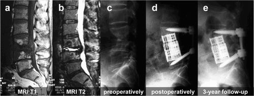
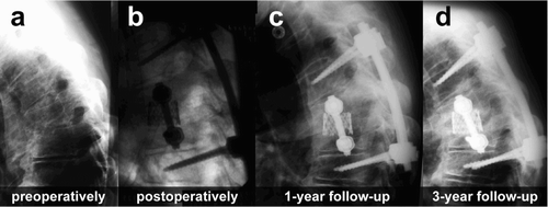
At the final follow-up, mean 36 months after the operation, there was a mean segmental loss of correction of 3.7 (0–6) degrees compared to early postoperative radiographs ( and ). The mean VAS improved from 9.1 (6–10) preoperatively to 2.6 (0–6) at the final follow-up, and the ODI was 17 (0–76) at the final follow-up. Healing of inflammation was confirmed in all cases from clinical, radiographic, and laboratory parameters (C-reactive protein, white blood cell count).
Discussion
Patients with spondylodiscitis often present with multiple co-morbidities. In our patients, septic infection, diabetes mellitus, coronary heart disease, and chronic hemodialysis were prevalent, making the patients highly susceptible to perioperative complications. Indications for operative treatment are large paraspinal abscesses, sepsis, deformities due to destruction of the endplates, progressive neurological impairment, and resistance to non-surgical treatment for more than 6 weeks (Schinkel et al. Citation2003). In all other patients a non-operative approach, including long-term antibiotic treatment after CT-guided specimen acquisition, and immobilization in a rigid brace should be tried (Grados et al. Citation2007). Spondylodiscitis with multiple abscesses causing severe sepsis nevertheless requires early surgical intervention (Robinson et al. Citation2007).
The primary goal of surgical therapy is complete debridement with radical removal of any infectious or necrotic tissue (Heyde et al. Citation2006). Then adequate stabilization is necessary, which mostly implies a 360-degree fusion including bone grafting (Ruf et al. Citation2007). For optimal long-term results, prevalent deformities must be corrected with regard to the mechanical properties of the spine. The tension-band principle of the posterior structures requires a solid reconstruction of the anterior column to take up the load without loss of correction (Stoltze and Harms Citation1999).
Titanium cages in combination with posterior instrumentation achieve stability similar to that of the intact spine (Oda et al. Citation1999). Thus, they are an excellent means of bridging large defects and taking the anterior load until the bony fusion of the cancellous bone graft is completed. However, several pathogens tend to adhere to metallic surfaces (Ha et al. Citation2004). Asymptomatic bacterial biofilm on implants has been found even months after successful metal implantation in spinal osteomyelitis (Shad et al. Citation2003). Even so, several authors have reported favorable results using titanium cages in spondylodiscitis.
In a staged anterior-posterior procedure, Fayazi et al. (Citation2004) used titanium-mesh cages in 11 patients with spondylodiscitis. One patient had to be reoperated because of hardware failure. One patient developed pseudarthrosis. None of the patients had recurrence of infection. Lerner et al. (Citation2006) investigated the use of anterior titanium ring cages in 43 patients with a mean loss of correction of 1.4 degrees at a mean follow-up of 2.5 years. Except for 1 patient with anterior revision, all infections were eradicated.
Titanium-mesh cages were also implanted by Korovessis et al. (Citation2006a, Citationb) in 14 and 17 patients with pyogenic spondylodiscitis. After a mean follow-up of 45 months, no loss in correction was found. Revision was necessary in 1 patient.
Kuklo et al. (Citation2006) implanted titanium-mesh cages in 22 patients with spondylodiscitis. All patients with additional posterior instrumentation had only minimal settling of the cage. Revision without implant removal was necessary in only 2 patients.
Ruf et al. (Citation2007) investigated the use of anterior titanium-mesh cages in combination with posterior instrumentation in spinal reconstruction after spondylodiscitis in 88 patients. In 3 cases, even 3 vertebrae had to be replaced. The authors found healing in all patients, and adequate correction of the sagittal profile.
Even though metal implants should be avoided in the infected bone, they obviously can be used successfully in spinal infections. Prerequisites are a radical debridement and additional bony fusion by use of cancellous bone grafting (Robinson et al. Citation2007). Our findings highlight the high healing rate, stability, and the low rate of complications when titanium implants are used. The increased stability with facilitated patient mobilization and the little loss of correction using anterior and posterior implants are of considerable advantage in the treatment of such patients, who often have co-morbidities.
Contributions of authors
YR and TF designed the study, performed the data analysis, participated in the sequence alignment, and drafted the manuscript. SKT, WE, and RK participated in the sequence alignment and in revision of the manuscript. CEH conceived the study and participated in its design and coordination, and helped to draft the manuscript. All authors read and approved the final manuscript.
No competing interests declared.
- Fayazi A H, Ludwig S C, Dabbah M, Bryan Butler R, Gelb D E. Preliminary results of staged anterior debridement and reconstruction using titanium mesh cages in the treatment of thoracolumbar vertebral osteomyelitis. Spine J 2004; 4: 388–95
- Grados F, Lescure F X, Senneville E, Flipo R M, Schmit J L, Fardellone P. Suggestions for managing pyogenic (non-tuberculous) discitis in adults. Joint Bone Spine 2007; 74(2)133–9
- Ha K Y, Chung Y G, Ryoo S J. Adherence and biofilm formation of staphylococcus epidermidis and mycobacterium tuberculosis on various spinal implants. Spine 2004; 30: 38–43
- Ha K Y, Shin J H, Kim K W, Na K H. The fate of anterior autogenous bone graft after anterior radical surgery with or without posterior instrumentation in the treatment of pyogenic lumbar spondylodiscitis. Spine 2007; 32: 1856–64
- Hadjipavlou A G, Mader J T, Necessary J T, Muffoletto A J. Hematogenous pyogenic spinal infections and their surgical management. Spine 2000; 25: 1668–79
- Heyde C E, Boehm H, El Saghir H, Tschoke S K, Kayser R. Surgical treatment of spondylodiscitis in the cervical spine: a minimum 2-year follow-up. Eur Spine J 2006; 15: 1380–7
- Korovessis P, Petsinis G, Koureas G, Iliopoulos P, Zacharatos S. One-stage combined surgery with mesh cages for treatment of septic spondylitis. Clin Orthop 2006a, 444: 51–9
- Korovessis P, Petsinis G, Koureas G, Iliopoulos P, Zacharatos S. Anterior surgery with insertion of titanium mesh cage and posterior instrumented fusion performed sequentially on the same day under one anesthesia for septic spondylitis of thoracolumbar spine: is the use of titanium mesh cages safe?. Spine 2006b; 31: 1014–9
- Kuklo T R, Potter B K, Bell R S, Moquin R R, Rosner M K. Single-stage treatment of pyogenic spinal infection with titanium mesh cages. J Spinal Disord Tech 2006; 19: 376–82
- Lerner T, Schulte T, Bullmann V, Schneider M, Hackenberg L, Liljenqvist U. Anterior column reconstruction using titanium ring cages in severe vertebral osteomyelitis. Eur J Trauma 2006; 32: 227–37
- Oda I, Cunningham B W, Abumi K, Kaneda K, McAfee P C. The stability of reconstruction methods after thoracolumbar total spondylectomy. An in vitro investigation. Spine 1999; 24: 1634–8
- Pee Y H, Park J D, Choi Y G, Lee S H. Anterior debridement and fusion followed by posterior pedicle screw fixation in pyogenic spondylodiscitis: autologous iliac bone strut versus cage. J Neurosurg Spine 2008; 8: 405–12
- Robinson Y, Reinke M, Kayser R, Ertel W, Heyde C E. Post-operative multisegmental lumbar discitis treated by staged ventrodorsoventral intervention. Surg Infect (Larchmt) 2007; 8: 529–34
- Robinson Y, Tschoeke S K, Kayser R, Boehm H, Heyde C E. Reconstruction of large defects in vertebral osteomyelitis with expandable titanium cages. Int Orthop 2008, Jul 5. (Epub ahead of print) DOI 10.1007/s00264-008-0567-2
- Ruf M, Stoltze D, Merk H R, Ames M, Harms J. Treatment of vertebral osteomyelitis by radical debridement and stabilization using titanium mesh cages. Spine 2007; 32: E275–80
- Schinkel C, Gottwald M, Andress H J. Surgical treatment of spondylodiscitis. Surg Infect (Larchmt) 2003; 4: 387–91
- Shad A, Shariff S, Fairbank J, Byren I, Teddy P J M A D, Cadoux-Hudson T A D. Internal fixation for osteomyelitis of cervical spine: the issue of persistence of culture positive infection around the implants. Acta Neurochir 2003; 145: 957–60
- Stoltze D, Harms J. Correction of posttraumatic deformities. Principles and methods. Orthopäde 1999; 28: 731–45

