List of papers
This thesis is based on the following papers:
Weidow J, Pak J, Kärrholm J. Different patterns of cartilage wear in medial and lateral gonarthrosis.
Acta Orthop Scand 2002; 73(3): 326-9.
Weidow J, Mars I, Cederlund CG, Kärrholm J. Standing radiographs underestimate joint width: comparison before and after resection of the joint in 34 total knee arthroplasties.
Acta Orthop Scand 2004; 75(3): 315-22.
Weidow J, Cederlund CG, Ranstam J, Kärrholm J. Ahlbäck grading of knee osteoarthritis. Reproducibility and validity based on visual inspection of the joint.
Acta Orthop 2006; 77.
Weidow J, Holmén A, Kärrholm J. Working conditions resulting in increased need for surgical treatment of knee osteoarthritis.
In manuscript.
Weidow J, Mars I, Kärrholm J. Medial and lateral osteoarthritis of the knee is related to variations of hip and pelvic anatomy.
Osteoarthritis Cartilage 2005; 13(6): 471-7.
Weidow J, Tranberg R, Saari T, Kärrholm J. Hip and knee joint rotations differ between patients with medial and lateral knee osteoarthritis. Gait analysis of 30 patients and 15 controls.
J Orthop Res 2006. Accepted.
Weidow J, McPherson A, Saari T, Kärrholm J. Changes of knee joint kinematics caused by lateral osteoarthritis.
Clin Orthop Relat Res 2006. Conditional acceptance.
Abbreviations
| ACL: | = | Anterior cruciate ligament |
| AP: | = | Anterior-posterior |
| BMD: | = | Bone mineral density |
| BMI: | = | Body mass index |
| DOF: | = | Degrees of freedom |
| FoB70: | = | Folkbokföringen (data on Swedish inhabitants collected by Sweden Statistics) 1970 |
| JSN: | = | Joint space narrowing |
| JSW: | = | Joint space width |
| OA: | = | Osteoarthritis |
| RR: | = | Relative risk (to normal) |
| OR: | = | Odds Ratio |
| TKA: | = | Total knee arthroplasty |
| TKR: | = | Total knee replacement. |
Summary
This thesis aimed to study osteoarthritis (OA) of the knee and its subgroup with lateral wear (valgus knees). Anatomy, kinematics and kinetics (movements and moments) of the hip and knee joint and the sensitivity and specificity of diagnostic procedures were evaluated. Our hypothesis was that there are biomechanical reasons for development of either lateral or medial OA.
The wear pattern of the tibial plateau and the femoral condyles was delineated in 42 patients. The diagnostic accuracy of standing knee radiographs was validated in 34 and reproducibility and validity of the Ahlbäck classification was studied in 48 patients. The Influence of working conditions or gender on the prevalence of uni or bilateral knee OA was evaluated in 990 patients from 2 hospitals (Varberg and Halmstad) operated during 1985–1994 with knee arthroplasty or proximal tibial osteotomy. For each patient, 3 age- and gendermatched controls were found. Studies of the anatomy of the hip and pelvic regions and the motions and moments in the hip and knee in medial and lateral OA were performed. The detailed kinematics of the knee during active extension in lateral OA were recorded using dynamic radiostereometry (RSA).
In both medial and lateral OA the central part of the tibial plateau showed the most pronounced wear (p<0.001), followed by the anterior part in medial OA (p=0.02) and the posterior part in lateral OA (p=0.001).
In medial OA the observed difference between the 2 radiographic methods was small and acceptable (median; p=0.05; 0–0.5 mm). In lateral OA there was no consistent underestimation, but larger scatter (median; p=0.04; −0.1–1.2 mm) suggesting less precise determination.
The repeatability of the Ahlbäck classification for one observer was fair (kappa: medial OA: 0.15–0.65; lateral OA: 0.59–0.76), but between observers it was poor (kappa: 0.1). The validity revealed an acceptable sensitivity in both medial (67–95%) and lateral (43–-86%) OA but the specificity was low (medial: 11–67%; lateral: 25–75%).
Farmers (RR: 1.7; p<0.0005) and building workers (RR: 1.4; p=0.047) run increased risk to undergo surgical treatment because of OA of the knee. Unilateral disease was 3.7 times more common among men.
Patients with lateral knee OA had a 14 mm wider pelvis (p=0.001) and those with medial knee OA an 11 mm higher offset (p=0.005). In the gait analysis they showed more outward rotation of the hip (p=0.001) and more inward rotation of the tibia than did patients with medial OA (p=0.001).
In lateral OA, the medial femoral condyle translated 7–8 mm forward with 45° flexion whereas controls translated 4 mm less (p=0.03), without any difference of the lateral femoral condyle.
Conventional radiographs do not give sufficient information for correct grading, especially in lateral OA where the scatter is high. The joint space can often be seen on radiographs despite presence of bone attrition as observed on the preparations. Increased incidence of unilateral disease in men and building workers suggests that this joint disease more commonly originates from previous trauma. Our findings suggest that the occurrence of medial or lateral OA has a biomechanical background originating from pelvis and hip anatomy.
Introduction and background
The word ‘arthrosis’ has been used in Scandinavia to describe radiographic changes of a joint with a narrowing or obliteration of the joint space associated with subchondral sclerosis and presence of osteophytes. Clinical symptoms in terms of pain, swelling, stiffness and instability may vary over time. Occasionally they can be minimal or even absent, but more often moderate to severe with a variable tendency to progression. The term ‘arthritis’ has been used for chronic inflammatory joint disorders such as rheumatoid/psoriatic arthritis and acute joint disorders such as reactive and septic arthritis. I have chosen to use the international term ‘osteoarthritis’. I personally think that arthrosis is more descriptive since it questions the inflammatory origin of the disease.
Osteoarthritis (OA) of the knee has been used in many studies in reference to the medial variety of the disease. Cases with lateral knee OA have often been either excluded or considered the same as those with medial OA. However, it seems reasonable to suppose that because of the differences in both clinical appearance and location, lateral knee OA may have another origin. This thesis aimed to evaluate diagnostic problems related to knee OA and especially the location of the disease. Studies of prevalence, anatomy, biomechanics and factors related to the location of OA were also conducted to evaluate whether or not these variables could indicate etiological differences between the medial and lateral varieties of the disease.
Epidemiology
OA is more common in women. One study shows a much worse functional status prior to total knee arthroplasty in women than in men (Katz et al. Citation1994). Because not all people have symptoms, the prevalence of radiographic knee OA is higher than those who seek medical attention. The definition of OA is not clear. Different radiographic criteria may be used in addition to other methods. Clinical symptoms may or may not be recorded. In the ages 55–64 years, Bagge et al. (Citation1991) found a prevalence of radiographic knee OA of 41% in the women and 30% in the men (Kellgren-Lawrence grade 2 and larger: definite osteophytes and possible joint narrowing). In the age range 75–79 years, these figures became equal (55%). In the Chingford study, Spector et al. (Citation1991) found symptomatic radiographic knee OA in 2.9% of the women aged 45–65 years. All epidemiological studies have shown a higher prevalence of osteoarthritis among Caucasian than in other ethnic groups, but despite claims that these studies are biased, it seems that the difference is real. In a study comparing a Chinese population in Beijing with a corresponding white population in Framingham with adjustment for age and frequency of squatting (a risk factor for developing knee OA) the prevalence was 9.5% higher among the Chinese women and 7% lower in Chinese men (Zhang et al. Citation2004).
Etiology
Some authors have suggested positive correlations between knee OA and hypertension, hypercholesterolemia and blood glucose (Hart et al. Citation1995) as well as free estrogens (Spector et al. Citation1991), whereas others found no such relationships (Bagge et al. Citation1991, Samanta et al. Citation1993). Obesity is a recognized risk factor for development of knee OA in women (Bagge et al. Citation1991, McAlindon et al. Citation1992, Hart and Spector Citation1993, Cicuttini et al. Citation1997). In men, a high body mass index (BMI) does not seem to have the same influence on the risk of developing knee OA (Felson et al. Citation1988, Citation1997b). Even normal BMI (20–24) seems to double the risk of developing knee OA compared to low BMI (17–19) in men (Järvholm et al. Citation2005). BMI during the second decade of life (at 20 years of age) has been found to relate to the severity of OA in women with varus deformity, but not in those with valgus knees (Sharma et al. Citation2000). High BMI at the age of 40 increases the relative risk (RR) with 9 in women and 4 in men (Sandmark et al. Citation1999). Obesity has been described as a stronger predictor than knee injury (Odds Ratio (OR): 6 vs. 3) in cases with the bilateral disease, but not in those with unilateral OA (OR: 3.4 vs. 16 in the right knee, and 2.4 vs. 11 in the left knee) (Davis et al. Citation1989). A stronger association has been found in men than in women between knee injury and OA (McAlindon et al. Citation1996). Previous tear of the anterior cruciate ligament (ACL) and meniscectomy increase the risk of development of OA (Kannus and Järvinen Citation1989, Roos et al. Citation1995, Citation1998). The mechanisms behind this development are not completely understood. Altered joint kinematics (Jonsson and Kärrholm Citation1994, Brandsson et al. Citation2001) absent or deformed menisci, changed contact area (Maquet et al. Citation1975) and instability cause altered load distribution, increase of peak pressures and joint subluxations, all of which may initiate and/or accelerate the disease.
Some authors have found an association between bone mineral density (BMD) and knee OA (Sowers et al. Citation1996). High BMD might be associated with increased risk of developing knee OA, at the same time as the risk of progression of a radiographic knee OA may decrease (Zhang et al. Citation2000). Increased BMD of the spine (Hordon et al. Citation1993, Hart et al. Citation1994) and the femur (Hannan et al. Citation1993) has been found in women with knee OA. High density of the bone is not equal to optimum quality. Ding et al. (Citation2001) studied the mechanical properties of post-mortem bone in tibias with early-stage osteoarthritis. They observed that increase in bone tissue did not compensate for loss of mechanical properties, which indicates deterioration of bone quality in OA. In the Chingford study, women with a previous fracture (mainly in the distal forearm and vertebrae) had reduced risk of developing knee OA independent of BMD status. These results suggest a possible common role of bone turnover and bone repair in the early manifestation of OA (Hart et al. Citation2002).
One study (Terauchi et al. Citation1998) has discussed the idea that osteoporosis in the lower extremity results in angular deformity of the knee because of structural changes in the proximal tibia. Low intake of calcium and D-vitamin as well as smoking, lowers the quality of the bone. Smokers have lower risk of knee OA (Felson et al. Citation1989, Bagge et al. Citation1991, Felson et al. Citation1997b). It has been suggested that environmental causes in the first year of life may influence the development of OA later in life. Fonnebo (Citation1995) proposed that sunlight exposure prior to a crucial period in skeletal development could be a possible cause. A study from Tromsö, Norway found a higher risk for April compared to November births (1.5/0.4), but only in women, who subsequently developed both hip and knee OA.
Exposure to long lasting and heavy work especially including stair climbing and knee bending (agricultural workers, fireman, post clerks, blue collar workers, building workers) increases the risk of degenerative changes on plain radiographs (Lindberg and Montgomery Citation1987, Vingård et al. Citation1991, Coggon et al. Citation2000, Sandmark et al. Citation2000). The risk of knee OA is elevated if a person has a job that entails more than 30 minutes of squatting per day (OR: 7), kneeling (OR: 3) or climbing more than 10 flights of stairs per day (OR: 3) (Cooper et al. Citation1994a). It was also elevated in men, whose jobs required knee bending and at least medium physical demands (OR: 2.2) (Felson et al. Citation1991). Zhang et al. (Citation2004) found that prolonged squatting in daily life at age 25 was more strongly associated with medial knee OA than with lateral disease in men, but had a similar effect on both knee compartments in women.
Sports, but only highly demanding activities such as weightlifting and soccer at competition level, are positively correlated to OA (Kujala et al. Citation1994, Citation1995, Vingård et al. Citation1995, Spector et al. Citation1996b). Lane et al. (Citation1986) could not show any certain association between knee OA and long distance running.
In a twin study, Spector et al. (Citation1996a) showed a genetic influence in hand and knee OA in women ranging from 39–65%, independent of known environmental or demographic confounders. In a review the same authors (Cicuttini and Spector Citation1996) suggested an association with the HLAA-A1B8 and HLA-B8 and co-inheritance with primary generalised OA and the gene for type II pro-collagen (CAOL2A1) on chromosome 12. They discuss the genetic influence in OA, which may involve a structural defect (collagen) or alterations in cartilage or bone metabolism for development OA.
Recently Lohmander et al. (Citation2005) studied subjects with high (upper tertile) plasma stromelysin (MMP-3: matrix metalloproteinase 3) at baseline. They had 4-fold increased risk of developing progression of joint space narrowing (JSN) after 16 months follow up compared to those with low (lower tertile) mmP-3.
Anatomical factors such as skeletal and joint morphology and leg alignment will have influence on the gait and thereby the moments acting over the joint and the load on the articulating cartilage. Varus and valgus alignment at baseline predicts progression of knee OA (Sharma et al. Citation2001, Cerejo et al. Citation2002) as well as knee adduction moment (Sharma et al. Citation1998, Miyazaki et al. Citation2002).
Morphology
Osteoarthritis is a degenerative disease of the joint where the cartilage thickness is decreased. In addition, there are often osteophytes as well as subchondral sclerosis and cysts. Ahlbäck (Ahlbäck Citation1968, Ahlbäck and Rydberg Citation1980) used the decreased joint width for staging, whereas Kellgren and Lawrence focused on the other criteria.
Keyes et al. (Citation1992) showed an anterior and middle part tibial cartilage lesion in early OA of the knee. In more advanced disease when the anterior cruciate ligament was invariably damaged the lesion extended to the posterior margin of the medial tibial plateau. White et al. (Citation1991) showed that in cases selected for unicompartmental arthroplasty with intact ACL the degenerative changes were located in the anterior part of the medial compartment. In varus knees with deficient ACL selected for TKR, Harman et al. (Citation1998) found maximum wear to be positioned 4 mm more posterior than in OA knees with intact ACL (48% the distance from the posterior edge compared to 55% in the anterior cruciate ligament intact varus knee). Lateral plateau wear was located an average of 1 mm posterior to the centre of the lateral plateau (40% the distance from the posterior edge), independent of anterior cruciate ligament integrity.
These observations are in line with studies where the distribution of bone strength between the medial and lateral condyles was found to be dependent on knee alignment (Hvid and Hansen Citation1985, Hvid Citation1988). In varus OA knee, maximum tibial bone strength was recorded in the medial compartment centrally. Anteriorly, tibial bone strength was intermediate and the bone was weakest posteriorly. In valgus OA knee the corresponding order was posterior, central and anterior in the lateral compartment. As expected, bone attrition was associated with high strength of the bone (Hvid and Hansen Citation1986).
It has been suggested that the relative contribution of the mechanical and constitutional factors in the pathogenesis of knee OA varies depending on compartment involved. Obesity and meniscectomy might be stronger risk factors for the development of medial tibiofemoral disease, while Heberden's nodes and family history were more closely associated with patellofemoral OA (Cooper et al. Citation1994b). In contrast, Cicuttini et al. (Citation1997) showed a strong correlation (OR: 7) between arthrosis in the distal interphalangeal joint and isolated tibiofemoral OA and a positive correlation between high BMI and all types of OA, which was further supported by McAlindon et al. (Citation1996).
Biomechanics
The kinematics of the normal knee during different types of activities reflects the anatomy of the knee joint including multiple factors such as the configuration of the joint area, the position and anatomy of the ligaments, the joint capsule and tendons and muscles acting over the knee joint. The forces acting over the joint are also influenced in a complex way by the anatomy of more proximal and distal parts of the body.
The three major joints of the lower extremity represent three different kinds of joints. The hip is a “ball-in-socket joint” with 3 degrees of freedom (DOF). The knee joint is a “condylar joint” with essentially 2 DOF. It moves mainly in one plane (flexion/extension) and permits translatory movements in another plane (translation). The ankle joint is closer to a hinge with mainly 1 DOF. In reality these joints allow other types of movements, but these motions are normally small.
The contact areas in the hip joint and ankle joint are comparatively large. Most of the cartilage is in contact during the flexion/extension, abduction/ adduction and rotation of the hip joint, as well as in flexion/extension in the ankle joint. In the knee joint the rather small cartilage contact is expanded with the menisci, which act as a shock absorber and transmit the load to a larger area. In normal knees the contact area ranges from about 20 to 12 cm2 from extension to flexion, and from 12 cm2 to 6 cm2 after removal of the menisci (Maquet et al. Citation1975).
The moments acting over the hip and knee joint are 4–5 times higher than the ground reaction force (i.e. the body weight) during walking (Andriacchi Citation1994, Vaughan et al. Citation1999). One reason is that the joint is not in line with the perpendicular axis of the body mass centre. Muscular moments are acting on the joint to stabilize the pelvis and the thigh and shank segments, and the moment arms partly depend on the anatomy of the pelvis, the hip and the HKA-angle (hip-knee-ankle-angle).
The forces are passing through the centre of the ankle joint, which may explain why this joint develops osteoarthritis less frequently. The rather big joint area (10 cm2) and its configuration (hinge joint with small rotations) are other explanations. At the end of the gait cycle (close to toe-off), the force in the joint rises beyond the weight of the body. However, this moment increases more slowly than in the knee during early stance/midstance, after the foot has hit the ground.
Efforts have been made to define the axial lower limb alignment (Wevers et al. Citation1982, Moreland et al. Citation1987, Cooke et al. Citation1989, Hsu et al. Citation1990, Cooke et al. Citation1994, Citation1997). In addition, Cooke et al. (Citation1997) identified differences in knee joint anatomy between medial and lateral OA. In medial OA of the knee (varus) there was abnormal femoral geometry in terms of less valgus inclination, whereas the tibial surface geometry was the same as in the normal knees. In lateral OA (valgus) the situation was reversed with abnormal tibial geometry (lesser plateau varus), but normal femoral geometry. Similar findings have been made by us (unpublished observations).
Anatomical measurements may be used for the calculation of static forces over the joint. Such forces are defined by a specific moment, where all the forces acting over a joint are in equilibrium when the joint does not move. Calculation of forces at one-legged stance is such an example (McLeish and Charnley Citation1970). Non-invasive recordings of dynamic forces during activity can be obtained by measuring of joint angles and ground reaction forces in gait analysis.
Studies performed in gait laboratories have identified no (Harrington Citation1983) or small (Weidenhielm et al. Citation1994) correlation between angular deformity of the knee measured on radiographs and the force distribution in the knee joint. Dynamic measurements have indicated a higher load on the medial side, even though the angle of the knee was more valgus than neutral (Johnson et al. Citation1980). The adduction moment over the knee joint has been found to be the best single predictor of the mediallateral ratio of proximal bone mineral content (bone distribution), except for cases with valgus malalignment (Hurwitz et al. Citation1998). In the valgus knees the medial/lateral distribution of strength was unpredictable. Higher medial strength was occasionally found even with severe valgus malalignment (Hvid and Hansen Citation1985, Citation1986). Presence of knee adduction moment during gait has been frequently reported in the literature. Its magnitude in midstance (Weidenhielm et al. Citation1994) as well as its peak value (Andrews et al. Citation1996, Baliunas et al. Citation2002, Hurwitz et al. Citation2002) seem to positively correlate to the degree of OA.
In addition to varus/valgus malalignment there may also be a rotational malalignment. Men have been observed to have inward tibial torsion (-5°) compared to women (+11° outward rotation) (Yoshioka et al. Citation1989). Patients with medial OA have been claimed to have 12° more internal tibial torsion than those without knee OA, and the difference seems to increase in cases with more severe disease (Yagi Citation1994). According to Takai et al. (Citation1985) lateral OA is associated with 5° more outward tibial torsion than observed in cases with medial OA. Reduced femoral torsion has also been observed in cases with knee OA (Moussa Citation1994), but without any difference related to the primary location of the disease (Takai et al. Citation1985).
Different methods have been used to study the kinematics of the knee. At 20° of knee flexion ultrasonic investigations have reported reduced internal tibial rotation of about 3° in early grade 1 OA compared to controls (Nagao et al. Citation1998). This abnormality was the initial pathological rotational change in osteoarthritis of the knee joint. The external rotation at maximal extension and the rotation of the screw-home movement proportionately decreased with progression of medial compartment osteoarthritis. Iwaki et al. (Citation2000) used magnetic resonance imaging (MRI) to study the kinematics of unloaded cadaver knees without any previous injury or disease. They found that the femur rotated externally with increasing flexion corresponding to a posterior translation of the lateral femoral condyle, whereas the medial condyle showed almost no anterior-posterior translation at all. About 5° of this external femoral rotation occurred from 0° to 10° flexion, thereafter little rotation occurred to at least to 45°.
In an in vivo study based on orthogonal fluoroscopic images and MRI-based computer models the medial tibiofemoral contact points remained within the central portion of the tibial plateau in the anterior/posterior direction. Both the medial and lateral tibiofemoral contact points were located on the inner portions of the tibial plateau and femoral condyles. The total translation of the medial tibiofemoral contact point from full extension to 90° flexion was less than 1.5 mm in the anterior/posterior direction, whereas the medial/lateral movement was more than 5 mm. On the lateral side the AP movement was 9 mm with 4 mm in medial/lateral direction (Li et al. Citation2005). Kärrholm et al. (Citation2000) used dynamic radiostereometry to study knee kinematics during active extension. They could confirm that knee flexion was associated with internal/external tibiofemoral rotations and posterior translation of the lateral femoral condyle. Contrary to the findings in the cadaveric knee, the medial condyle displaced anteriorly with flexion up to 50°. In addition these authors also found that the internal tibial rotation during knee flexion disappeared if the foot was placed in maximum external rotation. These observations were partially confirmed by Hill et al. (Citation2000). Nakagawa et al. (Citation2000) studied the kinematics of the maximally flexed and unloaded knee with MRI. Flexion over 90° was accompanied by 15 mm posterior translation of the lateral femoral condyle corresponding to internal tibial rotation. At 162° the lateral femoral condyle had reached a position posterior to the tibia. This was confirmed in a cadaveric study (McPherson et al. Citation2005) on 3 specimens, where an internal rotation of approximately 25° was observed at 120° of flexion.
In medial OA, dynamic radiostereometry (Saari et al. Citation2005a) has shown decreased or absent internal tibial rotation corresponding to less posterior displacement of the lateral femoral condyle, a kinematic abnormality which also has been observed in knees with anterior cruciate ligament rupture (Kärrholm et al. Citation1988). The centre of the tibia plateau (relative to a fixed femur) had a more posterior position from 50° to 20° of knee flexion, during active extension.
A number of studies have shown that the kinematics of the knee is altered after insertion of a total knee arthroplasty (D'Lima et al. Citation2000, Uvehammer et al. Citation2001, Catani et al. Citation2003, Dennis et al. Citation2004, Komistek et al. Citation2004). According to some studies (Uvehammer et al. Citation2000a, Uvehammer et al. Citation2000b, Saari et al. Citation2003), there is a variation between different joint area designs, but so far there is no total knee arthroplasty which has been found to articulate as the normal knees.
Diagnostics
Conventional radiography
In an epidemiological study Spector et al. (Citation1993) found that the best predictors of pain caused by knee OA were the presence of osteophytes and the grade of OA according to Kellgren and Lawrence. It has been suggested (Felson et al. Citation1997a) that a knee should be characterized as having radiographic OA if there are either grade 2–3 osteophytes or moderate to severe joint space narrowing (≥2 on a 0–3 scale; Kellgren-Lawrence scale used).
Much effort has been spent on evaluating and optimizing the radiographic procedure and joint positioning to facilitate the diagnosis of knee OA (Wevers et al. Citation1982, Cooke et al. Citation1991, Siu et al. Citation1991, Buckland-Wright Citation1994, Buckland-Wright et al. Citation1996, Ravaud et al. Citation1996, Buckland Citation1999). In 1968 Ahlbäck described the advantage of performing knee radiography during weight bearing, especially when the patient was asked to walk for some time immediately before examination to prevent the patient from active avoidance of articulation of the most worn and painful parts of the joint. Ahlbäck worked as a radiologist at the Lund University Hospital. In this hospital further measures were taken to improve the detection of cartilage wear, but these were probably never published. A pillow was placed either between the knees to detect medial OA or between the ankles to detect lateral OA. In addition a strap was placed around the ankle or the knee to force the knee joints into varus or valgus for optimum visualization of any joint space narrowing.
The advantage of weight-bearing radiographs was further confirmed in 1970 by Leach et al. Later, Marklund and Myrnerts (Citation1974) recommended examination in the semiflexed position to ascertain detection of cartilage wear. In 1991 Dacre et al. found no significant difference in joint space size between weight bearing and non-weight bearing healthy women. This finding supported Ahlbäck's criteria for knee OA in terms of decreased joint width with weight-bearing. Recently, Deep et al. (Citation2003) observed up to 2 mm wider joint space at weight-bearing extension compared with 30° flexion view in healthy subjects. Mazzuca et al. (Citation2002) observed that reduced pain (after resumption of analgesic/nonsteroidal anti-inflammatory drugs after a washout period) increases the joint space narrowing (JSN) on weight-bearing and extendedview anterior-posterior (AP) radiographs, but not in the semiflexed position. They interpreted this finding as the avoidance of extension when pain increased and concluded that longitudinal variations in pain may confound the diagnosis of changes in the apparent thickness of the articular cartilage.
Several attempts have been made to further improve the radiographic diagnosis, mainly by variations of the degree of knee flexion at exposure. Messieh et al. (Citation1990) recommended weightbearing radiographs at 28°, others 45° or 60° (the so-called weight bearing tunnel view) (Resnick and Vint Citation1980, Rosenberg et al. Citation1988, Buckland-Wright et al. Citation1994). Today weight-bearing examination in the semiflexed position (20°–30°) is commonly used, and has shown high reproducibility (0.1–0.2 mm) (Peterfy et al. Citation2003).
Reliability
In a presentation of the Ahlbäck classification (Ahlbäck and Rydberg Citation1980) postulated that their staging not was based on cases with lateral OA. Nonetheless, today it is the dominating classification not only for medial, but also for lateral OA in the Nordic countries. The posterior/anterior weight-bearing radiographs of the knee have been shown to be reproducible. According to Boegård et al. (Citation1997) radiographic joint-space narrowing of 3 mm or less should be present to diagnose OA based on comparison with cartilage defects visible on MR-images.
However, the reliability of radiographic diagnosis of lateral OA has been poor (Spector et al. Citation1993). Measurements of joint space width of cartilage thickness in knees with OA on plain films compared with double contrast macroradiography have revealed a highly significant correlation on the medial side, but not on the lateral side (Buckland-Wright et al. Citation1995).
Validity
Many studies have observed insensitivity of radiography to detect early articular cartilage loss in knee OA (DeHaven and Collins Citation1975, Lysholm et al. Citation1987, Brandt et al. Citation1991). Comparison with other more sensitive, but less specific methods such as scintigraphy has shown correlation with different radiographic features (McCrae et al. Citation1992). Increased uptake can be observed if the joint space is reduced by 75% or more (Egund et al. Citation1988). According to Dieppe et al. (Citation1993) scintigraphy can predict progression of knee OA, even if joint pain and knee radiographic severity (Kellgren-Lawrence grade 3 and greater) is a better predictor (Mazzuca et al. Citation2005).
Aims of the study
This thesis had two overall aims. The first was to study diagnostic problems and epidemiological aspects of knee OA with special focus on lateral compartment disease. The second aim was to study anatomic, morphologic, kinematic and kinetic changes in patients with lateral or medial knee OA and controls to evaluate any biomechanical reasons for the location of the primary disease.
Study 1
We analyzed the joint area morphology (i.e. the wear of the tibia plateau and the femoral condyles) in medial and lateral OA to map out any specific wear patterns depending on the primary location of the disease. Our hypothesis was that there was more anterior wear in medial knee OA and a more posterior wear in lateral knee OA, reflecting the motions of the femoral condyles during flexion/extension.
Study 2
We assessed the diagnostic accuracy of standing knee radiographs to detect knee OA. Our hypothesis was that standing knee radiographs underestimate the joint width, particularly in lateral knee OA.
Study 3
We evaluated the reproducibility and validity of the Ahlbäck classification of knee OA. Our hypothesis was that the Ahlbäck classification had poor reproducibility and validity.
Study 4
We evaluated whether or not working conditions influenced the prevalence of knee OA requiring surgical treatment and if there was a sex related difference in presence of uni- or bilateral disease, which could reflect work related exposure to trauma. Our hypothesis was that occupations with high physical demands increased the risk of OA requiring surgical treatment and that men with unilateral disease were more common in this group.
Study 5
We evaluated whether or not any differences in anatomy of the hip and pelvic regions could support the theory that there are biomechanical reasons behind evolution of medial and lateral knee OA. Our hypothesis was that an increased risk of combined hip and lateral knee OA could be attributed to coxa valga and reduced lever arm in the hip resulting in increased abductor moment over the knee.
Study 6
We evaluated the motions and moments in the hip and knee in medial and lateral knee OA. Our hypothesis was that not only the kinetics and kinematics of the knee but also of the hip would differ between patients with medial and lateral knee OA and controls.
Study 7
We measured the detailed kinematics of the knee during active extension in lateral OA using dynamic radiostereometry. Our hypothesis was that the lateral femoral condyle would occupy a more posterior position in knees with lateral knee OA.
Patients and methods
Patients
Study 1–3
All patients (63 knees in 62 patients) were operated on at the Department of Orthopaedics, Halmstad Hospital from February 1998 to May 2003.
Study 1
42 knees (median age 73 (53–89) years, 30 women) were included. 32 knees (21 women) had medial (Group 1) and 10 lateral OA (Group 2; 9 women).
Study 2
34 knees were included: 29 patients from study 1 (corresponding to the last consecutive patients in study 1) and a further 5 patients with lateral OA. 22 knees had medial (14 women: median age: 73 (55–83) years; 8 men, 70 (55–89) years) and 12 women lateral OA (75 (59–83) years). The first 13 patients in study 1 were not available because of the later start of this study.
Study 3
33 of the 34 knees from study 2 were included. In a further 15 knees (7 with lateral, 8 with medial OA) only the maximum wear was recorded. In total, there were 48 knees: 30 knees with medial OA (18 women, median age: 73 (55–85) years; 12 men, median age: 71 (59–84) years) and 18 knees with lateral OA (all women, median age 74 (59–86) years).
Study 4
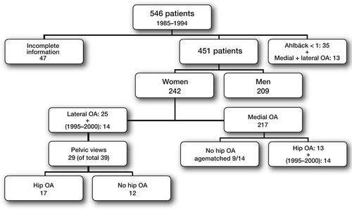
We identified 990 patients operated with knee arthroplasty or proximal tibial osteotomy at two hospitals (Varberg and Halmstad) 1985–1994. For each patient, 3 age- and sex-matched controls were identified. Information from 1970 about socio-economic grouping and work was obtained from the National statistical register (FoB 1970, Statistics, Sweden). In patients at one of the hospitals (Halmstad, n=546), the presence of unilateral or bilateral disease could be recorded in 451 patients based on information in case records and re-evaluation of radiographic examinations ().
Figure 1. There were 25 women with lateral OA (11 unilateral, 14 bilateral). Further 14 consecutive women operated during 1995–2000 with lateral OA (6 unilateral and 8 bilateral) were included to increase the number of observations. In the primary material we identified further 13 women with medial OA, who also had been operated because of OA of the hip. Further 5 consecutive patients with hip OA and medial OA of the knee from the period 1995–2000 were added to increase sample size resulting in a total of 27 cases to be studied (9 without and 18 with simultaneous hip and knee OA).
Study 5
Presence of bilateral knee OA was known in 451 patients from study 4 and were included in this study. Sequence of operations (left/right), presence of unilateral/bilateral and medial/lateral OA were recorded. The presence of OA was defined as joint space narrowing exceeding 50% (Ahlbäck Stage 1 or higher; Ahlbäck Citation1968). Bilateral OA was defined as the presence of at least Grade 1 OA on both sides. Lateral OA on both sides was only found in women. Therefore, we restricted our study to female sex (n=242), so as not to be biased by anatomical gender differences. Demographic data and grouping of this material is presented in () and ().
Table 1. Demographic data (Study 5)
Pelvic radiographs of 14 women admitted during 2000–2001, because of failed femoral neck fracture on the left side, were used as controls to measure hip and pelvis distances and hip angles on the intact side (right side).
Study 6
15 women with lateral (unilateral/bilateral: 9/6) and 15 women with medial (unilateral/bilateral: 4/11; Fisher's Exact Test: p=0.1) OA of the knee were identified on the waiting list for knee prosthesis surgery. Fifteen healthy women without any history of knee/hip pain or knee/hip trauma acted as controls (, page 25).
Study 7
Patients with lateral OA on the waiting list for total knee prosthesis surgery were asked to participate. During a period of 18 months, 5 patients (4 women and 1 man; 70 (62–74) yerars, Ahlbäck 3; (3–4) of 10 agreed to participate. The intact knee in 11 patients (8 men and 3 women; 26 (16–41) years) with anterior cruciate ligament rupture in the opposite knee acted as controls.
Methods
Study 1–3
The resected parts of the distal femur and proximal tibia were collected during operations with tricompartmental Freeman-Samuelson total knee replacements (Zimmer, USA – former Sulzer Company, Switzerland). Specimens were collected consecutively based on the willingness of the individual surgeons to participate. Standard instrumentation was used with intramedullar guide on the femoral and extramedullar guide on the tibial side. In all cases the cut could be placed a few millimetres below the level for the location of the most worn part of the medial or lateral compartment. The tibial part of the joint could always be obtained in one piece. The cartilage/bone pieces were marked for orientation and were stored at −70°C.
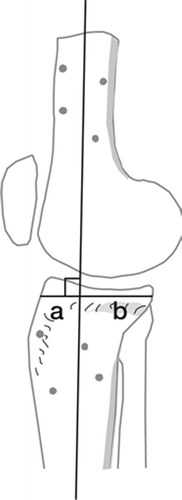
Analysis of joint area morphology (, page 19) was performed after a median time of 6 months (maximum 12 months). When the pieces had thawed the medial and lateral parts of the femoral and tibial joint area were divided into 6 squares. In each of the 4 articulations a line was drawn from anterior to posterior in the middle of each area. Further two lines perpendicular to the first one separated each articulation into six regions of almost equal size. The degenerative changes were divided into 5 Grades: no visible changes (0), cartilage fibrillation (1), cartilage destruction without visible bone (2), bone without cartilage and no or minor attrition of bone (3) and obvious attrition of bone (4). In regions with varying types of degeneration, the dominating type of lesion was reported.
To study interobserver reliability two observers independently graded the cartilage/bone pieces from the femur and tibia in 11 patients. One of these observers was not informed about the purpose of the study. The specimens were oriented in a standardized way by marking the medial and lateral side. This marking was done during the operation and by the surgeon. The wear in each region was recorded using a standardized template printed on a paper. The observer estimated each region using the central line. Because of individual variations in the configuration of the removed articulation areas a standardized transparent template could not be used. Intraobserver variability could not be studied in an unbiased way because of dehydration of the specimens. 2–3 hours after thawing the specimens showed macroscopic changes, which made proper evaluation impossible.
Study 2–3
All measurements on radiographs were done with a magnifying lens (×10) fitted with 0.1 mm divisions. The minimum joint space width was measured on 3 radiographic examinations (weight-bearing, HKA, experimental set-up). The degree of magnification was evaluated by exposure of a steel ball (Ø = 10.0 mm). All values were corrected for magnification (6% on standing radiographs, 14% on HKA radiographs and 14% in the experimental set-up).
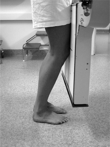
Weight-bearing radiographs were performed in all patients median 4 (1–29) days before surgery.The patients were standing with equal weight on both legs, and with their knees semi-flexed using the standard technique employed at the Department of Radiology, Halmstad hospital (). The patients were told to stand with their toes approximately at a vertical line from the roentgen film and flex their knees until they touched the film cassette. The amount of knee flexion was estimated at 20–30 degrees.
Figure 2. Weight-bearing radiographs with knees in semi- flexed position.
Joint width measurement. The width of the affected joint was measured according to Buckland-Wright et al. (Citation1994). On the femoral side we used the distal convex margin of the condyles. On the medial tibial side we used a line extending from near the tibial spine to the medial or outer margin, across the centre of the floor of the articular fossa in the midcoronal plane of the joint. This line was defined by the superior margin of the bright radiodense band of the subchondral cortex, and appeared below the anterior and posterior articular margins of the tibial plateau. On the lateral side we used the proximal margin of the articular surface, defined by the superior margin of the bright radio dense band in the subchondral cortex extending from near the tibial spine to the lateral or outer margin.
Study 3
A standing radiographic view of the whole leg with full extension of the knee (Hip-Knee-Ankle or HKA examination) was exposed at the same time as the semi-flexed AP view. This image was used to evaluate the joint space at full extension.
Testing equipment. The bone/cartilage pieces were thawed and mounted anatomically in a jig (Plexiglas box) () adapted to the equipment used at our mammography laboratory. High density mammography films were used to obtain maximum resolution. The size of the box corresponded to the size of the envelopes to roentgen films to facilitate reproducible positioning of the specimens and constant distances between the xray tube, the object and the roentgen film.
Figure 3. Experimental setup. Radiographic examination of specimen during a continuous force of 330 N.
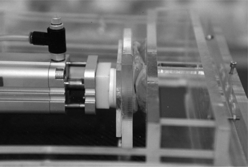

A pneumatic piston designed for industrial use was employed. Its performance was controlled and found to be correct within ±0.2%. This device produced a constant pressure of 400 kPa = 4 Bar on a piston with a diameter of 32 mm and act with a pressure of 324 N (approx equivalent with the force acting from a weight of 33 kg). The femoral bone pieces were allowed to slip into the deepest portion of the tibial plateau. An anterior-posterior view of the specimen was exposed.
Study 4
The socio-economic grouping and type of work were recorded for each patient using information from Statistics Sweden (SCB, Statistiska centralbyrån) collected in 1970 (FoB 1970). Socio-economic grouping is constructed by Statistics Sweden based on type of work, education and income. It is classified into 10 groups in terms of e.g. self-employed farmer, farm worker, industry businessmen and entrepreneur in other activity, employed businessmen, office staff, laborer, service employees, military personal and unidentified work.
Work is further differentiated and embraces several hundreds of different items coded into a number. A number of occupations have been identified (Vingård et al. Citation1991) (, page 22), which theoretically could be associated with increased loading of the knee. Those occupations have been condensed into 17 groups, each representing a number of occupations, where the knee is supposed to be exposed to an equal amount of load. All other works could be classified as a “low-risk” group according to Vingård et al.
In our material most persons were (or had retired as) office workers (27%), blue-collar workers (25%) or farmers (13%). The high-risk-groups (nr 1–17) included mainly farmers (group nr 1, n=136) or building workers (group nr 7, n=57).
In the subgroup of 451 patients any relationship between gender, uni-/bilateral OA involvement and work was studied.
Study 5 ()
All pelvic views were exposed supine with a fixed patient-film distance resulting in a standardised magnification of about 20%. At the radiographic evaluation we did not correct for magnification since only relative lengths and angles were measured. A small pillow was placed beneath the knees to obtain as standardised position of the pelvis as possible. All measurements were made using templates, pencil, set square and a ruler.
Figure 4. Reconstruction of angles and distances reconstructed in the pelvic and hip regions.
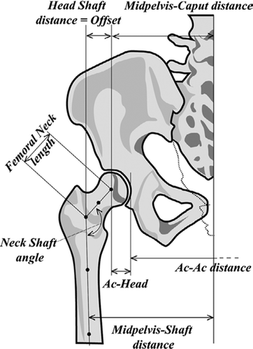
Hip angles. Two lines were constructed, one in the centre of the femoral neck and one in the centre of the femoral shaft. These lines were used to construct the neck shaft angle. The distance between the centre of the femoral head and the point where these two lines intersected represented the femoral neck length (femoral neck length). The horizontal distance between the femoral head centre and the line through the centre of the femoral canal represented offset (head–shaft distance).
Pelvic distances. Three distances were measured. The first was the distance between the medial border of the right and left acetabulum (Ac–Ac distance). The second distance corresponded to the sum of the Ac–Ac distance divided by 2 added to the distance between the medial border of the acetabulum and the femoral shaft (midpelvis–shaft distance). The third distance was the sum of the Ac–Ac distance divided by 2 and the distance between the medial border of the acetabulum and the centre of the femoral head (midpelvis–caput distance).
To estimate the length of the lever arm a functional offset was calculated. This parameter was defined as the quotient between the midpelvis– caput distance and the distance between the femoral head centre and a central line in the proximal femur (offset).
Study 6
6 infrared cameras recording at 240 Hz (MCU 240 Qualisys Medical AB, Göteborg, Sweden) were used. The camera system was calibrated to a measurable volume of 9.2 m3 (2 m × 2 m × 2.3 m). 18 reflective spherical markers were attached to the skin over bony landmarks (acromion, 12th thoracic vertebra, sacrum, anterior superior iliac spine, lateral knee joint line, proximal to the superior border of the patella, tibial tubercle, heel, lateral malleolus and between the second and third metatarsals). Markers were placed bilaterally. The position of each marker was detected by the system corresponding to a resolution of 0.2 mm. We used a modification of the Helen Hayes marker set (Davis et al. Citation1991), which has been used previously by our group (Saari et al. Citation2004a, Saari et al. Citation2004b, Saari et al. Citation2005b). The positions of the markers were partly chosen to limit the effect of subcutaneous fat thickness.
The room coordinate system was defined with one axis pointing in the direction of walking and parallel to the floor. Its direction was thus based on the first and the last “frame” in each test (i.e. the first to the last position in the gait cycle). The next axis was perpendicular to the first one and the floor.
The relationship between the marker positions on the skin surface and the joint centres was based upon data published by Vaughan et al. (Citation1992). Thus, the joint centres of interest were mathematically reconstructed based on marker positions. The obtained laboratory or technical coordinate system was transferred to a clinical coordinate system, which was aligned to palpable or indirectly identified skeletal landmarks within each body segment, i.e. hip, thigh, shank and foot (Davis Citation1997).
This implied that each segment had its own individual coordinate system. The marker set attached to the pelvis (sacrum and 2 markers on each of the two anterior superior iliac spines) was used to calculate the hip joint centres, as well as to align the coordinate system to the pelvis. The markers on the shank were used for detection of the knee joint as well as the ankle joints. The remaining body segments were defined using the computed joint centres as landmarks and as follows; the foot segment was identified using the heel and toe markers and the centre of the ankle joint. The anterior-posterior axis of the foot segment was pointing from the heel marker towards the toe marker. The shank segment was defined by the reconstructed knee and ankle joint centres with the longitudinal axis pointing from the ankle to knee joint (positive direction).
The reconstructed centres of the knee joint, the hip joint and the marker placed on the superior border of patella, determined the thigh segment. The thigh segment had its longitudinal axis pointing from the knee joint centre to the hip joint centre (positive direction). The anterior-posterior axis was perpendicular to the longitudinal one and passed through the suprapatellar marker (positive anterior direction). The transverse axis was perpendicular to these axes with positive medial direction. Corrections were made for marker offset, i.e. the distance from the centers of the markers to the skin surface. Zero degree position (start position) was defined when the patient is standing upright. In this position the coordinate systems are fixed to the different segments before any motion is initiated. The motion is presented as relative motions of the distal segment using the proximal segment as a fixed reference. The description of moments is calculated in the same way.
Two force plates (Kistler 9281C, Kistler Instruments AG, Winterthur, Switzerland) were used to record ground reaction forces during level walking. Eight piezoelectric sensors in each force plate recorded forces 3-dimensionally.
The ground reaction forces were calculated from the input of the 8 channels of each force plate. The standard equations provided from Kistler™ were used to compute force vectors, the moment about the normal axis of the force plate surface, and the location of the ground reaction force vector. The force vectors were then transferred from the internal coordinate system of each force plate into the global coordinate system for calculations of joint moments.
The joint moments were calculated using an inverse dynamics approach. The mass of each segment is calculated as a percentage of body weight. The moments over each joint were expressed as applied to the distal segment of each joint. The data from the 6 cameras and the forces were recorded synchronically.
Recordings of motion were achieved with the software, QtracC™ version 2.51 (Qualisys Medical AB, Göteborg, Sweden). Reconstruction from 2-dimensional into three-dimensional data was made with QtracV™ version 2.60 (Qualisys Medical AB, Göteborg, Sweden). QGait 2.0™ (Qualisys Medical AB, Göteborg, Sweden) was used for calculations of rotations in relation to the three cardinal axes.
The patients and the healthy controls were asked to walk without targeting on the force plates at a self-selected speed. Several trials preceded the actual measurements to define a start line, which facilitated stepping on both force plates according to the step length of the individual patient. Patients had the opportunity to test the walkway several times before recording was done. When patients felt that they were familiar with the situation and were able to perform the test without targeting the force plates, two measurements were completed. The most representative measurement was chosen for further analysis. This selection was made visually by comparing the walk tests prior to recording and the two recorded tests. In this study we evaluated flexion/extension, adduction/abduction and external/internal rotation of the hip joint (thigh vs. pelvis) and knee joint (shank vs. thigh). The 3 dimensional angles were calculated according to the Euler-angle method (Kadaba et al. Citation1990, Davis et al. Citation1991).
Internal moments are presented. Flexion, internal rotation and adduction have positive values. Moments were normalized to body weight (Nm/BW). The time between two consecutive heelstrikes was normalized for inter-subject comparison. Data were transferred into SPSS 11.5 (SPSS Inc. Chicago, Illinois, USA) for statistical analysis.
Information from both the right and left side were collected. Each patient contributed with one knee; the side with symptoms or in cases with bilateral symptoms the one with most symptoms. When comparing the left and right side in controls we found no differences. In this study only the right side was used.
Study 7
Our set-up for dynamic radiography during active extension of the knee has been presented previously (Kärrholm et al. Citation2000, Uvehammer et al. Citation2000b, Brandsson et al. Citation2001, Saari et al. Citation2005a). 2 to 4 weeks before the examination, spherical tantalum markers (0.8 mm) were inserted using local anaesthesia. 4 to 8 markers were placed in the proximal tibia and the distal femur respectively. In the controls, markers were inserted bilaterally during a preoperative arthroscopy of the injured knee.
To obtain a standardized starting position a pair of stereoradiographs were exposed supine at 0° extension with the knee inside a biplanar cage (RSA Biomedical, Umeå, Sweden). At this exposure the tibia was aligned with the longitudinal axis of the cage and the posterior cortex of the femoral condyles were positioned to project over each other. This meant that the transverse axis of the cage, which defined the reference coordinate system, was parallel with the posterior condylar plane.
At the recordings of the knee kinematics at active extension the patients mounted a 16 cm high platform after several trial procedures. The goal was to obtain a standardized speed during a period of 3–4 seconds. The exposures were done with two x-ray tubes designed for simultaneous and continuous exposures. They were placed at a 90° angle in relation to each other. We used a speed rate of 2–4 frames/s. The patients and controls started the active extension at median 57° flexion and ended at 2° hyperextension. During this motion 7 to 13 (median 10) exposures were available for analysis. Observations from all subjects were available between 45° and 15°. The median mean errors of rigid body fitting (marker stability indicator) and condition numbers (marker scatter indicator) were 0.14 and 0.09 mm (range 0.05–0.26, 0.05–0.22) and 83 and 82 (range 29–136, 39–166) for the femoral and tibial markers, respectively.
We used film-exchangers designed for conventional roentgen films. The films were scanned and measured digitally using our standard equipment (Bragdon et al. Citation2002, Bragdon et al. Citation2004).
At the evaluation we recorded the relative tibial rotations using the femoral markers as a fixed reference. Translations of the flexion facet centres in the medial and lateral femoral condyles and of a point localised in between the two tips of the tibial eminence were measured using the “point transfer technique” (Brandsson et al. Citation2001, Saari et al. Citation2005a). In order to reduce the amount of data, we focused on 7 parameters. Relative tibial rotations with fixed femur were recorded as flexion (+) / extension (−), internal (+) / external (−) rotation, varus (+) / valgus (−) angulation. Recordings of tibial translations were restricted to anterior (+) / posterior(−) motions of a midpoint between the two tips of the tibial intercondylar eminence relative to a fixed femur. We also recorded femoral translations with the tibia fixed at the flexion facet centres of the two condyles. Proximal (+) / distal (−) and anterior (+) / posterior (−) translations are accounted for.
To evaluate the relative tibial position at the reference (starting) position corresponding to the stereoradiographic examination at 0° we used the lateral view according to Nilsson et al (Nilsson et al. Citation1991). The quotient a/a+b x 100 () did not differ between the study group and controls (lateral OA: 23, 0–42; controls: 22, 10–25, median, range, p=0.8).
Figure 5. The relative anteroposterior position of the tibia was measured on the lateral view of the reference position. The reconstructed central femoral line divided the tibia into one anterior (distance a) and one posterior (distance b) part. The quotient a/a+b x 100 was computed.
In a previous study in the same laboratory (Uvehammer et al. Citation2000b) the repeatability (1 SD) between two series in the same patient was found to be 1.6°–2.3° and 1.2–2.2 mm. This is about up to ten times the resolution of radiostereometry and means that despite a number of trials the patients were not be able to exactly reproduce the same knee motion between two step ups.
Statistics
Study 1
Different patterns of cartilage wear in medial and lateral gonarthrosis. Friedman's test and Mann-Whitney U-test were used. Values are median and range. The weighted kappa was calculated to determine interobserver reliability.
Study 2
Standing radiographs underestimate joint width: comparison before and after resection of the joint in 34 total knee arthroplasties. Non-parametric tests (Wilcoxon sign rank test, Kendall tau) were used.
Study 3
Ahlbäck grading of knee osteoarthritis. Reproducibility and validity based on visual inspection of the joint. The concordance between the different observations and observers regarding the radiographic classification was evaluated using Kappa statistics. In addition the sensitivity and specificity were determined using visual inspection of the resected joint as reference. Cases with medial and lateral OA were studied separately.
Study 4
Working conditions resulting in increased need for surgical treatment of knee OA. Independent-sample t-test, Chi-Square, logistic and linear regression analysis (SPSS 10.0.5 for Windows, MedCalc 5.00.019) were used.
Study 5
Medial and lateral osteoarthritis of the knee is related to variations of hip and pelvic anatomy. When comparing the values between the left and right sides, Wilcoxon Signed Ranks Test, and for demographic data Mann Whitney U test were used. The values are presented as median and range. For all other statistics T-test and Fisher's Exact test were used and values are presented as mean and SD.
Study 6
Hip and knee joint rotations differ between patients with medial and lateral knee osteoarthritis. Gait analysis of 30 patients and 15 controls. Non-parametric tests were used. The two groups of patients with medial and lateral OA and the control group were analyzed using non-parametric ANOVA (Kruskal-Wallis test). If this test revealed the presence of a difference, further analyses between the groups were done using Mann-Whitney test. To reduce the risk of spuriously occurring significances, the significance level was set at p < 0.025.
Study 7
Changes of knee joint kinematics caused by lateral osteoarthritis. The observed motions were interpolated at 5° intervals in flexion/extension. Observations were collected at 10° to 50°. Mann-Whitney U-test was used for comparison at 45° and 15° of flexion. P-values less than 0.05 were regarded as significant.
Results
Study 1– Different patterns of cartilage wear in medial and lateral gonarthrosis
Studies on 11 tibial plateau specimens by 2 observers revealed complete agreement in 55%, difference with 1 Grade in 43% and with 2 Grades in 2% of the 132 regions studied (weighted kappa = 0.52).
Tibia
The most pronounced changes in medial OA were located peripherally in the central part (centralperipheral- medial: cpm) followed by the anterior part (apm) (median values, cpm: 3.0, 2–4; apm: 1.0, 0–4; p<0.001, Friedman's test including all regions medially) ().
Figure 6. Grading of cartilage destruction/ bone attrition in medial and lateral arthrosis of the tibia plateau. Median values and range. Template used to separate the 6 different areas on the tibia plateau is illustrated above. The darker the area the more pronounced the degenerative changes observed (median value). a = anterior, c = central, p = posterior in first position, peripheral in second position, m = medial, l = lateral.
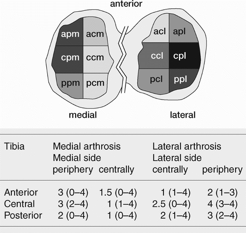

In lateral OA the most degenerative changes were also located in the central-peripheral region (cpl: 4.0, 3–4). The next region in term of severity of the changes was located posteriorly (ppl: 3, 2–4; p<0.001, Friedman's test including all regions laterally).
Medial compartments in medial OA vs. lateral compartment in lateral OA: There were more degenerative changes in the anterior part in medial OA compared to lateral OA (regions apm vs. apl: p=0.02, Mann-Whitney U-test). In lateral OA there were more degenerative changes both peripherally and centrally than in medial OA (regions pcm vs. pcl: p=0.03; ppm vs. ppl: 0.001, Mann-Whitney Utest). The attrition of bone tended to be more pronounced in the peripheral and central region in the group with lateral arthrosis (cpm vs. cpl: p=0.04).
Femur
In medial OA the degenerative changes were rather evenly distributed (p=0.3, Friedman's test including all regions medially). In lateral OA, the central and posterior regions showed more pronounced changes than the other regions (p=0.001, Friedman's test including all regions laterally).
Comparison between the affected sides in both groups on femur: No obvious differences were seen (p=0.07–0.7, Mann-Whitney u-test).
No difference were found between men and women either on the tibia or the femur in medial OA (p=0.07–0.9, Mann-Whitney u-test). Any influence of gender could not be studied in lateral OA, because of too few observations (1 male). On the non-affected sides there was a macroscopically intact cartilage layer in all cases (median values = 0).
Study 2 – Standing radiographs underestimate joint width: comparison before and after resection of the joint in 34 total knee arthroplasties
The intra- and interobserver variability on the films on standing radiographs were ±0.20 mm (1 SD) and –0.25 ±0.40 mm (mean difference ±1 SD). The intraobserver variability on the films from the experimental set-up was 0.54 mm (1 SD).
Evaluation of test equipment. When the 2 series were compared the compartment with degenerative changes showed smaller variability than the opposite with no or less degenerative changes (nonaffected) using the same or different loads. Within the ranges of loads used the magnitudes of the errors were the same as the intraobserver variability ().
Table 2. Difference in joint space width (mm) between measurements using different and same loads.
Standing radiographs vs. experimental set-up. On the affected side 17 cases had a joint space width less than 1 mm on standing radiographs, 10 cases had 1–2 mm and 7 cases more than 2 mm.
Medial OA: On the medial affected side weightbearing radiographs revealed a wider joint width than in specimen (median difference=0.3 mm, p=0.05). In 10 of 22 cases the absolute difference exceeded 0.5 mm, with a wider joint line in 8 of these cases. On the nonaffected lateral side the difference was larger (median difference=1.8 mm, p<0.001).
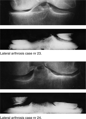
Lateral OA: Standing radiographs also overestimated the joint width on the affected lateral side (0.2 mm, p=0.04). In 4 of 12 cases the absolute differences exceeded 0.5 mm, and in 3 cases the joint space width was 1.3 to 2.9 mm wider than observed on the radiographs of the specimens (). On the nonaffected medial side there were a wider joint space on clinical radiographs in all but one of the 12 observations (median difference: 0.4 mm, p=0.003).
Figure 7. Standing radiographs and corresponding radiographs from experimental setup.
The differences in joint space width tended to vary more in cases with lateral arthrosis (95% CI: lateral: 0.1 to −1.2 mm; medial: 0 to −0.5 mm). ()
HKA radiographs vs. experimental set-up. HKA radiographs overestimated the joint width on the most worn side of the joint compared to the experimental set-up (median 0.5 mm in medial; p=0.002 and 1.5 mm in lateral OA; p=0.01).
HKA vs. standing radiographs. In the compartment with the most pronounced changes radio-graphic examination on the extended knee (HKAradiographs) showed median 0.5 mm wider joint than on standing radiographs in cases with medial OA (p=0.02). The difference was more pronounced in cases with lateral OA (1.1 mm, p=0.02; medial vs. lateral differences: p=0.05) ().
Table 3. Comparison of joint width (mm) between HKA (Hip-Knee-Ankle, knee in full extension) and standing radio-graphs (with semiflexed knee) and between standing radiographs and radiographs of specimens
In the compartment with no or less pronounced changes the measurement on the HKA radiographs did not differ from those obtained in the semi flexed position in either medial or lateral arthrosis (p≥0.2).
Study 3 – Ahlbäck grading of knee osteoarthritis. Reproducibility and validity based on visual inspection of the joint
Ahlbäck classification
The intraobserver reproducibility when one of the 4 observers classified the radiographs 2 times, varied from high to comparatively low (0.15), but was in most instances acceptable or high (kappa: medial OA: 0.15–0.65, mean 0.46; lateral OA: 0.59–0.76, mean 0.67).
The interobserver variability was consistently low (combined kappa for all 4 observers: medial: 0.11; lateral: 0.12).
The sensitivity (comparison between radiographs and visual inspection of resected bone ends) was acceptable both in medial (67–95%) and in lateral (50–86%) OA.
The specificity for most comparisons was lower (medial: 11–67%; lateral: 25–75%).
Bone attrition (radiographic diagnosis vs. visual inspection of resected bone)
In medial OA the sensitivity to detect any tibial and femoral attrition varied between 53–94% and 63–95%, respectively. The corresponding variation of specificity for the different combinations of observers varied between 8–77% (tibia) and 36–73% (femur).
In lateral OA the sensitivity to detect tibial and femoral attrition varied between 38–92% and 25–87% (only one observer had higher value than 50%). The specificity varied between 40–80 % (tibia) and 20-60 % (femur) ().
Table 4. The two values for sensitivity and specificity refer two first and second observation, respectively
Study 4 – Working conditions resulting in increased need for surgical treatment of knee OA
Sex, age and civil status. In the total material more female (n=540) than male (n=446) patients had received surgical treatment because of OA (p= 0.002). The male patients were at an average 3.6 years younger age than the females: 65 (11) vs. 69 (8.6) years (p<0.001). In the Hospital A material (256 men and 286 women) the difference was slightly higher: 66 (12) vs. 70 (8.6) years (p<0.001). This was mainly an effect of lower average age in men operated with osteotomy. The mean age in men (n=137) and women (n=217) operated with uni-, bi- or tricompartmental prosthesis was the same: 73 (6.2) vs. 73 (5.9) years.
Work. 262 patients (638 controls) belonged to high-risk groups (). In two groups (farmers, farm workers and building-, piping-, bricklayer-, metal workers) we found statically significant differences. People who in 1970 were reported to work in a farm (RR: 1.7, p=0.001) or as building worker group (RR: 1.4, p=0.047, ) were 15 to 24 years later more often subjected to surgical treatment of their knee OA. However, the surgical intervention was obtained at a higher age in farm workers than in those who had other types of job: 69 vs. 67 years (p=0.003)
Table 5. The number of patients and controls based on figures reported to Sweden Statistics in 1970. For each patient, 3 age- and gender-matched controls were identified by Sweden Statistics.
Table 6. Observed risk ratio to receive surgical treatment of knee arthrosis in relation to occupation in 1970
Socio-economic grouping (). Overall, patients who in 1970 had been employees in an office work or as businessmen had a lower risk to be operated due to knee OA (RR: 0.7, p=0.002). Farmers who owned their farm had an increased risk (1.4, p=0.004), whereas employees in a farming enterprise did not show any overrepresentation of surgical procedures (RR: 0.9, p=0.6).
Table 7. Observed risk ratio to receive surgical treatment of knee arthrosis in relation to socioeconomic belonging in 1970.
Uni- vs. bilateral OA. There were 20% more women than men with bilateral OA (p<0.001). Men treated with surgery because of knee OA had about 3 times more often unilateral OA (33/199) than had women (11/217) (p<0.001). In a logistic regression analysis with uni- and bilateral OA as a dependent variable we found a stronger correlation with gender (p<0.001) than high-risk occupation (p=0.7). Unilateral OA tended to be more common in building workers than in the other occupational groups (RR: 3.2, CI: 0.96–11, p=0.06). A corresponding analysis comparing all high risk groups (14/102) with low risk groups (13/145) did not reach statistical significance (RR: 1.5, CI: 0.76–3.0, p=0.2).
Study 5 – Medial and lateral osteoarthritis of the knee is related to variations of hip and pelvic anatomy
Intra-interobserver variability. 17 radiographic examinations of the pelvis were used. Intraobserver variability for the neck shaft angle was 1.7° (SD), and for the femoral neck length and head–shaft distance 4.2 and 3.1 mm, respectively. The interobserver error (mean (1 SD)) for neck shaft angle, femoral neck length and head–shaft distance was–0.3 (2.4°), 1.1 (4.5) mm and 1.6 (2.2) mm.
The intraobserver variability for Ac-Ac distance and midpelvis–shaft distance was 1.8 and 3.2 mm. The interobserver error (mean (1 SD)) for the same distances was –1.0 (6.6) and –0.2 (3.5) mm.
Left vs. right side (). The femoral neck length was 4 mm longer (p=0.03) and the head–shaft distance was 3 mm longer (p=0.03) on the right side in patients with lateral OA and the head–shaft distance 1 mm longer in medial OA (p=0.03). Therefore, further comparisons between patients with medial and lateral OA were restricted to the right side.
Table 8. Anatomic hip angles and distances. Comparison between right and left side.
OA of the hip. In the first series of patients, operated during1985–1994, those with lateral OA had been operated more often with hip arthroplasty (13 of 25) than those with medial OA (13 of 217) (p<0.001).
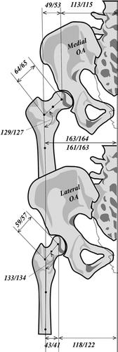
Hip angles and distances (). Patients with medial knee OA (without hip OA) revealed a greater offset than controls (+11.4 mm; p=0.005), but there was no difference in pelvis width. The femoral neck in patients with medial OA was also longer (64 vs. 53 mm; p=0.001).
Table 9. Pelvis and hip distances and angles. Lateral and medial arthrosis of the knee with and without coxarthrosis versus controls. Hip values on the right side (missing observations are due to discarded radiographs)
Patients with lateral knee OA (without hip OA) did not differ in offset (+4.7 mm, p=0.11) from controls, but had a wider pelvis (Ac-Ac distance + 13.7 mm; p=0.001). The femoral neck was longer than in controls (59 vs. 53 mm, p=0.02) but 5 mm shorter than in patients with medial knee OA (p=0.02).
The wider pelvis in lateral knee OA was compensated for by shorter offset on the AP radiographs resulting in about equal shaft-to-shaft distances (midpelvis–shaft distance) in the two groups (lateral vs. medial OA of the knee: −1.6 mm, p=0.7) ().
Figure 8. Schematic view of results presented as median values without/with hip OA. Observations at the hip region in patients with medial (top) and lateral (bottom) OA of the knee. In this example the midpelvis-shaft distances are the same. At the top the pelvis is more narrow, which has been compensated for by higher femoral offset. At the bottom the patient has a wide pelvis, but coxa valga resulting in a smaller femoral offset.
The influence of hip OA. With hip OA, the difference in offset between the medial and lateral knee OA groups increased to 12.2 mm (p=0.01), and the difference in pelvic width (Ac-Ac distance) decreased to 6.3 mm (p=0.15; medial OA: +3.9 mm, lateral OA: −2.0 mm).
Compared to cases without hip OA, the femoral neck angle decreases in the medial knee OA group with 2.2° and increased in lateral OA group with 1.2°. Comparison between the 2 groups revealed a difference of 7.8° (p=0.008).
In lateral knee OA the calculated lever arm (offset/midpelvis–caput distance) was 19% smaller in patient without and 30% smaller in patients with OA of the hip compared with medial knee OA (p=0.02 and 0.02). No differences were found compared to controls (p<0.3).
Study 6 – Hip and knee joint rotations differ between patients with medial and lateral knee osteoarthritis. Gait analysis of 30 patients and 15 controls
Walking speed and stride length (). There was a tendency to reduced speed in the two patient groups (p=0.03). The stride length was shorter in medial OA (p=0.001) and lateral OA (p=0.02) than in controls. These parameters did not differ between the two patient groups (p=0.8/0.7).
Table 10. Demographic data (Study 6), values are median (range).
Flexion/extension – hip (). Both patient groups had smaller maximum hip extension angle than controls. In the group with medial OA it was reduced with 5° (p=0.02) and in the lateral OA group with 11° (p=0.008). The flexion angles did not differ (p=0.8).
Table 11. Hip or relative femoral motions and hip angle and moments in all three study groups. Maximum values are presented, and adduction motion at 40% of gait cycle (midstance). Values are median (range)
Table 12. Knee motions (tibial motions relative a fixed femur) and moments for all three study groups. Maximum values are presented and rotation at 40% of Gait Cycle (midstance). Values are median (range)
Table 13. Statistical overview for all parameters in the total study popula-tion. Mann-Whitney U Test was used for comparison between the individual groups in pairs. Two digits are used for values below 0.05.
In the early swing phase the peak flexion moment was higher in controls than in patients with medial OA (p=0.004). A similar tendency was observed in cases with lateral OA (p=0.03).
Flexion/extension – knee. Decreased flexion was shown in lateral knee OA (5°; p=0.006), but not in medial knee OA (8°; p=0.03), but both groups walked with less maximum knee extension (4–5°; p=0.004) than controls. The knee extension moments were smaller in both patient groups (p<0.001), but their flexion moments did not differ from normal (p=1).
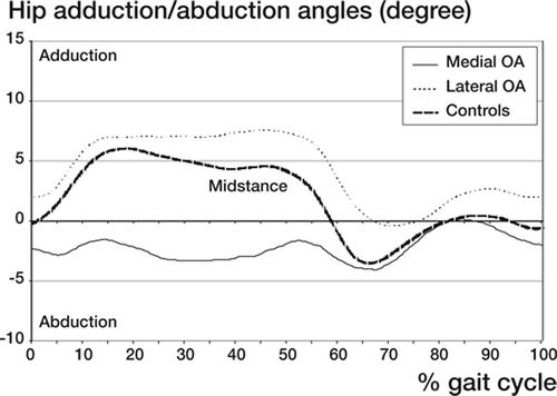
Adduction/ abduction – hip. At midstance patients with lateral OA had 2° more adduction of their hip joints (p=0.02), and patients with medial OA 7° more abduction than controls (p<0.001), but at the end of the cycle (past 60%) the pattern became closer to normal ()
Figure 9. Mean value of hip or relative femoral adduction/abduction angles related to a standardized gait cycle in women with medial or lateral OA and in the controls (normal). Data represent mean values based on division of the gait cycle into 200 intervals for each patient, independent of the individual time period for each cycle. In the figures the mean value for each of the 3 groups (n = 3 x 15) at each time interval is presented. Thus, each curve is based on 200 subsequent mean values. No further filtering of data was done.
Patients with OA walked with reduced abduction moments throughout stance. The maximum values were reduced in medial OA by 13% (p=0.007) and lateral OA with 8% (p<0.001). The minimum values (adduction moments) did not differ.
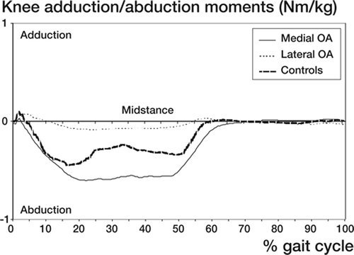
Adduction/ abduction – knee. In controls the maximum adduction angle reached 4° and the maximal abduction angle 3°. In medial OA the knee was in 8° more adduction (max value) through the gait cycle, and in lateral OA in 8° more abduction (max value) than in controls.
The maximum internal knee abduction moments were 52% higher in medial OA (p=0.009) and 63% less in lateral OA (p<0.001) than observed in the controls ().
Figure 10. Mean value of knee or relative tibial adduction/ abduction moments related to a standardized gait cycle in women with medial or lateral OA and in the controls. See also legend to .
Rotations – hip. In patients with lateral OA the femur was positioned in about 7° more external rotation (maximum value, p=0.01) than in controls, but seemed to otherwise follow the variations observed in the normal hip during the gait cycle. Patients with medial OA did not differ from controls (p=0.8) ().
Figure 11. Mean value of hip or relative femoral internal/ external rotation angles related to a standardized gait cycle in women with medial or lateral OA and in the controls. See also legend to .
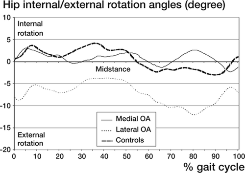
The internal rotation moment in lateral OA was greater than in controls (p=0.021). The outward rotation moments were small without difference (−0.2 to 0.1 Nm/Kg; p≥0.04).
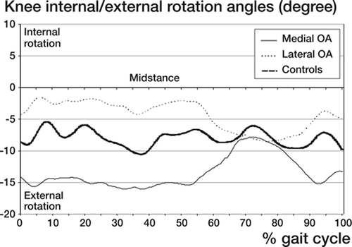
Rotations – knee. At midstance, cases with medial OA had 9° more external tibial rotation than controls and cases with lateral OA 6° less. These differences did not reach significance (p<0.06), but comparison between the OA groups revealed a significant difference (p=0.001) (). The moments were small and did not differ (p≥0.08).
Figure 12. Mean value of knee or relative tibial internal/ external rotation angles related to a standardized gait cycle in women with medial or lateral OA and in the controls. Rotation of tibia vs. femur. See also legend to .
Study 7 – Changes of knee joint kinematics caused by lateral osteoarthritis
Rotations. At 45° of flexion the tibia had rotated inwards whether the knee had lateral OA or not. Both groups showed a tendency to valgus angulation during active extension compared to the supine reference position, without significant difference at 45° (p=0.3) (). With increasing extension knees with lateral OA tended to rotate into slightly more valgus, whereas the normal knees showed slight varus angulation resulting in a mean / median difference of about 3 / 4.5° at 15° of flexion (p=0.001) ().
Figure 13. Tibial rotations around the sagittal axis (varus/ valgus rotation) are illustrated. In the group with lateral osteoarthritis the tibia rotated into valgus as an effect of load-bearing. In both groups there were small changes during active motion, but in opposite directions. Numbers of observations are placed at the bottom. Mean and standard error of the mean (SE) are shown.
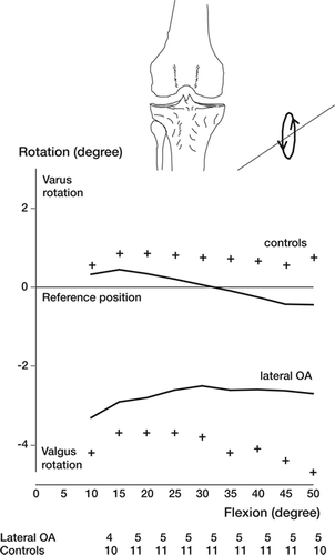
Table 14. Kinematic recordings at 15 and 45 degrees of knee flexion. Values are median (range)
Translations. The centre of the tibial plateau had displaced slightly more than 1 cm posteriorly at 45°. This displacement decreased to slightly less than 2 mm at 15° still without any difference between the two groups.
At 45° the medial flexion facet centre had displaced 3–4 mm distally in both groups. It moved proximally and to about the same amount with proceeding extension (p<0.7).
In both groups the internal tibial rotation resulted in a relative anterior displacement of the medial condylar centre. At 45° and in knees with lateral OA the medial condyle had translated 7–8 mm forwards. The corresponding displacement in normal knees was about 4 mm smaller (p=0.03) (). At 15° there was still a mean/median anterior displacement in the OA group, whereas normal knees showed a corresponding minimum posterior displacement (p=0.05).
Figure 14. Anteroposterior translations of the medial flexion facet center are illustrated. In lateral osteoarthritis the medial condyle had a more anterior position. Numbers of observations are according to . Mean and standard error of the mean (SE) are shown.
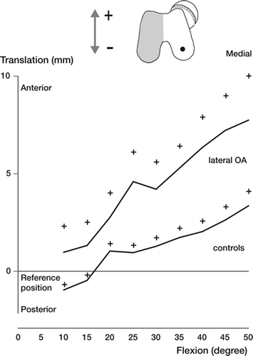
In both groups the lateral flexion facet centre had displaced distally at 45° (3–4 mm more in lateral OA than controls; p=0.05). In the group with lateral OA this distal displacement was 2–3 mm higher than observed on the medial side. In contrast, the control group was 2–3 mm smaller laterally than medially resulting in a difference of about 4–5 mm between the groups.
The lateral flexion facet centre translated in a proximal direction with proceeding extension in both groups (lateral OA/ controls; 1.2/ 0.6 mm), with a 3.2 mm more proximal position in lateral OA compared to controls at 15° of flexion (p=0.001) ().
Figure 15. Proximal-distal translation of the lateral condylar flexion facet center. The lateral condyle had a more distal position and especially up to 20° probably because of loss of cartilage and bone. Numbers of observations are placed according to . Mean and standard error of the mean (SE) are shown.
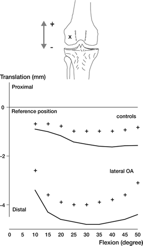
At 45° this centre had translated posteriorly mean / median −0.9 /-3.4 mm in the OA group and −4.5 / −2.7 mm in the controls (p=0.7). At 15° these positions had changed with about 1–2 mm, still without any differences (p=0.6) ( and ).
Figure 16. Anteroposterior translations of the lateral flexion facet center are illustrated. These motions were small without any substantial difference between the two groups. Numbers of observations are according to . Mean and standard error of the mean (SE) are shown.
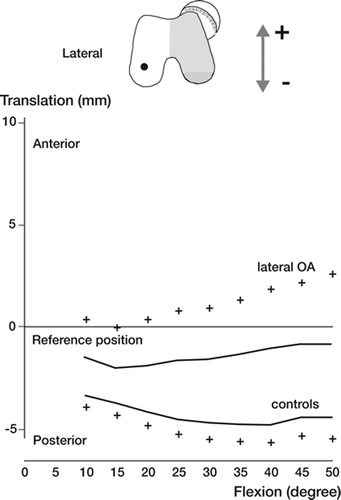
Figure 17. Reconstruction of the tibial plateau and the projections of the flexion facet centers between the reference position (x) and 45° (dot or square) are shown. The tibial plateau was reconstructed from CT data of a normal knee. In addition to data from control subjects and knees with lateral osteoarthritis (OA) the corresponding mean translations in 14 knees with medial OA (from Saari et al. 2005 are illustrated. As an effect of internal tibial rotation at 45° the medial femoral condylar center displaced anteriorly and the lateral one posteriorly (mean values) in control subjects and knees with lateral OA. In medial OA, the mean AP translations were small and in the opposite directions (posteriorly in medial compartment and anteriorly in the lateral one).
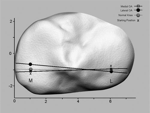
Discussion
The location of wear on the tibial joint surface may reflect the position of maximum load and the location of primary failure of the cartilage during the initiation of the disease. The locations of the wear in study 1 are consistent with our primary hypothesis with more anterior wear in medial OA and a more posterior wear in lateral OA. Findings of a stiffer bone in these regions also support these observations (Hvid and Hansen Citation1985). Vande Berg et al. (Citation2002) used multidetector spiral CT arthrography to study lesions of the femoro-tibial cartilage in patients with knee pain but without OA changes on conventional radiographs. They used a detailed grid with 16 areas and the cartilage defects found on the tibial plateau were located posterior on the lateral side and in the bare area (without meniscus) on the medial side. We chose to divide each articular surface into only 6 parts, because we thought that a more detailed division would jeopardize the reproducibility of this procedure. Tibial plateaus are more or less different, not only in size but also regarding other anatomical features and particularly if there are degenerative changes. Division of the articular surface into 6 regions was recently used in the previously quoted study by Moschella et al. (Citation2006), who compared degenerative changes in cases with and without old tears of the ACL.
Our observations may explain some of the difficulties to obtain an accurate diagnosis of OA and especially in cases with primary involvement of the lateral compartment. The degree of tension in the knee ligaments is probably important. When the tension of the ligament decreases the femoral condyle may more easily slip into the most worn part of the tibia. However, even if this often occurs it might be that the most worn part of the two joint surfaces does not contact because the instantaneous moment over the knee joint counteracts such a position. Unevenness of the surface or active avoidance of the patient to obtain such a position due to pain are further possible explanations. In study 2, the standing radiographs overestimated the joint space width, with an increased scatter in lateral OA, probably because the wear is often located in the posterior part on the femoral condyle (Boegård et al. Citation1997). This, together with a frequently occurring spherical wear on the tibial plateau implies that the most worn part is not visualized unless weightbearing radiographs are exposed in a more flexed position than usually practiced. In these instances the so called tunnel view has been recommended (Resnick and Vint Citation1980). This is consistent with our own clinical experience that flexion up to 50–60° can sometimes be necessary to visualize the location of most pronounced wear. Our experimental setup allows precise positioning, but could not completely address the need for a more flexed position. Use of different pressures in our experimental model had little if any influence on the recorded joint space width indicating that this factor did not act as a confounder within the ranges of load used. The clinical implication of this study is that present routines do not fulfil the need for exact diagnosis in lateral OA.
The observations in studies 1-2 explains some of the problems with the Ahlbäck classification documented in study 3. Also a previous study has shown that the Ahlbäck classification has poor inter- and intra-observer reliability and is dependent on the experience of the individual observer (Galli et al. Citation2003). Contrary to these authors our intra-observer variability tended to be somewhat better than the inter-observer variation. Overall, we could confirm that the consistency of the Ahlbäck classification was poor.
When evaluating the radiographs we recognized 2 important problems. One was to determine if a visible joint space represents remaining cartilage. The other was to determine presence of bone attrition. In his Thesis, Ahlbäck (Citation1968) was aware of cases with obvious bone attrition despite presence of a visible joint opening, but did not comment on this problem in this or a later publication (Ahlbäck and Rydberg Citation1980). During our study we tried to improve the definition of the radiographic changes, but failed probably because the information on the radiographs about the true extension of the disease is insufficient. Lack of improvement after repeated analyses of the same observer supports this hypothesis. Changes of the radiographic technique and if possible improved radiographic classification are probably necessary to improve the diagnostic accuracy. However, other methods than conventional radiography might be required to achieve this goal. Recently, several articles have indicated that detecting cartilage loss on MRI is more sensitive than conventional radiography (Cicuttini et al. Citation2001, Citation2003, Jones et al. Citation2004, Amin et al. Citation2005). Loss of cartilage may become visible on MRI when no radiographic changes are detectable.
In study 4 we found an increased risk to undergo surgical treatment for knee OA in subjects who worked in a farm. Interestingly, farmers who owned their farm had an increased risk but not those who were employed in a farming enterprise. The reason for this difference is not clear, but could be related to the length of exposure to one or more risk factors associated with farming. Those who own their farm have often lived and worked there since young age, whereas persons employed at a farm may have a more variable background in terms of living conditions and earlier employments. One could expect that farmers and building workers who also underwent more frequent surgical treatment, were operated at a lower age. Instead, their age was higher suggesting that the onset of the disease was similar to the other groups. It is possible that these patient groups seek medical attention later or that they have a slower progression of OA or increased tolerance to this disease. Unilateral disease was more common in men, probably indicating that men more often had a traumatic origin, which previously has been documented in knees (Davis et al. Citation1989) and hips (Heliövaara et al. Citation1993). Our study supports the theory that uni- and bilateral OA of the knee, at least to certain extent, has different origin.
Standardized pelvic positioning is a prerequisite for reproducible radiographic measurements. Anterior/posterior tilting will change the angles of the hip joint (Jacobsen et al. Citation2004, Lembeck et al. Citation2005), but not the medial/lateral distances, nor the angle and rotation of the proximal femur. Tilting from side to side (i.e. rotation of the trunk/pelvis), will change the pelvic width. An angular rotation of e.g. 10° can be expected to reduce the width with 1.5% (cos 10° = 0.985) but at the same time the magnification is greater as the pelvis is closer to the beam source. The ratio between the distance between the centre of the pelvis and the centre of the femoral head and the corresponding distance to the centre of the femur may be subjected to such errors even if the femur is not rotated. Probably, this error has a smaller influence than the error caused by femoral rotation, which changes the apparent length of the offset. In our study a small pillow was routinely placed under the knees to prevent such rotation. Studies based on CT are necessary to further elucidate this issue.
The controls consisted of patients with a contralateral femoral neck fracture, which is another source of possible methodological error. Based on previous observations (Faulkner et al. Citation1993, Duboeuf et al. Citation1997, Gnudi et al. Citation1999, Gnudi et al. Citation2004), it could be expected that these patients have a longer femoral neck than normal, even if it still remains uncertain if patients with unilateral femoral neck fracture have a longer neck on both sides. Nonetheless in both OA groups the femoral necks were still longer than in our “normal” hips, indicating that our observation is valid.
Our hypothesis that patients with lateral OA had coxa valga could not be confirmed in our study. The neck shaft angle showed equal values in lateral OA and controls. However, we found a coxa vara in patients with medial OA, who also had a manifest osteoarthritis of the hip.
We found a wider pelvis in patients with lateral OA, than in those with medial OA and controls. The width of the pelvis (the distance between the medial borders of the acetabulum) was more than 1 cm larger in cases with lateral OA, regardless presence of osteoarthritis of the hip. These patients had smaller functional offset, which decreased the shaft to shaft distance resulting in the same total width from the centre of the pelvis to the femoral canal. In medial OA a wide pelvis was observed only in cases, which also had hip OA. Thus, it seems as that pelvic width could influence the location of the knee OA. Women, who have a short functional femoral neck, may have difficulties to maintain sufficient hip abductor strength due to a short lever arm. During gait the pelvic tilt might increase resulting in hip adduction and increased external abduction moment at the knee. This gait pattern might also predispose to development of hip OA resulting in further contracture of the hip adductors.
More than 20 years ago (Cooke Citation1983) proposed that the coexistence of knee disease in cases with hip OA was more common than the opposite. Almost half of his cases with hip OA had knee involvement, but only 1/10 with knee OA had hip involvement. The reason for this is not known, but our observations suggest that there could be biomechanical reasons
In study 6 we found less maximum abduction moment over the hip in both medial (13 %) and lateral OA (8 %), than in controls. These findings can not be explained by the seemingly smaller lever arm in lateral OA as observed in the radiographic study (−19% compared to medial knee OA, study 5). This reduced functional offset may be an effect of valgus position of the femoral neck. Our findings could also, at least partially, be an effect of increased outward rotation (7°) of the femur as observed in study 6. Reduced abductor moment at the hip may result in adduction of the hip joint and secondary displacement of the knee into valgus. Any increase of the activity of the hip abductors trying to compensate the small lever arm at the hip might also involve the tensor fascia lata muscle. This muscle may also influence the forces acting on the knee since its distal insertion is located on the tibia and passes the lateral side of the knee.
The outward rotation of the hip in lateral knee OA could result in changed pressure distribution in the hip joint (Genda et al. Citation2001). If so this could be a reason for the coexistence of hip and lateral knee OA in 50 % of the cases. In cases with medial knee OA only 5% of the patients had osteoarthritis in their ipsilateral hip joint. In those patients the rotational position of the femur did not differ from controls.
Our hypothesis that patients with lateral OA of the knee showed hip biomechanics different from those with medial OA could be verified in study 6. Both the hip and knee angles and moments differed between patients with medial and lateral OA of the knee during gait. In the knee the difference in abduction/adduction angles and moments were expected.
Walking speed and stride length may influence the gait parameters and especially the moments in the sagittal plane (Andriacchi et al. Citation1977, Vaughan et al. Citation1992, Holden et al. Citation1997, Lelas et al. Citation2003). The difference between patients and controls could partly be an effect of different walking speed. We think that the angular velocity has a higher influence on the hip than on the knee because of the difference in mass of the thigh and shank segment. Healthy adults raised their external rotational moment with ¼ in early stance and the hip internal rotation moment in late stance with increased speed (Draganich and Kuo Citation2004). In our study patients and controls walked with a self-selected speed which probably is influenced by various degrees of pain. Standardization of walking speed, if possible, may represent a walking pattern never used by the patient. Even if such an observation could have some interest, the clinical relevance could be questioned. In a recently performed study, self-selected normal walking speed explained only 9 % of the variation in maximum knee adduction moment in patients with knee OA (Mazzuca et al. Citation2002). Patients with less-severe OA adapt a walking style that differs from that of patients with more-severe OA and controls. This walking style is associated with the potential to reduce the adduction moment when walking at slower speeds. However, the walking speed between the two OA groups was equal and we do not think that the observed difference could be explained by any demographic difference.
In our study the diverging pattern of femoral and tibial rotations about the longitudinal axis was not expected. Anatomical torsion of the tibia itself (between the distal and the proximal part) has been described with inward anatomical torsion of tibia in men (mean −5°) compared to women (+11°) (Yoshioka et al. Citation1989). In medial OA the ankle region is further internally rotated (12° more than normal) with increasing values in severe OA (Yagi Citation1994). 5 degrees of increased outward tibial torsion has been described in the lateral compared to medial OA but any gender related differences were not accounted for (Takai et al. Citation1985). The rotation of the entire tibia relative to the thigh segment found in study 6 could to some extent explain these differences in tibial torsion. In our gait analysis we found 15° more internal tibial rotation in lateral OA than in medial OA (medial OA 9° more external tibial rotation; lateral OA 6° less external rotation than controls). With RSA, external tibial rotation relative to a fixed femur in medial OA has been confirmed (Saari et al. Citation2005a). This reduction of external rotation was mirrored by reduced posterior translation of the lateral femoral facet centre. Saari et al also described a small (1 mm) anterior movement of the medial facet centre up to 25° of flexion followed by a small posterior displacement in the more flexed positions. In normal knees the medial facet centre showed increasing anterior translation with flexion and 3 mm difference between 20° and 50° of flexion.
The tibial internal rotation found in lateral OA could be an effect of the external rotation of the hip where rotational malalignment of the femur may influence the position of the knee. The internal tibial rotation will neutralize the position of the lower leg and the foot. Because of this and compared to medial OA, the medial femoral condyle may contact the tibia more anteriorly or the lateral femoral condyle more posteriorly or both. This was partly evaluated in study 7, in which we chose to illustrate tibial rotations around the longitudinal axis computing anterior-posterior translations of the flexion facet centres. We found increased anterior translation of the medial facet centre which at 45° was 4–5 mm more than in the normal knees. With extension this condyle displaced posteriorly but remained more anteriorly displaced than normal up to at least 15° of flexion. Hereafter the number of observations became too few for relevant analysis. A small posterior dislocation on 1 mm of the lateral facet centre of the femoral condyle was found. These observations indicate that the centre of rotation is displaced laterally in cases with lateral OA.
Study 7 has limitations with a small patient group and the limited arc of flexion studied. Our hypothesis with a more posterior location of femur condyle could not be verified. Studies beyond 50° are necessary, especially since the location for maximum wear in lateral OA often is located posteriorly. In some cases it might be that these areas may not contact until flexion has passed this limit.
Future
Our studies have indicated that a more accurate diagnosis of lateral knee OA can be expected if the knee is examined in a more flexed position. Further studies including radiographic exposure at increasing degrees of flexion and dynamic RSA studies including more pronounced knee flexion are desirable. Anatomical hip abnormalities may, to a certain extent, cause the development of knee OA. Our observations support that this is the case in lateral OA. Anatomical abnormalities of the pelvis and the hip may result in rotational malalignment and an increased external abduction moment over the knee. The changes of the hip anatomy also seem to be associated with development of hip OA, but these theories need documentation in future prospective studies. To evaluate the anteversion of the hip in lateral knee OA, more detailed measurements based on CT evaluations of patients with lateral as well as medial knee OA and age-matched controls are necessary. To evaluate whether or not the anatomical changes in our studies are primary or secondary would require a prospective radiographic study preferably including gait analysis starting in the second decade of life.
It is not clear whether the gait abnormalities observed in this thesis are secondary to wear/pain of the joint or are primary and causing degenerative changes and development of OA. We found that presence of lateral OA is associated with specific changes of the pattern of knee motion. We speculate that the abnormal rotation of the tibia with translation of the medial femoral condyle with flexion in an anterior position is one reason for the development of wear.
One could speculate that the abnormal knee motion observed in cases with lateral and medial OA was already present to a greater or lesser extent before any degenerative changes had developed. The observation of changes in knee joint kinematics similar to those observed in medial OA after tear of the anterior cruciate ligament supports this theory. If so prediction and perhaps also prevention of degenerative knee disease would be facilitated.
We have shown altered kinetics of the knee as well as the hip in lateral knee OA, compared to medial knee OA. Further studies of the anatomy of knees with lateral knee OA, together with more comprehensive investigations of the kinetics and kinematics of the knee could open up for improvements of knee prosthesis design. Different configuration based on knee joint anatomy and function reflecting the primary location of the disease might be beneficial to optimize the clinical results.
Conclusions
Study 1 – Different patterns of cartilage wear in medial and lateral gonarthrosis
The hypothesis of more anterior wear in medial knee OA and more posterior wear in lateral knee OA could be verified. In both groups, the central part of the tibial plateau showed the most pronounced wear, followed by the anterior part in medial OA and the posterior part in lateral OA.
Study 2 – Standing radiographs underestimate joint width: comparison before and after resection of the joint in 34 total knee arthroplasties
In medial OA standing radiographs underestimated joint width, but the observed difference between the 2 methods was small and acceptable. The more pronounced discrepancy in lateral OA motivates the use of other diagnostic measurements. In lateral OA there was no consistent underestimation, but larger scatter suggesting less precise determination, which motivates development of improved diagnostics.
Study 3 – Ahlbäck grading of knee osteoarthritis. Reproducibility and validity based on visual inspection of the joint.
The reproducibility and validity of Ahlbäck classification was poor. When the same observer classified the radiographs according to Ahlbäck twice, the repeatability was fair in both medial and lateral OA, but between different observers it was poor. Comparison between the radiographic classification and classification based on visual inspection of the bone pieces removed during arthroplasty revealed an acceptable sensitivity in both medial and lateral OA but the specificity was low. The main problem with the Ahlbäck classification is that the joint space can often be seen on radiographs despite the presence of bone attrition as observed on the preparations. According to our study conventional radiographs do not give sufficient information for correct grading.
Study 4 – Working conditions resulting in increased need for surgical treatment of knee OA
Our study supports the theory that individuals expected to have had high physical demands on their knees during work, such as farmers and building workers, run increased risk to undergo surgical treatment because of OA of the knee. Increased incidence of unilateral disease in men and building workers suggests that this joint disease more commonly originates from previous trauma and supports the hypothesis that primary knee OA is a bilateral disease.
Study 5 – Medial and lateral osteoarthritis of the knee is related to variations of hip and pelvic anatomy
The hypothesis that patients with lateral OA of the knee had reduced lever arm in the hip compared to those with medial OA could be verified. In presence of hip OA this was an effect of coxa valga in cases with lateral knee OA and coxa vara in those with medial knee OA. In the subgroups without hip OA the Femoral Neck length was shorter in presence of lateral than in presence of medial OA of the knee. Our findings suggest that occurrence of medial or lateral OA has a biomechanical background originating from pelvis and hip anatomy.
Study 6 – Hip and knee joint rotations differ between patients with medial and lateral knee osteoarthritis. Gait analysis of 30 patients and 15 controls
According to our hypothesis we found an association between presence of lateral OA of the knee and changed biomechanics of the hip joint with the femur positioned in more external rotation, compared to medial OA and controls. Patients with medial OA had more and those with lateral OA less external tibial rotation than the controls had. If the changed biomechanics of the hip joint is a reason for or a result of lateral OA is still unclear.
Study 7 – Changes of knee joint kinematics caused by lateral osteoarthritis
The hypothesis that the lateral femoral condyle would occupy a more posterior position in knees with lateral knee OA could not be verified. Instead and compared to controls, the medial femoral condyle occupied a more anterior position consistent with a more internally rotated tibial position as observed during the gait analysis (study 6).
Summary in Swedish—svensk sammanfattning
I avhandlingen studeras om patienter med lateral knäledsartros (förlitning i den yttre delen av knäleden) företer speciella karaktäristika beträffande anatomi, kinematik/kinetik (rörelser och moment) av höftoch knäled som skulle kunna ha samband med sjukdomens uppkomst.
Studie 1. För att utvärdera om det finns systematiska skillnader av slitage mönstret vid medial respektive lateral artros undersöktes tibiaoch femurplatån på preparat från 42 patienter som genomgått knäproteskirurgi. Slitaget definierades efter en 5-gradig skala och noterades i ett rutmönster. Maximalt slitage observerades i mitten av tibiaplatån oberoende av artrosens primära lokalisering. Vid medial artros var slitaget mer uttalat i den främre (p=0.02) medan knäleder med lateral artros uppvisade ett större slitage i den bakre 1/3-delen (p=0.001). Det olika slitage mönstret talar för att knäleden rör sig på olika sätt beroende på artrosens primära lokalisation.
Studie 2. För att utvärdera validiteten av konventionell röntgenundersökning av knäledsartros under belastning bedömdes 34 undersökningar som jämfördes med motsvarande röntgenundersökning av de skelettdelar avlägsnade vid operation med totalprotes. Dessutom graderades brosk och benslitagets omfattning visuellt. Benpreparaten monterades och belastades under kontinuerligt tryck och röntgades i en apparat som konstruerats för detta projekt. Vi använde röntgenfilm med hög densitet för optimal bildkvalité. De preoperativa röntgenundersökningarna visade en vidare ledspringa jämfört med preparatbilder. Vid medial respektive lateral artros var skillnaderna 0,3 mm (median; p=0,05; 0–0,5 mm) samt 0,2 mm (median; p=0,04; −0,1–1,2 mm). Den större spridningen vid lateral artros beror sannolikt på att patientens knäled vid klinisk undersökning inte ledar mot platsen för maximalt slitage, som vid lateral artros ofta finns långt bak på femurkondylen.
Studie 3. För att utvärdera reproducerbarhet vid gradering av knäledsartros enligt Ahlbäck bedömdes 48 belastade röntgenundersökningar av knäleden 2 gånger av 4 observatörer. Grad av artros (medial; n=30, lateral: n=18) bedömdes enligt Ahlbäck. Jämförelse mellan röntgenbilder och visuell gradering av benpreparat visade en acceptabel sensitivitet vid medial (67–95%) och lateral (43–86%) artros, men låg specificitet särskilt vid den mediala varianten (medial: 11–67%; lateral: 25–75%). Reproducerbarheten mellan observatörer var låg (kappa: 0,1) men inom samma observatör bättre (medial: kappa = 0,15–0,65; lateral: kappa = 0.59–0,76).
Studie 4. För att undersöka om socioekonomisk tillhörighet och typ av arbete påverkade uppkomsten av knäledsartros studerades 990 patienter som opererats med knäledsprotes och proximal tibiaosteotomi. För varje patient identifierade 3 åldersoch könsmatchade kontroller. Med hjälp av folkboksdata om yrke och sociala förhållanden under 1970 fann vi att jordbrukare hade en 70% ökad risk (p<0,0005) och byggnadsarbetare en 40% ökad risk (p=0,047). Personer med kontorsarbete visade en riskreduktion med 30% (p=0,001). I en undergrupp (n=451) kunde vi visa att ensidig förslitning var 3,7 gånger vanligare hos män än hos kvinnor och hos byggnadsarbetare 3,2 gånger vanligare än hos andra yrkesgrupper. Detta kan tolkas så att tidigare knäledskada är en viktig orsak till utveckling av ensidig knäledsartros även om bevis saknas.
Studie 5. För att undersöka om biomekaniska faktorer relaterade till bäcken och höftregionen kan spela roll för utveckling av medial respektive lateral knäledsartros studerades röntgenbilder av bäckenet på 29 kvinnor med lateral och jämfördes med 27 kvinnor med medial knäledsartros. Patienter med lateral knäledsartros hade ett 13,7 mm bredare (p=0,001) bäcken än kontroller och de med medial knäledsartros 11,4 mm längre (p=0,005) avstånd från acetabulum till femur (offset). Hos patienter utan höftartros (lateral/medial = 12/9) hade de med lateral knäartros ett bredare bäcken (p=0,009), kortare femurhalslängd (p=0,02) och offset (p=0,04) än medial artros. I gruppen med höftartros (lateral/ medial: 17/18) fann vi ökad femurhalsvinkel (coxa valga: p=0,008), medan skillnaden i bäckenvidden försvann (p=0,15). Vi fann en kortare hävarmöverhöften vid lateral jämfört med medial knäledsartros (p=0,02), men inte jämfört med kontroller. Denna studie visade att bäcken och höftanatomin skiljer sig fast på olika sätt från det normala beroende på förekomst av medial eller lateral knäledsartros.
Studie 6. För att undersöka om rörelser och moment över höft och knä skiljer sig mellan medial och lateral knäledsartros undersöktes 15 patienter ur vardera gruppen och 15 kontroller med rörelseanalys där ledvinklar och ledmoment uppmättes vid gång. Vår hypotes var att förutom förväntade skillnader av knäledskinetik och kinematik mellan grupperna, skulle det även finnas skillnader av motsvarande parametrar i höftleden. Patienter med lateral knäledsartros hade mer höftadduktion (2°, p=0,015) och 7 graders ökad utåtrotation (p=0,001) jämfört med kontroller. Patienter med medial knäledsartros hade 7 graders ökad höftabduktion (p<0,001). Det maximala abducerande momentet över knäleden var 52% högre vid medial OA (p=0,005) och 63% lägre vid lateral artros (p<0,001). Tibia utåtroterades mer vid medial artros och inåtroterades mer vid lateral artros.
Studie 7. Dynamisk radiostereometri användes för att studera knäledens detaljerade rörelsemönster (kinematik) på 5 patienter med lateral knäledsartros. Vid undersökningen utförde patienterna en aktiv sträckning av leden när de steg upp på en plattform. Data jämfördes mot 11 friska kontroller. Vid lateral artros noterades att den mediala femurkondylen förflyttades längre fram än hos kontroller. Vid jämförelse med tidigare studier av patienter med medial artros förelåg ökad inåtrotation av tibia, vilket kan relateras till nedsatt rörlighet av framför allt den mediala femurkondylen hos patienter med medial artros.
Acknowledgements
I want to express my gratitude to those who with their work, influence, patience, and enthusiasm guided and supported me through this work. I want to thank:
Johan Kärrholm, my tutor, for your enormous patience with this work, as well as your patience with me. Thank you for your humble attitude to science and humans.
Eva Fermén, for all your professional help as secretary, acting as an important link between me and my tutor and most of all for your personal help and guidance during these years.
Tuuli Saari, for all support, collaboration and patience when reading manuscripts and with all my questions.
Roy Tranberg, for all discussions about kinetics and kinematics, lasting for hours.
Lennart Jonsson and his colleagues at Medical Technical Department at Halmstad hospital, for technical knowledge and support.
Andy McPherson for language correction.
Anders Holmén, FoU Halmstad, for strenuous support regarding statistics, references, computer programs. You always solved the problems.
Marie Hultgren for your invaluable help with patient administration.
My colleagues at the Department of Orthopaedics and the personal at the Department of Surgery, Halmstad Hospital during the collection of specimens from knee arthroplasty surgery.
My colleagues Ingvar Mars, Claes-Göran Cederlund and Björn Flodmark at the Radiographic Department, Halmstad Hospital for your practical help and discussions.
The Department of Radiology, Halmstad Hospital for use of radiographic laboratories.
Vetenskapliga rådet, Hallands county, for economic support.
References
- Ahlbäck S. Osteoarthrosis of the knee. A radiographic investigation. Acta Radiol [Diagn] (Stockh) 1968, Suppl (277): 7–72
- Ahlbäck S., Rydberg J. Röntgenologisk klassifikation och undersökningsteknik vid gonartros. Läkartidningen 1980; 77(22)2091–6
- Amin S., LaValley M. P., Guermazi A., Grigoryan M., Hunter D. J., Clancy M., Niu J., Gale D. R., Felson D. T. The relationship between cartilage loss on magnetic resonance imaging and radiographic progression in men and women with knee osteoarthritis. Arthritis Rheum 2005; 52(10)3152–9
- Andrews M., Noyes F. R., Hewett T. E., Andriacchi T. P. Lower limb alignment and foot angle are related to stance phase knee adduction in normal subjects: a critical analysis of the reliability of gait analysis data. J Orthop Res 1996; 14(2)289–95
- Andriacchi T. P., Ogle J. A., Galante J. O. Walking speed as a basis for normal and abnormal gait measurements. J Biomech 1977; 10(4)261–8
- Andriacchi T. P. Dynamics of knee malalignment. Orthop Clin North Am 1994; 25(3)395–403
- Bagge E., Bjelle A., Eden S., Svanborg A. Factors associated with radiographic osteoarthritis: results from the population study 70-year-old people in Goteborg. J Rheumatol 1991; 18(8)1218–22
- Baliunas A. J., Hurwitz D. E., Ryals A. B., Karrar A., Case J. P., Block J. A., Andriacchi T. P. Increased knee joint loads during walking are present in subjects with knee osteoarthritis. Osteoarthritis Cartilage 2002; 10(7)573–9
- Boegård T., Rudling O., Petersson I. F., Sanfridsson J., Saxne T., Svensson B., Jonsson K. Postero-anterior radiogram of the knee in weight-bearing and semiflexion. Comparison with MR imaging. Acta Radiol 1997; 38(6)1063–70
- Bragdon C. R., Malchau H., Yuan X., Perinchief R., Kärrholm J., Borlin N., Estok D. M., Harris W. H. Experimental assessment of precision and accuracy of radiostereometric analysis for the determination of polyethylene wear in a total hip replacement model. J Orthop Res 2002; 20(4)688–95
- Bragdon C. R., Estok D. M., Malchau H., Kärrholm J., Yuan X., Bourne R., Veldhoven J., Harris W. H. Comparison of two digital radiostereometric analysis methods in the determination of femoral head penetration in a total hip replacement phantom. J Orthop Res 2004; 22(3)659–64
- Brandsson S., Karlsson J., Eriksson B. I., Kärrholm J. Kinematics after tear in the anterior cruciate ligament. Acta Orthop Scand 2001; 72(4)372–378
- Brandt K. D., Fife R. S., Braunstein E. M., Katz B. Radiographic grading of the severity of knee osteoarthritis: relation of the Kellgren and Lawrence grade to a grade based on joint space narrowing, and correlation with arthroscopic evidence of articular cartilage degeneration. Arthritis Rheum 1991; 34(11)1381–6
- Buckland-Wright J. C. Quantitative radiography of osteoarthritis. Ann Rheum Dis 1994; 53(4)268–75
- Buckland-Wright J. C., Macfarlane D. G., Jasani M. K., Lynch J. A. Quantitative microfocal radiographic assessment of osteoarthritis of the knee from weight bearing tunnel and semiflexed standing views. J Rheumatol 1994; 21(9)1734–41
- Buckland-Wright J. C., Macfarlane D. G., Lynch J. A., Jasani M. K., Bradshaw C. R. Joint space width measures cartilage thickness in osteoarthritis of the knee: high resolution plain film and double contrast macroradiographic investigation. Ann Rheum Dis 1995; 54(4)263–8
- Buckland-Wright J. C., Lynch J. A., Bird C. Microfocal techniques in quantitative radiography: measurement of cancellous bone organization. Br J Rheumatol 1996; 35(Suppl 3)18–22
- Buckland W. Radiographic assessment of osteoarthritis: comparison between existing methodologies. Osteoarthritis Cartilage 1999; 7(4)430–3
- Catani F., Benedetti M. G., De Felice R., Buzzi R., Giannini S., Aglietti P. Mobile and fixed bearing total knee prosthesis functional comparison during stair climbing. Clin Biomech 2003; 18(5)410–8, (Bristol, Avon)
- Cerejo R., Dunlop D. D., Cahue S., Channin D., Song J., Sharma L. The influence of alignment on risk of knee osteoarthritis progression according to baseline stage of disease. Arthritis Rheum 2002; 46(10)2632–6
- Cicuttini F. M., Spector T. D. Genetics of osteoarthritis. Ann Rheum Dis 1996; 55(9)665–7
- Cicuttini F. M., Spector T., Baker J. Risk factors for osteoarthritis in the tibiofemoral and patellofemoral joints of the knee. J Rheumatol 1997; 24(6)1164–7
- Cicuttini F. M., Wluka A. E., Stuckey S. L. Tibial and femoral cartilage changes in knee osteoarthritis. Ann Rheum Dis 2001; 60(10)977–80
- Cicuttini F. M., Wluka A. E., Forbes A., Wolfe R. Comparison of tibial cartilage volume and radiologic grade of the tibiofemoral joint. Arthritis Rheum 2003; 48(3)682–8
- Coggon D., Croft P., Kellingray S., Barrett D., McLaren M., Cooper C. Occupational physical activities and osteoarthritis of the knee. Arthritis Rheum 2000; 43(7)1443–9
- Cooke D., Scudamore A., Li J., Wyss U., Bryant T., Costigan P. Axial lower-limb alignment: comparison of knee geometry in normal volunteers and osteoarthritis patients. Osteoarthritis Cartilage 1997; 5(1)39–47
- Cooke T. D. The polyarticular features of osteoarthritis requiring hip and knee surgery. J Rheumatol 1983; 10(2)288–90
- Cooke T. D., Pichora D., Siu D., Scudamore R. A., Bryant J. T. Surgical implications of varus deformity of the knee with obliquity of joint surfaces. J Bone Joint Surg [Br] 1989; 71(4)560–5
- Cooke T. D., Scudamore R. A., Bryant J. T., Sorbie C., Siu D., Fisher B. A quantitative approach to radiography of the lower limb. Principles and applications. J Bone Joint Surg [Br] 1991; 73(5)715–20
- Cooke T. D., Li J., Scudamore R. A. Radiographic assessment of bony contributions to knee deformity. Orthop Clin North Am 1994; 25(3)387–93
- Cooper C., McAlindon T., Coggon D., Egger P., Dieppe P. Occupational activity and osteoarthritis of the knee. Ann Rheum Dis 1994a; 53(2)90–3
- Cooper C., McAlindon T., Snow S., Vines K., Young P., Kirwan J., Dieppe P. Mechanical and constitutional risk factors for symptomatic knee osteoarthritis: differences between medial tibiofemoral and patellofemoral disease. J Rheumatol 1994b; 21(2)307–13
- D'Lima D. D., Trice M., Urquhart A. G., Colwell C. W., Jr. Comparison between the kinematics of fixed rotating bearing knee prostheses. Clin Orthop Relat Res 2000, 380: 151–7
- Dacre J. E., Scott D. L., Da Silva J. A., Welsh G., Huskisson E. C. Joint space in radiologically normal knees. Br J Rheumatol 1991; 30(6)426–8
- Davis M. A., Ettinger W. H., Neuhaus J. M., Cho S. A., Hauck W. W. The association of knee injury and obesity with unilateral and bilateral osteoarthritis of the knee. Am J Epidemiol 1989; 130(2)278–88
- Davis R. B., Ounpuu S., Tyburski D., Cage J. R. A gait data collection and reduction technique. Human Movement Sciences 1991; 10: 575–587
- Davis R. B. Reflections on clinical gait analysis. J Electromyogr Kinesiol 1997; 7(4)251–257
- Deep K., Norris M., Smart C., Senior C. Radiographic measurement of joint space height in non-osteoarthritic tibiofemoral joints. A comparison of weight-bearing extension and 30 degrees flexion views. J Bone Joint Surg Br 2003; 85(7)980–2
- DeHaven K. E., Collins H. R. Diagnosis of internal derangements of the knee. The role of arthroscopy. J Bone Joint Surg [Am] 1975; 57(6)802–10
- Dennis D. A., Komistek R. D., Mahfouz M. R., Walker S. A., Tucker A. A multicenter analysis of axial femorotibial rotation after total knee arthroplasty. Clin Orthop Relat Res 2004, 428: 180–9
- Dieppe P., Cushnaghan J., Young P., Kirwan J. Prediction of the progression of joint space narrowing in osteoarthritis of the knee by bone scintigraphy. Ann Rheum Dis 1993; 52(8)557–63
- Ding M., Danielsen C. C., Hvid I. Bone density does not reflect mechanical properties in early-stage arthrosis. Acta Orthop Scand 2001; 72(2)181–5
- Draganich L. F., Kuo C. E. The effects of walking speed on obstacle crossing in healthy young and healthy older adults. J Biomech 2004; 37(6)889–96
- Duboeuf F., Hans D., Schott A. M., Kotzki P. O., Favier F., Marcelli C., Meunier P. J., Delmas P. D. Different morphometric and densitometric parameters predict cervical and trochanteric hip fracture: the EPIDOS Study. J Bone Miner Res 1997; 12(11)1895–902
- Egund N., Frost S., Brismar J., Gustafson T. Radiography and scintigraphy in the assessment of early gonarthrosis. Acta Radiol 1988; 29(4)451–5
- Faulkner K. G., Cummings S. R., Black D., Palermo L., Gluer C. C., Genant H. K. Simple measurement of femoral geometry predicts hip fracture: the study of osteoporotic fractures. J Bone Miner Res 1993; 8(10)1211–7
- Felson D. T., Anderson J. J., Naimark A., Walker A. M., Meenan R. F. Obesity and knee osteoarthritis. The Framingham Study. Ann Intern Med 1988; 109(1)18–24
- Felson D. T., Anderson J. J., Naimark A., Hannan M. T., Kannel W. B., Meenan R. F. Does smoking protect against osteoarthritis?. Arthritis Rheum 1989; 32(2)166–72
- Felson D. T., Hannan M. T., Naimark A., Berkeley J., Gordon G., Wilson P. W., Anderson J. Occupational physical demands, knee bending, and knee osteoarthritis: results from the Framingham Study [see comments]. J Rheumatol 1991; 18(10)1587–92
- Felson D. T., McAlindon T. E., Anderson J. J., Naimark A., Weissman B. W., Aliabadi P., Evans S., Levy D., LaValley M. P. Defining radiographic osteoarthritis for the whole knee. Osteoarthritis Cartilage 1997a; 5(4)241–50
- Felson D. T., Zhang Y., Hannan M. T., Naimark A., Weissman B., Aliabadi P., Levy D. Risk factors for incident radiographic knee osteoarthritis in the elderly: the Framingham Study. Arthritis Rheum 1997b; 40(4)728–33
- Fonnebo V. Arthrosis of the hip and knee: environmental causes in the first year of life? A study of 1405 cases of arthrosis in north Norway 1984–1989. Arctic Med Res 1995; 54(3)151–4
- Galli M., De Santis V., Tafuro L. Reliability of the Ahlback classification of knee osteoarthritis. Osteoarthritis Cartilage 2003; 11(8)580–4
- Genda E., Iwasaki N., Li G., MacWilliams B. A., Barrance P. J., Chao E. Y. Normal hip joint contact pressure distribution in single-leg standing—effect of gender and anatomic parameters. J Biomech 2001; 34(7)895–905
- Gnudi S., Ripamonti C., Gualtieri G., Malavolta N. Geometry of proximal femur in the prediction of hip fracture in osteoporotic women. Br J Radiol 1999; 72(860)729–33
- Gnudi S., Malavolta N., Testi D., Viceconti M. Differences in proximal femur geometry distinguish vertebral from femoral neck fractures in osteoporotic women. Br J Radiol 2004; 77(915)219–23
- Hannan M. T., Anderson J. J., Zhang Y., Levy D., Felson D. T. Bone mineral density and knee osteoarthritis in elderly men and women. The Framingham Study. Arthritis Rheum 1993; 36(12)1671–80
- Harman M. K., Markovich G. D., Banks S. A., Hodge W. A. Wear patterns on tibial plateaus from varus and valgus osteoarthritic knees. Clin Orthop 1998, 352: 149–58
- Harrington I. J. Static and dynamic loading patterns in knee joints with deformities. J Bone Joint Surg [Am] 1983; 65(2)247–59
- Hart D. J., Spector T. D. The relationship of obesity, fat distribution and osteoarthritis in women in the general population: the Chingford Study. J Rheumatol 1993; 20(2)331–5
- Hart D. J., Mootoosamy I., Doyle D. V., Spector T. D. The relationship between osteoarthritis and osteoporosis in the general population: the Chingford Study. Ann Rheum Dis 1994; 53(3)158–62
- Hart D. J., Doyle D. V., Spector T. D. Association between metabolic factors and knee osteoarthritis in women: the Chingford Study. J Rheumatol 1995; 22(6)1118–23
- Hart D. J., Cronin C., Daniels M., Worthy T., Doyle D. V., Spector T. D. The relationship of bone density and fracture to incident and progressive radiographic osteoarthritis of the knee: the Chingford Study. Arthritis Rheum 2002; 46(1)92–9
- Heliövaara M., Makela M., Impivaara O., Knekt P., Aromaa A., Sievers K. Association of overweight, trauma and workload with coxarthrosis. A health survey of 7,217 persons. Acta Orthop Scand 1993; 64(5)513–8
- Hill P. F., Vedi V., Williams A., Iwaki H., Pinskerova V., Freeman M. A. Tibiofemoral movement 2: the loaded and unloaded living knee studied by MRI. J Bone Joint Surg Br 2000; 82(8)1196–8
- Holden J. P., Chou G., Stanhope S. J. Changes in knee joint function over a wide range of walking speeds. Clin Biomech 1997; 12(6)375–382, (Bristol, Avon)
- Hordon L. D., Stewart S. P., Troughton P. R., Wright V., Horsman A., Smith M. A. Primary generalized osteoarthritis and bone mass. Br J Rheumatol 1993; 32(12)1059–61
- Hsu R. W., Himeno S., Coventry M. B., Chao E. Y. Normal axial alignment of the lower extremity and load-bearing distribution at the knee. Clin Orthop 1990, 255: 215–27
- Hurwitz D. E., Sumner D. R., Andriacchi T. P., Sugar D. A. Dynamic knee loads during gait predict proximal tibial bone distribution. J Biomech 1998; 31(5)423–30
- Hurwitz D. E., Ryals A. B., Case J. P., Block J. A., Andriacchi T. P. The knee adduction moment during gait in subjects with knee osteoarthritis is more closely correlated with static alignment than radiographic disease severity, toe out angle and pain. J Orthop Res 2002; 20(1)101–7
- Hvid I., Hansen S. L. Trabecular bone strength patterns at the proximal tibial epiphysis. J Orthop Res 1985; 3(4)464–72
- Hvid I., Hansen S. L. Subchondral bone strength in arthrosis. Cadaver studies of tibial condyles. Acta Orthop Scand 1986; 57(1)47–51
- Hvid I. Trabecular bone strength at the knee. Clin Orthop 1988; 227: 210–21
- Iwaki H., Pinskerova V., Freeman M. A. Tibiofemoral movement 1: the shapes and relative movements of the femur and tibia in the unloaded cadaver knee. J Bone Joint Surg Br 2000; 82(8)1189–95
- Jacobsen S., Sonne-Holm S., Lund B., Soballe K., Kiaer T., Rovsing H., Monrad H. Pelvic orientation and assessment of hip dysplasia in adults. Acta Orthop Scand 2004; 75(6)721–9
- Johnson F., Leitl S., Waugh W. The distribution of load across the knee. A comparison of static and dynamic measurements. J Bone Joint Surg [Br] 1980; 62(3)346–9
- Jones G., Ding C., Scott F., Glisson M., Cicuttini F. Early radiographic osteoarthritis is associated with substantial changes in cartilage volume and tibial bone surface area in both males and females. Osteoarthritis Cartilage 2004; 12(2)169–74
- Jonsson H., Kärrholm J. Three-dimensional knee joint movements during a step-up: evaluation after anterior cruciate ligament rupture. J Orthop Res 1994; 12(6)769–79
- Järvholm B., Lewold S., Malchau H., Vingård E. Age, bodyweight, smoking habits and the risk of severe osteoarthritis in the hip and knee in men. Eur J Epidemiol 2005; 20(6)537–42
- Kadaba M. P., Ramakrishnan H. K., Wootten M. E. Measurement of lower extremity kinematics during level walking. J Orthop Res 1990; 8(3)383–92
- Kannus P., Järvinen M. Posttraumatic anterior cruciate ligament insufficiency as a cause of osteoarthritis in a knee joint. Clin Rheumatol 1989; 8(2)251–60
- Katz J. N., Wright E. A., Guadagnoli E., Liang M. H., Karlson E. W., Cleary P. D. Differences between men and women undergoing major orthopedic surgery for degenerative arthritis [see comments]. Arthritis Rheum 1994; 37(5)687–94
- Keyes G. W., Carr A. J., Miller R. K., Goodfellow J. W. The radiographic classification of medial gonarthrosis. Correlation with operation methods in 200 knees. Acta Orthop Scand 1992; 63(5)497–501
- Komistek R. D., Dennis D. A., Mahfouz M. R., Walker S., Outten J. In vivo polyethylene bearing mobility is maintained in posterior stabilized total knee arthroplasty. Clin Orthop Relat Res 2004, 428: 207–13
- Kujala U. M., Kaprio J., Sarna S. Osteoarthritis of weight bearing joints of lower limbs in former elite male athletes [published erratum appears in BMJ 1994 Mar 26;308(6932):819]. Bmj 1994; 308(6923)231–4
- Kujala U. M., Kettunen J., Paananen H., Aalto T., Battie M. C., Impivaara O., Videman T., Sarna S. Knee osteoarthritis in former runners, soccer players, weight lifters, and shooters. Arthritis Rheum 1995; 38(4)539–46
- Kärrholm J., Selvik G., Elmqvist L. G., Hansson L. I. Active knee motion after cruciate ligament rupture. Stereoradiography. Acta Orthop Scand 1988; 59(2)158–64
- Kärrholm J., Brandsson S., Freeman M. A. Tibiofemoral movement 4: changes of axial tibial rotation caused by forced rotation at the weight-bearing knee studied by RSA. J Bone Joint Surg Br 2000; 82(8)1201–3
- Lane N. E., Bloch D. A., Jones H. H., Marshall W. H., Jr., Wood P. D., Fries J. F. Long-distance running, bone density, and osteoarthritis. Jama 1986; 255(9)1147–51
- Leach R. E., Gregg T., Siber F. J. Weight-bearing radiography in osteoarthritis of the knee. Radiology 1970; 97(2)265–8
- Lelas J. L., Merriman G. J., Riley P. O., Kerrigan D. C. Predicting peak kinematic and kinetic parameters from gait speed. Gait Posture 2003; 17(2)106–12
- Lembeck B., Mueller O., Reize P., Wuelker N. Pelvic tilt makes acetabular cup navigation inaccurate. Acta Orthop 2005; 76(4)517–23
- Li G., DeFrate L. E., Park S. E., Gill T. J., Rubash H. E. In vivo articular cartilage contact kinematics of the knee: an investigation using dual-orthogonal fluoroscopy and magnetic resonance image-based computer models. Am J Sports Med 2005; 33(1)102–7
- Lindberg H., Montgomery F. Heavy labor and the occurrence of gonarthrosis. Clin Orthop 1987, 214: 235–6
- Lohmander L. S., Brandt K. D., Mazzuca S. A., Katz B. P., Larsson S., Struglics A., Lane K. A. Use of the plasma stromelysin (matrix metalloproteinase 3) concentration to predict joint space narrowing in knee osteoarthritis. Arthritis Rheum 2005; 52(10)3160–7
- Lysholm J., Hamberg P., Gillquist J. The correlation between osteoarthrosis as seen on radiographs and on arthroscopy. Arthroscopy 1987; 3(3)161–5
- Maquet P. G., Van de Berg A. J., Simonet J. C. Femorotibial weight-bearing areas. Experimental determination. J Bone Joint Surg [Am] 1975; 57(6)766–71
- Marklund T., Myrnerts R. Radiographic determination of cartilage height in the knee joint. Acta Orthop Scand 1974; 45(5)752–5
- Mazzuca S. A., Brandt K. D., Lane K. A., Katz B. P. Knee pain reduces joint space width in conventional standing anteroposterior radiographs of osteoarthritic knees. Arthritis Rheum 2002; 46(5)1223–7
- Mazzuca S. A., Brandt K. D., Schauwecker D. S., Katz B. P., Meyer J. M., Lane K. A., Bradley J. D., Hugenberg S. T., Wolfe F., Moreland L. W., Heck L. W., Yocum D. E., Schnitzer T. J., Sharma L., Manzi S., Oddis C. V. Severity of joint pain and Kellgren-Lawrence grade at baseline are better predictors of joint space narrowing than bone scintigraphy in obese women with knee osteoarthritis. J Rheumatol 2005; 32(8)1540–6
- McAlindon T., Zhang Y., Hannan M., Naimark A., Weissman B., Castelli W., Felson D. Are risk factors for patellofemoral and tibiofemoral knee osteoarthritis different?. J Rheumatol 1996; 23(2)332–7
- McAlindon T. E., Snow S., Cooper C., Dieppe P. A. Radiographic patterns of osteoarthritis of the knee joint in the community: the importance of the patellofemoral joint. Ann Rheum Dis 1992; 51(7)844–9
- McCrae F., Shouls J., Dieppe P., Watt I. Scintigraphic assessment of osteoarthritis of the knee joint. Ann Rheum Dis 1992; 51(8)938–42
- McLeish R. D., Charnley J. Abduction forces in the one-legged stance. J Biomech 1970; 3(2)191–209
- McPherson A., Karrholm J., Pinskerova V., Sosna A., Martelli S. Imaging knee position using MRI, RSA/CT and 3D digitisation. J Biomech 2005; 38(2)263–8
- Messieh S. S., Fowler P. J., Munro T. Anteroposterior radiographs of the osteoarthritic knee. J Bone Joint Surg [Br] 1990; 72(4)639–40
- Miyazaki T., Wada M., Kawahara H., Sato M., Baba H., Shimada S. Dynamic load at baseline can predict radiographic disease progression in medial compartment knee osteoarthritis. Ann Rheum Dis 2002; 61(7)617–22
- Moreland J. R., Bassett L. W., Hanker G. J. Radiographic analysis of the axial alignment of the lower extremity. J Bone Joint Surg [Am] 1987; 69(5)745–9
- Moschella D., Blasi A., Leardini A., Ensini A., Catani F. Wear patterns on tibial plateau from varus osteoarthritic knees. Clin Biomech 2006; 21(2)152–8, (Bristol, Avon)
- Moussa M. Rotational malalignment and femoral torsion in osteoarthritic knees with patellofemoral joint involvement. A CT scan study. Clin Orthop 1994, 304: 176–83
- Nagao N., Tachibana T., Mizuno K. The rotational angle in osteoarthritic knees. Int Orthop 1998; 22(5)282–7
- Nakagawa S., Kadoya Y., Todo S., Kobayashi A., Sakamoto H., Freeman M. A., Yamano Y. Tibiofemoral movement 3: full flexion in the living knee studied by MRI. J Bone Joint Surg Br 2000; 82(8)1199–200
- Nilsson K. G., Kärrholm J., Gadegaard P. Abnormal kinematics of the artificial knee. Roentgen stereophotogrammetric analysis of 10 Miller-Galante and five New Jersey LCS knees. Acta Orthop Scand 1991; 62(5)440–6
- Peterfy C., Li J., Zaim S., Duryea J., Lynch J., Miaux Y., Yu W., Genant H. K. Comparison of fixed-flexion positioning with fluoroscopic semi-flexed positioning for quantifying radiographic joint-space width in the knee: test-retest reproducibility. Skeletal Radiol 2003; 32(3)128–32
- Ravaud P., Auleley G. R., Chastang C., Rousselin B., Paolozzi L., Amor B., Dougados M. Knee joint space width measurement: an experimental study of the influence of radiographic procedure and joint positioning. Br J Rheumatol 1996; 35(8)761–6
- Resnick D., Vint V. The “Tunnel” view in assessment of cartilage loss in osteoarthritis of the knee. Radiology 1980; 137(2)547–8
- Roos H., Adalberth T., Dahlberg L., Lohmander L. S. Osteoarthritis of the knee after injury to the anterior cruciate ligament or meniscus: the influence of time and age. Osteoarthritis Cartilage 1995; 3(4)261–7
- Roos H., Lauren M., Adalberth T., Roos E. M., Jonsson K., Lohmander L. S. Knee osteoarthritis after meniscectomy: prevalence of radiographic changes after twenty-one years, compared with matched controls. Arthritis Rheum 1998; 41(4)687–93
- Rosenberg T. D., Paulos L. E., Parker R. D., Coward D. B., Scott S. M. The forty-five-degree posteroanterior flexion weightbearing radiograph of the knee. J Bone Joint Surg Am 1988; 70(10)1479–83
- Saari T., Uvehammer J., Carlsson L. V., Herberts P., Regnér L., Kärrholm J. Kinematics of three variations of the Freeman-Samuelson total knee prosthesis. Clin Orthop 2003, 410: 235–47
- Saari T., Tranberg R., Zügner R., Uvehammer J., Kärrholm J. Total knee replacement influences both knee and hip joint kinematics during stair climbing. Int Orthop 2004a; 28(2)82–6
- Saari T., Tranberg R., Zügner R., Uvehammer J., Kärrholm J. The effect of tibial insert design on rising from a chair; motion analysis after total knee replacement. Clin Biomech 2004b; 19(9)951–6, (Bristol, Avon)
- Saari T., Carlsson L., Karlsson J. J., Kärrholm J. Knee kinematics in medial arthrosis. Dynamic radiostereometry during active extension and weight-bearing. J Biomech 2005a; 38(2)285–92
- Saari T., Tranberg R., Zugner R., Uvehammer J., Karrholm J. Changed gait pattern in patients with total knee arthroplasty but minimal influence of tibial insert design: gait analysis during level walking in 39 TKR patients and 18 healthy controls. Acta Orthop 2005b; 76(2)253–60
- Samanta A., Jones A., Regan M., Wilson S., Doherty M. Is osteoarthritis in women affected by hormonal changes or smoking?. Br J Rheumatol 1993; 32(5)366–70
- Sandmark H., Högstedt C., Lewold S., Vingård E. Osteoarthrosis of the knee in men and women in association with overweight, smoking, and hormone therapy. Ann Rheum Dis 1999; 58(3)151–5
- Sandmark H., Högstedt C., Vingård E. Primary osteoarthrosis of the knee in men and women as a result of lifelong physical load from work. Scand J Work Environ Health 2000; 26(1)20–5
- Sharma L., Hurwitz D. E., Thonar E. J., Sum J. A., Lenz M. E., Dunlop D. D., Schnitzer T. J., Kirwan-Mellis G., Andriacchi T. P. Knee adduction moment, serum hyaluronan level, and disease severity in medial tibiofemoral osteoarthritis. Arthritis Rheum 1998; 41(7)1233–40
- Sharma L., Lou C., Cahue S., Dunlop D. D. The mechanism of the effect of obesity in knee osteoarthritis: the mediating role of malalignment. Arthritis Rheum 2000; 43(3)568–75
- Sharma L., Song J., Felson D. T., Cahue S., Shamiyeh E., Dunlop D. D. The role of knee alignment in disease progression and functional decline in knee osteoarthritis. Jama 2001; 286(2)188–95
- Siu D., Cooke T. D., Broekhoven L. D., Lam M., Fisher B., Saunders G., Challis T. W. A standardized technique for lower limb radiography. Practice, applications, and error analysis. Invest Radiol 1991; 26(1)71–7
- Sowers M. F., Hochberg M., Crabbe J. P., Muhich A., Crutchfield M., Updike S. Association of bone mineral density and sex hormone levels with osteoarthritis of the hand and knee in premenopausal women. Am J Epidemiol 1996; 143(1)38–47
- Spector T. D., Perry L. A., Jubb R. W. Endogenous sex steroid levels in women with generalised osteoarthritis. Clin Rheumatol 1991; 10(3)316–9
- Spector T. D., Hart D. J., Byrne J., Harris P. A., Dacre J. E., Doyle D. V. Definition of osteoarthritis of the knee for epidemiological studies. Ann Rheum Dis 1993; 52(11)790–4
- Spector T. D., Cicuttini F., Baker J., Loughlin J., Hart D. Genetic influences on osteoarthritis in women: a twin study [see comments]. Bmj 1996a; 312(7036)940–3
- Spector T. D., Harris P. A., Hart D. J., Cicuttini F. M., Nandra D., Etherington J., Wolman R. L., Doyle D. V. Risk of osteoarthritis associated with long-term weight-bearing sports: a radiologic survey of the hips and knees in female exathletes and population controls. Arthritis Rheum 1996b; 39(6)988–95
- Takai S., Sakakida K., Yamashita F., Suzu F., Izuta F. Rotational alignment of the lower limb in osteoarthritis of the knee. Int Orthop 1985; 9(3)209–15
- Terauchi M., Shirakura K., Katayama M., Higuchi H., Takagishi K. The influence of osteoporosis on varus osteoarthritis of the knee. J Bone Joint Surg Br 1998; 80(3)432–6
- Uvehammer J., Kärrholm J., Brandsson S. In vivo kinematics of total knee arthroplasty. Concave versus posterior-stabilised tibial joint surface. J Bone Joint Surg Br 2000a; 82(4)499–505
- Uvehammer J., Kärrholm J., Brandsson S., Herberts P., Carlsson L., Karlsson J., Regner L. In vivo kinematics of total knee arthroplasty: flat compared with concave tibial joint surface. J Orthop Res 2000b; 18(6)856–64
- Uvehammer J., Kärrholm J., Regnér L., Carlsson L., Herberts P. Concave versus posterior-stabilized tibial joint surface in total knee arthroplasty: randomized evaluation of 47 knees. J Arthroplasty 2001; 16(1)25–32
- Vande Berg B. C., Lecouvet F. E., Malghem J. Frequency and topography of lesions of the femoro-tibial cartilage at spiral CT arthrography of the knee: a study in patients with normal knee radiographs and without history of trauma. Skeletal Radiol 2002; 31(11)643–9
- Vaughan C. L., Davis B. L., O'Connor J. C. Dynamics of Human Gait. First ed. Human Kinetic Publishers, Champaign, IL, USA 1992
- Vaughan C. L., Davis B. L., O'Connor J. C. Dynamics of Human Gait. Second ed. Kiboho Publisher, Western CapeSouth Africa 1999
- Weidenhielm L., Svensson O. K., Broström L. A., Mattsson E. Adduction moment of the knee compared to radiological and clinical parameters in moderate medical osteoarthrosis of the knee. Ann Chir Gynaecol 1994; 83(3)236–42
- Wevers H. W., Siu D. W., Cooke T. D. A quantitative method of assessing malalignment and joint space loss of the human knee. J Biomed Eng 1982; 4(4)319–24
- White S. H., Ludkowski P. F., Goodfellow J. W. Anteromedial osteoarthritis of the knee. J Bone Joint Surg Br 1991; 73(4)582–6
- Vingård E., Alfredsson L., Goldie I., Högstedt C. Occupation and osteoarthrosis of the hip and knee: a register-based cohort study. Int J Epidemiol 1991; 20(4)1025–31
- Vingård E., Sandmark H., Alfredsson L. Musculoskeletal disorders in former athletes. A cohort study in 114 track and field champions. Acta Orthop Scand 1995; 66(3)289–91
- Yagi T. Tibial torsion in patients with medial-type osteoarthrotic knees. Clin Orthop Relat Res 1994, 302: 52–6
- Yoshioka Y., Siu D. W., Scudamore R. A., Cooke T. D. Tibial anatomy and functional axes. J Orthop Res 1989; 7(1)132–7
- Zhang Y., Hannan M. T., Chaisson C. E., McAlindon T. E., Evans S. R., Aliabadi P., Levy D., Felson D. T. Bone mineral density and risk of incident and progressive radiographic knee osteoarthritis in women: the Framingham Study. J Rheumatol 2000; 27(4)1032–7
- Zhang Y., Hunter D. J., Nevitt M. C., Xu L., Niu J., Lui L. Y., Yu W., Aliabadi P., Felson D. T. Association of squatting with increased prevalence of radiographic tibiofemoral knee osteoarthritis: the Beijing Osteoarthritis Study. Arthritis Rheum 2004; 50(4)1187–92