
1. Introduction
In the pharmaceutical industry, the process of drug development is very expensive and time-consuming. Typically, the US $1–1.5 billion and several years are estimated to be needed for the development of a new drug before extensive clinical examinations [Citation1]. Such a cost and time only contribute to about a quarter of the total costs and time needed for the entrance of a new drug into the markets. Also, such an investment in early-stage drug discovery determines the quality of the final compound and its success rate in clinical trial phases. It is critical to control and manage the costs per an FDA-approved drug so that the R&D output in the pharmaceutical industry can be optimized. Molecular imaging (MI) techniques are of great assistance to reduce the costs of drug discovery processes [Citation2]. To select a compound for clinical development, its pharmacokinetic and pharmacodynamic properties must be optimized so that the best candidate can be selected. Pharmacokinetics and pharmacodynamics imply what the body does to the drug and what the drug does to the body. These two components are quite critical to be known in the process of drug discovery. It is also essential to precisely determine the actions of a new drug, from the molecular target to the clinical effects. In the area of drug discovery, in vivo MI techniques allow us to address two important and crucial queries [Citation1]: how much drug gets a specific site in the body and [Citation2] what does drug do? To answer these questions, a huge amount of money and time must be consumed if the conventional methods are applied. Recently, an enormous growing body of MI techniques such as Magnetic Resonance Imaging (MRI), Positron Emission Tomography (PET), Single Photon Emission Computed Tomography (SPECT), Computed Tomography (CT), and Optical Imaging modalities has been examined to accelerate the drug discovery process and keep the R&D costs under control. Due to a great deal of capabilities from viewpoints of speed, cost, availability, and efficiency, micro-CT based MI techniques need to receive greater attention. In this opinion, a critical perspective on the importance of molecular micro-CT imaging methods and their impact on early-stage drug discovery process is provided. On the other hand, with the emergence of nanotechnology and its potentials in production of all-in-one theranostic nanoparticles, the impact of CT imaging technique or its related hybrid imaging modalities (such as MR/CT or SPECT/CT) on drug discovery process is being further highlighted [Citation3–Citation6]. We expect that the integration of molecular micro-CT scan technique and all-in-one theranostic nanoparticles can revolutionize the future procedures of drug discovery. In the rest of the current editorial, we separately provide a short introductory overview of molecular micro-CT imaging and all-in-one theranostic nanoparticles. Finally, we discuss our visions in the opinion section.
2. Molecular micro-CT scan
Imaging techniques are of critical importance in disease management and play an essential role in the process of disease diagnosis and staging, treatment planning, and evaluation of therapeutic efficiency. Typical structural imaging modalities such as CT, MRI, and ultrasound are able to provide anatomical information and allow us to find the location of an abnormality inside the body. However, such imaging techniques are not adequately efficient in identifying the lesions or abnormalities that are smaller than 0.5 cm, and they can barely report the function of a specific organ. Molecular imaging (MI) is an emerging arena that combines the concepts of molecular biology with in vivo imaging so that a great deal of information regarding various biological processes can be obtained. Presently, nuclear medicine (NM) modalities such as PET and SPECT are the main MI modalities widely used in both clinical and investigational fields. However, NM modalities offer only functional information regarding biological processes, molecules, and metabolites. Therefore, it is obvious that NM modalities have poor ability to provide anatomical information.
Today, micro-CT is one of the most useful diagnostic tools capable of providing anatomical information. Micro-CT scan is a powerful technique capable of scanning a sample at various degrees using an x-ray to form 2D projections of the sample. Furthermore, a 3D volume can be generated using the collected 2D projections with the help of computer-based reconstruction techniques [Citation7]. Therefore, the final output of micro-CT imaging technique is a high-resolution 3D volume. Interestingly, in this technique, manipulation and slicing of the obtained 3D volume in any orientation are easily feasible. 2D or 3D resolution implies the detail a slice or a volume holds. Higher resolution reflects more details. Resolution is usually described in many various ways such as pixel resolution and spatial resolution. More details about these terms may be found in other references [Citation8]. shows a typical manipulable 3D volume obtained from a rat brain and the feasibility to characterize a brain lesion through micro-CT imaging. Similar potential is available to study various abnormalities in other organs such as liver, spleen, and kidneys. It can be also stated that the micro-CT imaging technique is of great assistance to study the structural effects of a new drug on various organs, but it cannot provide any information regarding functional effects.
Figure 1. Feasibility of brain lesion characterization in small animals (such as rat) through micro-CT imaging technique. (a) A typical manipulable 3D volume of a rat brain. (b, c) Processed micro-CT images showing a lesion in the context of a rat brain from two viewpoints. Reprinted from open access Ref. [Citation7]. Copyright 2018 Nature Publishing Group.
![Figure 1. Feasibility of brain lesion characterization in small animals (such as rat) through micro-CT imaging technique. (a) A typical manipulable 3D volume of a rat brain. (b, c) Processed micro-CT images showing a lesion in the context of a rat brain from two viewpoints. Reprinted from open access Ref. [Citation7]. Copyright 2018 Nature Publishing Group.](/cms/asset/16c7ea50-0a7e-483d-acb4-98ba8bebcd31/iedc_a_1623203_f0001_oc.jpg)
On the other hand, micro-CT scan is not considered as an MI modality since appropriate targeted and molecularly specific contrast agents have not yet been introduced into the clinics. Current CT contrast agents are mainly based on iodine which is effective in absorbing x-rays. Iodine-based CT contrast agents can be conjugated to various biomolecules, but it is very hard for such conjugates to be detected because of low sensitivity of conventional CT scanners. Therefore, in addition to the micro-CT imaging technique development, there exists an essential need to develop some new multifunctional complexes containing the new drug which is under investigation, CT contrast agent, and targeting/biomarker ligand [Citation9–Citation11]. By the emergence of nanotechnology, development of such multifunctional complexes has been rapidly grown. Such multifunctional complexes produced by nanotechnology-based procedures are usually named as theranostic nanoparticles. With fabricating and taking advantages of theranostic nanoparticles and high-resolution 3D images provided by micro-CT scan, a great deal of information regarding drug actions is expected to be provided.
3. Theranostic nanoparticles and their impact on molecular micro-CT scan
The idea of production of theranostic nanoparticles is originated from the integration of three various concepts, as shown in . Theranostic nanoparticles are generally made for concurrent diagnostic and therapeutic uses. As shown in , an ideal theranostic nanoparticle includes three main features [Citation1]: it contains a targeting ligand and can selectively and rapidly accumulate in diseased tissue [Citation2]; it usually has a central imaging core capable to report biochemical and morphological characteristics of the target area [Citation3]; it can also deliver a therapeutic material to diseased tissue [Citation12].
Figure 2. The origin of theranostic nanoparticles and their essential components.
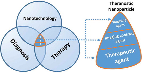
Nanoparticles made of high Z materials (such as gold, bismuth, barium, and tungsten) are the main components of theranostic nanoparticles which can be used as the CT scan contrast agents [Citation13]. Such nanoparticles can be easily entrapped into a nanocarrier to be accompanied by drugs or genes [Citation9–Citation11]. They can be also conjugated with various targeting agents [Citation14]. With regard to the mentioned potentials of theranostic nanoparticles and the capabilities of CT scan imaging, we can determine the biodistribution profile of new drug formulations [Citation13,Citation15]. This is also a good idea for longitudinal studies with the minimum level of invasiveness and without the need to sacrifice the animals which are under examinations. To describe this scenario better, two important examples are presented. The first example is related to taking advantage of the potentials of both theranostic nanoparticles and micro-CT scan. The second one is related to only taking advantage of the capability of high-resolution images provided by micro-CT scan.
Example 1. Suppose that the pharmacokinetic and pharmacodynamic investigations for a new drug are of interest. Routinely, we characterize the time-course and distribution of a drug in the liver, spleen, kidneys, and blood after injection to the animals in a period of time (for example; in a month). This action gives us valuable information, but we need to sacrifice huge numbers of animals, consume much time, and pay much money for investigators and technicians. Compare this situations with the below story:
At first, we design a theranostic nano-system made of gold nanoparticles (as the CT contrast agent) and the new drug molecules. Various designations can be made to produce a theranostic nano-system which are out of the scope of this editorial. However, after producing theranostic nano-system, it can be injected to animals and various organs (such as liver and spleen) can be scanned longitudinally in a period of time (for example; every day or every week). In fact, we can easily characterize the time-course and distribution of a new drug inside the animal body using micro-CT imaging. In this method, we don’t need to sacrifice the animals at various stages of the study and they can be kept alive, after CT scan, for further examinations.
Example 2. Suppose that the toxic effects of a new drug on liver, spleen and kidneys are of interest to be precisely determined. Toxicity is usually developed gradually or immediately following drug injection which can be detected by imaging studies. The structural integrity in the liver, spleen and kidneys can be easily evaluated using micro-CT imaging in a period of time. Once endocytosed by the cells of mononuclear phagocytes, the mechanism of drug molecules distribution from circulating blood leads to an accumulation in the Kupffer cells of the liver and in the macrophages of the spleen. When we profile the x-ray attenuation in the liver and spleen with micro-CT imaging, we can assess the possible damages induced in these organs. Such an assessment is simply feasible by comparing the pre-injection images with post-injection ones. This is certainly of high interest to be mentioned scientists have recently reported scanning resolution of ~5μm for micro-CT imaging [Citation7]. This is incredibly good resolution to investigate the changes of surface area, density and volume of the liver, spleen, kidneys, or even brain for a long period of time post dosing. Also, micro-CT imaging seems to be adequately efficient for liver and spleen evaluation in sub-acute/chronic pharmacology and toxicology studies [Citation16].
4. Conclusion
Micro-CT imaging can revolutionize the quality of current drug discovery procedures, while it requires less labor and expertise. Scientists continuously attempt to make micro-CT imaging more useful for quantification and visualization of a new drug effects in a rapid and non-invasive manner. With regard to the mentioned potentials of theranostic nanoparticles and molecular micro-CT scan, we can design a new scenario in the field of drug discovery. It appears safe to conclude, based on what presented in this editorial, the use of theranostic nanoparticles and micro-CT imaging has a great potential to build a non-invasive imaging modality for various organs macro-morphology examinations. Moreover, such a new rapid and non-invasive imaging modality is of great assistance in longitudinal pharmacology and toxicology studies up to 1 to 6 months [Citation16].
5. Expert opinion
To study the action of a new drug, the conventional methods in the field of drug discovery are traditional histological examination and blood sampling. For example, histological examination is conducted when we need to precisely determine if a synthesized compound causes a lesion in important organs such as brain or liver. Lesion detection and quantification through histological examination of a sectioned organ is a time-consuming process and greatly depends on manual estimation. Particularly, sectioning is difficult and challenging because it may lead to significant damage and distortion of tissue. Therefore, the requirement for sectioning in histological investigations makes the process slow and imprecise. Accordingly, it can be stated there exists an essential need to develop other techniques for quantification and visualization of a new drug effects. Micro-CT imaging can be considered as one of the best alternative solutions for this challenge because it is non-invasive, precise, accurate, rapid, less labor-intensive, and it can provide more detailed results than conventional methods. Furthermore, the micro-CT imaging can provide 3D reconstructions of an organ with various section thicknesses dependent on what we need to know (from 5 to 15 microns). The reconstructed images can be also segmented manually or automatically. These are some examples of the opportunities provided by micro-CT scan while the traditional methods of drug discovery are disabled to provide such a great deal of information.
Regardless of the well-known drawbacks of CT imaging (e.g. low sensitivity to conventional iodine-based contrast media or exposure to ionizing radiation), the key challenge against further development of micro-CT imaging is that most organ tissues are generally transparent to x-rays. Fortunately, a variety of drug and radiopaque containing nanocarriers exist that can adequately overcome soft tissues contrast problems in a wide range of drug discovery studies [Citation9,Citation17–Citation21]. Another alternative method for 3D imaging and drug effects visualization is microscopic magnetic resonance imaging [Citation7]. Although micro-MRI, compared to micro-CT, has greater soft tissue contrast sensitivity, the resolution of micro-MRI is poorer (~25μm). Also, the micro-MRI technique is much more expensive because the micro-MRI machine is much more costly to be obtained and maintained [Citation7]. Therefore, micro-CT scan has its own priority to be utilized in new drug discovery procedures.
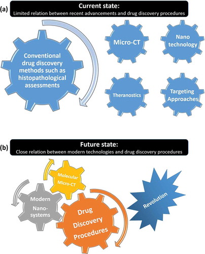
Considering the potentials of theranostic nanoparticles, another possible application of micro-CT scan in drug discovery is to precisely determine the pharmacological indices of a new synthesized compound. We can also target a drug towards an organ and study the drug effects, specifically. What stated here shows that we advocate for synergies between the recent advancements in micro-CT imaging and theranostic nanoparticles so that some new drug discovery procedures can be developed. However, one of the major obstacles that we see is the disconnection between recent advancements reported in the field of micro-CT scan, nanotechnology, theranostics, and targeting approaches ()). This is while the conventional drug discovery procedures are still used extensively, and they can be revolutionized if the concepts of molecular CT-scan, and modern nano-systems are effectively combined. Such a wise combination can make a rapid movement for the gear of drug discovery procedures in the near future ()).
Figure 3. (a) The current and (b) future states of relations between various modern disciplines, micro-CT imaging, and drug discovery procedures.
We believe that drug discovery procedures will be modified and experienced even more progress by effective integration of the molecular micro-CT scan and theranostic nanoparticles benefits. Such a combination definitely has a great potential to accelerate drug discovery procedures and reduce the costs needed for the development of a new drug before extensive clinical examinations. While the interest in micro-CT imaging methods is experiencing an explosion, we also see a high chance for quick improvement of micro-CT based MI methods. We expect further qualitative changes in drug discovery procedures if micro-CT imaging based MI methods are located at their real site. Further, into the future, we expect that the CT imaging based MI methods will be of great assistance to develop image-guided drug delivery methods which they can provide more quantitative estimations.
Declaration of interest
The authors have no other relevant affiliations or financial involvement with any organization or entity with a financial interest in or financial conflict with the subject matter or materials discussed in the manuscript apart from those disclosed.
Reviewer disclosures
Peer reviewers on this manuscript have no relevant financial or other relationships to disclose.
Additional information
Funding
References
- DiMasi JA, Grabowski HG, Hansen RW. Innovation in the pharmaceutical industry: new estimates of R&D costs. J Health Econ. 2016;47:20–33.
- Beik J, Jafariyan M, Montazerabadi A, et al. The benefits of folic acid-modified gold nanoparticles in CT-based molecular imaging: radiation dose reduction and image contrast enhancement. Artif Cells Nanomed Biotechnol. 2017;46(8):1993–2001.
- Wen S, Zhao L, Zhao Q, et al. A promising dual mode SPECT/CT imaging platform based on 99m Tc-labeled multifunctional dendrimer-entrapped gold nanoparticles. J Mat Chem B. 2017;5(21):3810–3815.
- Li X, Xiong Z, Xu X, et al. 99mTc-labeled multifunctional low-generation dendrimer-entrapped gold nanoparticles for targeted SPECT/CT dual-mode imaging of tumors. ACS Appl Mater Interfaces. 2016;8(31):19883–19891.
- Wen S, Li K, Cai H, et al. Multifunctional dendrimer-entrapped gold nanoparticles for dual mode CT/MR imaging applications. Biomaterials. 2013;34(5):1570–1580.
- Chen Q, Li K, Wen S, et al. Targeted CT/MR dual mode imaging of tumors using multifunctional dendrimer-entrapped gold nanoparticles. Biomaterials. 2013;34(21):5200–5209.
- Masís J, Mankus D, Wolff SB, et al. A micro-CT-based method for quantitative brain lesion characterization and electrode localization. Sci Rep. 2018;8(1):5184.
- Gonzalez RC, Woods RE. Digital image processing. USA: Addison-Wesley. 1993.
- Zhu J, Zheng L, Wen S, et al. Targeted cancer theranostics using alpha-tocopheryl succinate-conjugated multifunctional dendrimer-entrapped gold nanoparticles. Biomaterials. 2014;35(26):7635–7646.
- Zhu J, Wang G, Alves CS, et al. Multifunctional dendrimer-entrapped gold nanoparticles conjugated with doxorubicin for pH-responsive drug delivery and targeted computed tomography imaging. Langmuir. 2018;34(41):12428–12435.
- Li D, Wen S, Shi X. Dendrimer‐entrapped metal colloids as imaging agents. Wiley Interdiscip Rev Nanomed Nanobiotechnol. 2015;7(5):678–690.
- Jokerst JV, Gambhir SS. Molecular imaging with theranostic nanoparticles. Acc Chem Res. 2011;44(10):1050–1060.
- Si-Mohamed S, Cormode DP, Bar-Ness D, et al. Evaluation of spectral photon counting computed tomography K-edge imaging for determination of gold nanoparticle biodistribution in vivo. Nanoscale. 2017;9(46):18246–18257.
- Brannon-Peppas L, Blanchette JO. Nanoparticle and targeted systems for cancer therapy. Adv Drug Deliv Rev. 2012;64:206–212.
- Wang L, Xing H, Zhang S, et al. A Gd-doped Mg-Al-LDH/Au nanocomposite for CT/MR bimodal imagings and simultaneous drug delivery. Biomaterials. 2013;34(13):3390–3401.
- Liu C-N, Morin J, Dokmanovich M, et al. Nanoparticle contrast-enhanced micro-CT: A preclinical tool for the 3D imaging of liver and spleen in longitudinal mouse studies. J Pharmacol Toxicol Methods. 2019;96:67–77.
- Lu N, Huang P, Fan W, et al. Tri-stimuli-responsive biodegradable theranostics for mild hyperthermia enhanced chemotherapy. Biomaterials. 2017;126:39–48.
- Mirrahimi M, Hosseini V, Kamrava SK, et al. Selective heat generation in cancer cells using a combination of 808 nm laser irradiation and the folate-conjugated Fe2O3@ Au nanocomplex. Artif Cells Nanomed Biotechnol. 2018;46:241–253.
- Eyvazzadeh N, Shakeri-Zadeh A, Fekrazad R, et al. Gold-coated magnetic nanoparticle as a nanotheranostic agent for magnetic resonance imaging and photothermal therapy of cancer. Lasers Med Sci. 2017;32(7):1469–1477.
- Ghaznavi H, Hosseini-Nami S, Kamrava SK, et al. Folic acid conjugated PEG coated gold–iron oxide core–shell nanocomplex as a potential agent for targeted photothermal therapy of cancer. Artif Cells Nanomed Biotechnol. 2018;46(8):1594–1604.
- Beik J, Abed Z, Ghadimi-Daresajini A, et al. Measurements of nanoparticle-enhanced heating from 1 MHz ultrasound in solution and in mice bearing CT26 colon tumors. J Therm Biol. 2016;62:84–89.