1. Introduction
What is visual acuity? Why do we measure it? How should we measure it?
I suspect if you were to ask these questions to a hundred different ophthalmic professionals, you would receive a hundred different answers. Since the advent of letter acuity charts by Snellen more than 150 years ago, their ease of understanding by patients, short test duration, and the fact that they actually measure residual visual function mean that letter acuity testing will likely continue to occupy a prime position in the increasing battery of tests available to assess ocular status.
But have we done all we can to optimize the diagnostic ability of this much-used test of visual function?
2. Background
The term acuity refers to sharpness or keenness of some kind. The problem with this definition is that, unlike a metric such as body weight, patients do not possess an intrinsic value for ‘acuity’ and the value is very much dependent on both the acuity target and task.
It is well accepted that acuity values are different for grating and letter acuity [Citation1–Citation3], with the number of cycles in a grating target [Citation4], between Snellen and logMAR acuity [Citation5], between Snellen and Landolt C acuity [Citation6], and with different number of alternative letter choices and what they are [Citation7]. Thus, when recording the ‘acuity’ value for a particular patient, it is essential to indicate that it refers to acuity for a particular acuity chart design, with any comparisons between acuity results from different charts not always being a trivial matter. This is commonly appreciated when changing from picture optotypes to letter optotypes in pediatric patients for example.
Thibos and Bradley [Citation8] defined three types of acuity task, forming different levels of an acuity pyramid. These they successively named detection acuity, resolution acuity, and recognition acuity. The most basic detection acuity task merely requires the patient to determine the presence or absence of some form of contrast against a background of the same average luminance. This type of task is employed in acuity tests such as preferential looking when the patient is perhaps asked, ‘Is the target on the right or the left of the chart?’ No further feedback about the stimulus is required. On the other hand, resolution acuity requires a higher level of discrimination in that the patient must, for example, report whether the bars of the grating are horizontal or vertical. This type of task usually employs a small number of choices (alternatives) between targets that are quite different from each other. The highest level of the Thibos and Bradley pyramid, recognition acuity, requires some form of identity determination by the patient, usually from a larger number of alternatives, and requiring more cognitive input in the task.
Thibos and Bradley pointed out that, in the normal visual system, foveal acuity of all three types is largely determined by optical factors (refractive error, diffraction, and higher order aberrations) affecting the base layer of the pyramid (detection) in that spatial frequencies that manage to pass through the optics of the eye are well within the resolving power of the retinal receptors. Only when retinal cell density declines significantly does recognition acuity switch to become dependent on neural rather than optical factors.
Thibos and Bradley point out that conventional letter acuity charts test at the top of the pyramid (recognition) in that they require the patient to identify the letter without any further information about previous stages. The advantage of this is that the test is quick, and a good performance means that the patient’s visual system is performing well as a ‘black box.’ However, when acuity declines it is not possible to determine at which level the system failed. Is it because declining optical quality renders the contrast too low for detection, or because the density of the retinal cells is too depleted to permit good resolution? Detection acuity may actually remain unaffected but cannot properly be measured with conventional black-on-white letter charts because of the large difference in mean luminance between the letters and the background. It is known that patients with age-related macular degeneration (AMD) display different detection and recognition thresholds [Citation9] but we have no way of knowing with conventional acuity charts.
3. The Moorfields Acuity Chart
The Moorfields Acuity Chart (MAC) was initially designed to improve the test-retest variability (TRV) that limits the clinical monitoring ability of conventional acuity charts, both in normal subjects [Citation10,Citation11] and especially in subjects with eye disease [Citation9,Citation12–Citation14]. Because of this variability, clinical trials typically require a change of three logMAR lines (15 letters) to indicate a significant change in visual acuity.
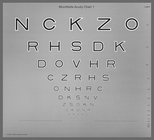
One factor that our group identified as a significant source of acuity measurement variability was the hugely varying spatial frequency content of the letters employed on conventional charts, particularly in the low frequencies that form the basic ‘shape’ of the letters. Thus, some letters remain easily identified by their overall shape long after the frequency of their individual strokes, commonly considered to be five strokes per letter, has exceeded the resolution limit [Citation2,Citation3]. We hypothesized that the large differences in letter legibility owing to the differing low frequency content often resulted in the within-line differences in legibility being greater that the between-line differences. If we removed the low frequency content of letters they should become more equally similar and TRV should reduce.
The MAC is the result of this idea. It employs ‘high-pass’ letters, preserving the high frequency content that forms the ‘edges’ of the letters, and removing the low frequency content where large between-letter differences lie. The letters, constructed from black and white lines on a gray background of the same mean luminance (see ), have also been labeled ‘vanishing optotypes’ in that, for normal foveal vision, the detection and recognition thresholds for these letters are very similar and the letter is observed to disappear soon after the recognition limit has been reached.
Interestingly, in peripheral vision, the thresholds display very different detection and recognition thresholds [Citation15] because recognition acuity outside the fovea is known to be determined by the previously mentioned lower retinal sampling density, rather than optical quality [Citation16–Citation18].
The MAC has been shown to not only reduce TRV over early treatment of diabetic retinopathy study charts [Citation7,Citation19] but also to possess higher sensitivity to early AMD [Citation9]. Based on a retinal sampling simulation, Shah et al. [Citation9] proposed that this is because recognition of the high-pass letters is more vulnerable to cell drop out than conventional black-on-white letters. Interestingly, the same simulation showed that cell under-sampling resulted in the letters remaining detectable long after they could no longer be recognized. The same study found that, as AMD progressed, the letters no longer displayed equal detection and recognition acuity but recognition acuity fell sharply while detection remained much more stable. This means that, for these letters, foveal vision in early AMD becomes more like peripheral vision which is known to be limited by retinal cell sampling density.
4. Discussion
So, what is the clinical relevance of these observations? For too long conventional visual acuity has acted as a ‘one size fits all’ test of functional vision, with little or no diagnostic ability in and of itself. Acuity using conventional letters is affected by both neural and optical losses of vision to differing degrees depending on the condition and its stage of development. This limits the diagnostic ability of the test in early stage disease and its power to differentiate the various factors contributing to loss of function. The MAC has been shown to display better repeatability and high sensitivity to conditions like early AMD but it may also hold potential to improve the separation of neural and optical losses of vision in a way that is quick and easy for the patient to understand. Modern number-of-letters scoring methods could be employed to separately measure the detection and recognition components of Bradley and Thibos’ acuity pyramid. In the former the patient is asked to count the number of letters s\he can ‘see.’ In the latter s\he is asked to (more conventionally) read the letters until errors are made. The ratio of ‘letters read’ to ‘letter detected’ is an indicator of the extent to which ‘acuity’ is limited at the optical contrast detection stage rather than the neural resolution stage. A normal visual system should display a ratio close to 1. A patient with early AMD would display a ratio significantly lower than 1. As disease progresses further both values may suffer more equally and the ratio change yet again. Patients with diseases that cause loss of contrast in the neural image (e.g. optic neuritis) may display a parallel decline in both measures indicating that recognition acuity is limited by the lower contrast detection base of the pyramid. Future electronic versions of the chart may better facilitate the separate measurement of these different thresholds and subsequent scoring, and this is under development.
Visual acuity measurement shows no signs of declining as a functional test of vision. It is perhaps time we sought to better exploit its diagnostic potential in the interests of better patient management.
Declaration of interest
RS Anderson has received funding from the NIHR, The Macular Society, Fight for Sight and Moorfields Eye Charity and he is a co-inventor of the Moorfields Acuity Chart. The authors have no other relevant affiliations or financial involvement with any organization or entity with a financial interest in or financial conflict with the subject matter or materials discussed in the manuscript apart from those disclosed. Peer reviewers on this manuscript have no relevant financial or other relationships to disclose.
Additional information
Funding
References
- Genter CR 2nd, Kandel GL, Bedell HE. The minimum angle of resolution vs angle of regard function as measured with different targets. Ophthalmic Physiol Opt. 1981;1:3–12.
- Anderson RS, Thibos LN. Sampling limits and critical bandwidth for letter discrimination in peripheral vision. J Opt Soc Am A. 1999;16:2334–2342.
- Bondarko VM, Danilova MV. What spatial frequency do we use to detect the orientation of a Landolt C? Vision Res. 1997;37:2153–2156.
- Anderson RS, Evans DW, Thibos LN. Effect of window size on detection acuity and resolution acuity for sinusoidal gratings in central and peripheral vision. J Opt Soc Am A. 1996;13:697–706.
- Falkenstein IA, Cochran DE, Azen SP, et al. Comparison of visual acuity in macular degeneration patients measured with snellen and early treatment diabetic retinopathy study charts. Ophthalmology. 2008;115:319–323.
- Grimm W, Rassow B, Wesemann W, et al. Correlation of optotypes with the Landolt ring–a fresh look at the comparability of optotypes. Optom Vis Sci. 1994;71:6–13.
- Shah N, Dakin SC, Redmond T, et al. Vanishing optotype acuity: repeatability and effect of the number of alternatives. Ophthalmic Physiol Opt. 2011;31:17–22.
- Thibos LN, Bradley A. New methods for discriminating neural and optical losses of vision. Optom Vis Sci. 1993;70:279–287.
- Shah N, Dakin SC, Dobinson S, et al. Visual acuity loss in patients with age related macular degeneration, measured using the Moorfields Acuity Chart. Br J Ophthalmol. 2016;100:1346–1352.
- Rosser DA, Cousens SN, Murdoch IE, et al. How sensitive to clinical change are ETDRS logMAR visual acuity measurements? Invest Ophthalmol Vis Sci. 2003;44:3278–3281.
- Rosser DA, Murdoch IE, Cousens SN. The effect of optical defocus on the test-retest variability of visual acuity measurements. Invest Ophthalmol Vis Sci. 2004;45:1076–1079.
- Blackhurst DW, Maguire MG. Reproducibility of refraction and visual acuity measurement under a standard protocol. The macular photocoagulation study group. Retina. 1989;9:163–169.
- Kiser AK, Mladenovich D, Eshraghi F, et al. Reliability and consistency of visual acuity and contrast sensitivity measures in advanced eye disease. Optom Vis Sci. 2005;82:946–954.
- Beck RW, Moke PS, Turpin AH, et al. A computerized method of visual acuity testing: adaptation of the early treatment of diabetic retinopathy study testing protocol. Am J Ophthalmol. 2003;135:194–205.
- Shah N, Dakin SC, Anderson RS. Effect of optical defocus on detection and recognition of vanishing optotype letters in the fovea and periphery. Invest Ophthalmol Vis Sci. 2012;53:7063–7070.
- Thibos LN, Cheney FE, Walsh DJ. Retinal limits to the detection and resolution of gratings. J Opt Soc Am A. 1987;4:1524–1529.
- Thibos LN, Walsh DJ, Cheney FE. Vision beyond the resolution limit: aliasing in the periphery. Vision Res. 1987;27:2193–2197.
- Demirel S, Anderson RS, Dakin S, et al. Detection and resolution of vanishing optotype letters in central and peripheral vision. Vision Res. 2012;59:9–16.
- Shah N, Dakin SC, Whitaker HL, et al. Effect of scoring and termination rules on test-retest variability of a novel high-pass letter acuity chart. Invest Ophthalmol Vis Sci. 2014;55:1386–1392.

