
ABSTRACT
Background
Clinical guidance on the identification and management of connective tissue disease-associated interstitial lung disease (CTD-ILD) is needed for optimal clinical practice. We aimed to develop clinical algorithms for identifying and managing three common CTD-ILDs: those associated with systemic sclerosis (SSc-ILD), rheumatoid arthritis (RA-ILD), and polymyositis/dermatomyositis (PM/DM-ILD).
Research design and methods
Meetings were held October–November 2023 to create consensus-based algorithms for identifying and managing SSc-ILD, RA-ILD, and PM/DM-ILD in clinical practice, based on expert consensus statements for identification and management of CTD-ILD previously derived from a Delphi process.
Results
We developed clinical algorithms for SSc-ILD, RA-ILD, and PM/DM-ILD that highlight both commonalities and differences in the identification and management of these CTD-ILDs. Importantly, ILD should be suspected in patients with SSc, RA, or PM/DM who have respiratory symptoms. Chest high-resolution computed tomography has utility for screening, diagnosis and assessment of severity. Furthermore, regular follow-up and multidisciplinary management are important. Disease-specific considerations include unique risk factors such as anti-topoisomerase I antibodies in SSc-ILD, high-titer cyclic citrullinated peptide antibodies in RA, anti-aminoacyl tRNA synthetase antibodies in PM/DM, and anti-melanoma differentiation-associated gene 5 antibody in DM.
Conclusions
These algorithms may help physicians to identify and manage patients with SSc-ILD, RA-ILD, or PM/DM-ILD.
1. Introduction
Interstitial lung disease (ILD) is the most common respiratory complication of connective tissue diseases (CTD) [Citation1], a large and heterogenous group of disorders characterized by inflammation and fibrosis, resulting in functional disability, in various organs including the lungs. CTD is frequently a consequence of systemic autoimmune disease, as is the case in systemic sclerosis (SSc), rheumatoid arthritis (RA), and polymyositis/dermatomyositis (PM/DM) – conditions in which ILD is associated with progressive dyspnea leading to end-stage respiratory failure and is the leading cause of premature mortality [Citation2–7].
Management of connective tissue disease-associated interstitial lung disease (CTD-ILD) to optimize patient outcomes requires appropriate diagnosis, evaluation, and treatment. However, there is a general lack of gold-standard data from randomized clinical trials to guide the identification and management of CTD-ILD. In its absence, expert consensus has been employed to develop consensus statements and clinical guidance for CTD-ILD in Europe, Japan, and the United States (US) [Citation8–15].
In particular, we recently developed comprehensive consensus statements and a clinical algorithm for identification and management of CTD-ILD, in general, using a modified Delphi process [Citation11]. In this follow-up study, we aimed to develop individual clinical algorithms for SSc-ILD, RA-ILD, and PM/DM-ILD.
2. Methods
As previously described in detail, we developed consensus statements for identification and management of CTD-ILD in general, based on a Delphi consensus process, as well as an algorithm to guide clinical decision-making [Citation11]. In brief, a statement-development committee (Y Kondoh, M Bando, Y Kawahito, S Sato, T Suda, M Kuwana) formulated 109 statements on managing CTD-ILD across six domains, based on a targeted literature review using Population/Intervention/Comparison/Outcomes/Study (PICOS) criteria of congress abstracts and manuscripts published in English or Japanese between 1 July 2018 and 5 October 2021; the review encompassed CTD-ILD risk factors, screening tools, diagnosis and severity assessment, treatment initiation, and disease progression (see the Supplemental Information). A modified Delphi process was then used to survey 22 physicians in Japan involved in managing CTD-ILD, who were specialists in pulmonology, rheumatology, pathology, and radiology (the CTD-ILD Delphi Collaborators). Two rounds of web-based survey of the CTD-ILD Delphi Collaborators were conducted to establish consensus statements (consensus defined as mean value ≥ 70 on a scale of 0 [strong disagreement] to 100 [strong agreement]). These statements were used by the statement-development committee to construct a general algorithm for identification and management of CTD-ILD. This study was conducted according to Japanese Ethical Guidelines for Medical and Biological Research Involving Human Subjects [Citation16] and approved by an institutional review board (NPO-MINS: http://www.npo-mins.com/).
To extend the clinical utility of this work, the statement-development committee was re-convened to develop specific clinical algorithms for identifying and managing SSc-ILD, RA-ILD, and PM/DM-ILD, based on expert consensus.
3. Results
Three meetings of the statement-development committee were held between 16 October 2023 and 27 November 2023 to create, discuss, and finalize expert consensus-based clinical algorithms for identification and management of SSc-ILD, RA-ILD, and PM/DM-ILD from the previously developed consensus statements for CTD-ILD (Supplemental Table S1 [Citation11]). The commonalities and differences across these algorithms are illustrated in and the individual algorithms are shown in .
Figure 1. Commonalities and differences in identification and management of SSc-ILD, RA-ILD, and PM/DM-ILD. ARS: aminoacyl tRNA synthetase; CRP: C-reactive protein; CT: computed tomography; CTD: connective tissue disease; DLco: diffusing capacity of the lungs for carbon monoxide; DM: dermatomyositis; ESR: erythrocyte sedimentation rate; FVC: forced vital capacity; HRCT: high-resolution computed tomography; ILD: interstitial lung disease; ILD-GAP: interstitial lung disease gender, age, physiology index score; KL-6: Krebs von den Lungen 6; MDA5: melanoma differentiation-associated gene 5; MRC: Medical Research Council dyspnea scale; mRSS: modified Rodnan total skin thickness score; PM/DM: polymyositis/dermatomyositis; QOL: quality of life; RA: rheumatoid arthritis; SADL: smoking, age, DLco; SGRQ: St George’s Respiratory Questionnaire; SPAR: SpO2 and arthritis; SP-D: surfactant protein D; SpO2: peripheral oxygen saturation; SSc: systemic sclerosis.
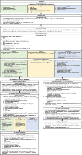
Figure 2. Clinical algorithm for identification and management of SSc-ILD. ![]()
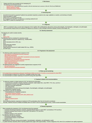
Figure 3. Clinical algorithm for identification and management of RA-ILD. ![]()
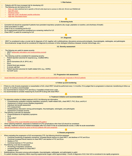
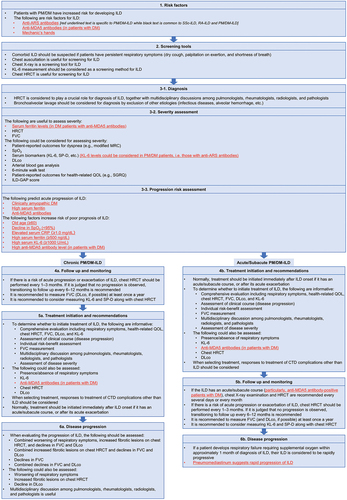
Figure 4. Clinical algorithm for identification and management of PM/DM-ILD. ![]()
3.1. Commonalities and differences in management of RA-ILD, SSc-ILD, and PM/DM-ILD
Several common approaches to identifying and managing CTD-ILD across SSc-ILD, RA-ILD, and PM/DM-ILD are noteworthy (). Firstly, CTD is clearly established to be a risk factor for ILD, including SSc, RA and PM/DM, and comorbid ILD in such patients could manifest as persistent respiratory symptoms such as dry cough, palpitation on exertion, and shortness of breath. Notably, chest high-resolution computed tomography (HRCT) is considered to be useful both as a screening tool and for assessing severity, and is essential for diagnosis. Chest X-ray may also be useful only for screening for ILD. For diagnostic purposes, bronchoalveolar lavage could be considered in order to exclude other causes such as infection and alveolar hemorrhage. In patients with ILD at risk of acute progression or exacerbation, chest HRCT should be performed every 1–3 months (except for those with chronic ILD, typically seen in SSc), with a transition to follow up every 6–12 months if no progression by respiratory symptoms or other tests is observed. It is also recommended to measure forced vital capacity (FVC) (and possibly diffusing capacity of the lungs for carbon monoxide [DLco]) at least once a year.
Several general approaches are potentially informative when deciding whether to initiate pharmacological and/or non-pharmacological treatment for ILD in patients with SSc-ILD, RA-ILD or PM/DM-ILD, including a comprehensive evaluation that assesses the patient’s respiratory symptoms and health-related quality of life, employs HRCT, and measures FVC, DLco, serum Krebs von den Lungen-6 (KL-6) levels, and autoantibodies. Assessing the severity and progression of the ILD is also informative, as is individual risk-benefit assessment and multidisciplinary team discussion (between pulmonologists, rheumatologists, radiologists, and pathologists).
The following should be assessed either separately or in combination when evaluating the progression of ILD: worsening of respiratory symptoms, increased fibrotic lesions on chest HRCT, and declines in FVC and/or DLco. Again, a multidisciplinary team discussion is useful for this purpose. Importantly, ILD is considered to be rapidly progressing if the patient develops respiratory insufficiency requiring supplemental oxygen within 1 month of diagnosis.
3.2. Algorithm for identification and management of SSc-ILD
In addition to the common approaches described above, there are several unique aspects to the management of ILD in patients with SSc ( and ). Specific risk factors include anti-topoisomerase I antibodies, while the smoking, age, DLco (SADL) model [Citation17] is useful to assess severity of ILD (this model predicts mortality risk based on smoking history, age, and DLco). Furthermore, there are several specific markers that predict progression and/or poor prognosis of ILD – including high serum KL-6 (≥1,000 U/mL), high modified Rodnan total skin thickness score, extent of disease on chest HRCT, low baseline FVC or DLco, and the SpO2 and arthritis (SPAR) model (this predicts progression based on ≤ 94% SpO2 after 6-minute walk test and arthritis) [Citation18] or SADL model scores – while old age and male sex may also be predictive. Concomitant pulmonary hypertension is identified as a predictor for poor survival. In addition to the general considerations for initiating ILD treatment described above, other factors to consider assessing in patients with SSc are the presence or absence of early diffuse cutaneous SSc, presence of extensive ILD [Citation10], and elevations in inflammatory markers such as C-reactive protein (≥1.0 mg/dL), erythrocyte sedimentation rate, and platelet count.
3.3. Algorithm for identification and management of RA-ILD
There are several specific risk factors for ILD in patients with RA, including both demographic (male sex, aged 60 years or older) and clinical characteristics (history of smoking history, high-titer cyclic citrullinated peptide [CCP] antibodies, high-titer rheumatoid factor) ( and ). For assessing severity, reticulation and honeycomb scoring on HRCT is particularly informative in those with RA, while presence of the usual interstitial pneumonia (UIP) pattern on HRCT is considered uniquely predictive of acute exacerbation of ILD and poor prognosis in RA patients. Thus, presence of the UIP pattern is a consideration for initiating treatment in RA-ILD patients, while those with poor prognosis should be given special consideration.
3.4. Algorithm for identification and management of PM/DM-ILD
A notable feature of the algorithm for PM/DM-ILD ( and ) is the difference in the order of monitoring and treatment between chronic disease and acute/subacute disease – this difference is based on the consensus to normally begin treatment immediately after ILD onset if it has an acute/subacute onset. The algorithm also notes the expert consensus that certain autoantibodies signify elevated risk of ILD in PM/DM patients, specifically anti-aminoacyl tRNA synthetase (ARS) antibody in patients with PM/DM and anti-melanoma differentiation-associated gene 5 (MDA5) antibody in patients with DM [Citation19], while mechanic’s hands (hyperkeratosis and dirty appearance of fingers and/or palms [Citation20]) may also signify risk. For assessing ILD severity, serum ferritin levels are useful (in DM patients with anti-MDA5 antibodies), while KL-6 levels should be considered in PM/DM-ILD patients, especially those with anti-ARS antibodies. Predictors of rapid progression or poor prognosis include high anti-MDA5 antibody level and high serum ferritin (≥500 ng/dL) and/or KL-6 levels (≥1,000 U/mL). Anti-MDA5 antibodies are also a consideration when deciding whether to initiate treatment in DM patients and strengthen the recommendation for repeated chest X-ray/HRCT in such patients where the ILD has an acute/subacute course.
4. Discussion
In this study, we have developed algorithms to guide the identification and management of SSc-ILD, RA-ILD, and PM/DM-ILD, given the general lack of comprehensive clinical guidance for these CTD-ILDs.
Among the key points in the algorithms are that SSc, RA, and PM/DM increase risk for ILD and the presence of respiratory symptoms in such patients should therefore arouse suspicion. Specific risk factors are identified (although not ranked in terms of strength). The algorithms also highlight the utility of chest HRCT (for screening, diagnosis, and assessment of disease severity) and the importance of a multidisciplinary team approach (involving pulmonologists, rheumatologists, radiologists, pathologists) for diagnosis, treatment, and assessing progression. Chest X-ray may be useful for screening, although its sensitivity and specificity are much lower than HRCT [Citation21].
When deciding whether to initiate treatment or not, the algorithms suggest considering the disease behavior of the ILD, individualized risk-benefit assessment, and tests such as chest HRCT, FVC, DLco, and serum biomarkers (e.g. KL-6) – and that treatment should be initiated immediately for acute/subacute ILD. Although specific treatment recommendations were beyond the scope of this work, options include pharmacotherapies, apheresis, oxygen supplementation, mechanical ventilation, and lung transplant (in patients with end-stage disease) [Citation10]. Pharmacotherapies include glucocorticoids, immunosuppressants such as mycophenolate and cyclophosphamide, nintedanib (an antifibrotic agent), and molecular-targeting drugs such as tocilizumab and rituximab. Although glucocorticoids and immunosuppressants are generally used for treatment of CTD-ILD [Citation10], randomized clinical trial data supports the therapeutic use of nintedanib in patients with SSc-ILD [Citation22] and progressive fibrosing ILD [Citation23], which is approved for these indications in the European Union (EU), Japan, and the US – specifically, for slowing the rate of decline in pulmonary function in SSc-ILD in the US. Pirfenidone, another antifibrotic agent, is approved for idiopathic pulmonary fibrosis (IPF) but not for other fibrotic ILDs. In the RELIEF trial in individuals with non-IPF fibrotic ILD – including CTD-ILD – there was a signal for slowed FVC decline with pirfenidone, but the trial was terminated prematurely because of slow recruitment [Citation24]. Tocilizumab is also approved in the US for slowing the rate of decline in pulmonary function in adults with SSc-ILD, based on a randomized clinical trial [Citation25]. Unlike the SENSCIS trial of nintedanib where change in FVC was the primary endpoint [Citation22], this was only a secondary endpoint in the focuSSced trial of tocilizumab, which failed the primary endpoint of change in modified Rodnan Skin Score [Citation25]. There are also data from randomized clinical trials supporting the use of cyclophosphamide, mycophenolate and rituximab for treatment of SSc-ILD [Citation26–29]; however, although these pharmacotherapies are licensed for this indication in Japan, they are not licensed for SSc-ILD in the EU or the US. Aside from choice of specific therapeutic agent, other important aspects of the treatment of these CTD-ILDs include the timing of treatment. For rapidly progressive ILD in DM patients, an early aggressive immunosuppressive approach has been recommended by others [Citation30], whereas for RA-ILD a more conservative ‘wait and watch’ approach may be warranted, depending on the disease course [Citation31]. These therapeutic options and strategies are reviewed in detail elsewhere [Citation10,Citation21,Citation32,Citation33]. A recent clinical guideline from the American Thoracic Society (ATS) discusses these and other treatment options for SSc-ILD [Citation13], while new guidelines from the American College of Rheumatology (ACR) discuss treatment options for ILD in systemic autoimmune rheumatic disease in general [Citation15]. For individuals with pulmonary hypertension, there are few specific treatments beyond supportive care [Citation21]. However, the inhaled prostacyclin analog treprostinil was approved by the FDA in April 2021 for treating pulmonary hypertension in ILD patients, based on the randomized INCREASE trial showing a significant increase in distance on the 6-minute walk test after 16 weeks [Citation34]. Additionally, supportive care with pulmonary rehabilitation may improve exercise capacity and reduce dyspnea in patients with ILD generally [Citation35,Citation36], and previous Japanese guidelines have noted its potential use for SSc-ILD patients [Citation10].
The algorithms also highlight factors specific to the individual diseases, including the presence of certain autoantibodies signifying risk for ILD – notably anti-topoisomerase I antibody in SSc, anti-ARS antibody in PM/DM, anti-MDA5 antibody in DM, and high-titer anti-CCP antibodies in RA. With regard to the latter, a study published after our Delphi process found that a 4-variable model including anti-CCP antibodies had predictive utility for assessing the risk of ILD in RA patients [Citation37]. Furthermore, the following can aid the assessment of severity: SADL model in SSc-ILD, reticulation and honeycombing scores on chest HRCT in RA-ILD, and KL-6 levels in SSc-ILD and PM/DM-ILD. Specific considerations for treatment initiation include early diffuse cutaneous disease in SSc-ILD, the UIP pattern in RA-ILD, and anti-MDA5 antibodies in DM-ILD.
Prior to our study, algorithms for management of these CTD-ILDs were limited to draft algorithms for SSc-ILD and PM/DM-ILD in Japan [Citation10] and a European algorithm for SSc-ILD [Citation8], while a US study made some specific recommendations for SSc-ILD [Citation12] and an Italian study developed checklists for SSc-ILD and RA-ILD [Citation9]. All these studies used similar expert consensus-based methods to our study. In general, our algorithms are broadly consistent with these studies; for example, in emphasizing the utility of chest HRCT for identification and management of these conditions. As mentioned above, the ACR recently published guidelines for screening, monitoring and treating ILD in systematic autoimmune rheumatic disease [Citation14,Citation15] and the ATS published its official clinical guideline for treating SSc-ILD [Citation13]. There are some differences worth highlighting between these guidelines and the Delphi-based studies, including the algorithms reported in our study. Notably, the ACR guidelines conditionally recommend to not use chest X-ray for either screening or monitoring; they also conditionally recommend HRCT for monitoring when clinically indicated, rather than at regular intervals. Furthermore, the ATS guideline recommends mycophenolate for treatment of SSc-ILD but only suggests the use of nintedanib, tocilizumab, and other medications. Some of the differences between the various guidelines and algorithms may reflect the experiences of clinicians around the world in these CTD-ILDs – for example, differences in genetic predisposition and pathophysiology (e.g. the prevalence of anti-MDA5-positive rapidly progressive ILD), cost and access to HRCT and DLco assessment, availability of biomarker testing (e.g. KL-6), and regulatory approval of therapeutic agents. Differences such as these suggest important research topics for future studies. Other topics of interest for future research include the potential roles of infection and drug-induced lung injury in acute exacerbation of ILD, especially in RA-ILD [Citation38–40].
The main limitation of our study is that the algorithms are based on expert opinion. However, they were derived from statements developed during a Delphi process, a technique that is increasingly used to robustly generate clinical guidance when gold-standard evidence from randomized clinical trials is lacking [Citation41–43]. It is also noteworthy that these algorithms and the Delphi consensus statements used to derive them were developed by physicians in Japan, so their generalizability to other countries has not yet been established. However, as noted above, the algorithms seem generally congruent with those developed in Europe and the US. In this regard, one important issue is the use of serum biomarkers such as SP-D and KL-6 for assessing ILD severity, predicting progression, and guiding treatment initiation, which is more firmly established in East Asia – although several studies have suggested their utility in Western populations [Citation44–50], further research is needed.
5. Conclusions
These expert consensus-based algorithms provide some guidance for identifying and managing ILD in patients with SSc, RA, and PM/DM. They may assist clinical decision-making for such patients until fully evidence-based guidance can be developed, which will require data from randomized clinical trials and other studies.
Declaration of interest
The authors received no direct compensation related to the development of the manuscript. All authors received honoraria for their participation in the study. Outside the submitted work, Y Kondoh has received consulting fees from Asahi Kasei Pharma Corp, Boehringer Ingelheim, Chugai Pharmaceutical Co., Ltd., Healios K.K., Janssen Pharmaceutical KK, Shionogi Co, Ltd., and Taiho Pharmaceutical Co., Ltd., and serves on speaker bureaus for Asahi Kasei Pharma Corp, Boehringer Ingelheim, Eisai Co, Ltd, Bristol Myers Squibb, Janssen Pharmaceutical K.K., KYORIN Pharmaceutical Co, Ltd, Mitsubishi Tanabe Pharma, NIPPON SHINYAKU CO., LTD, Novartis Pharma KK, Shionogi Co, Ltd., and Teijin Pharma Ltd. M Bando has received honoraria from Nippon Boehringer Ingelheim Co., Ltd. and Shionogi & Co., Ltd. Y Kawahito has received research grants from AbbVie GK, Asahi Kasei Pharma Corp., Astellas Pharma Inc., Ayumi Pharmaceutical Corp., Boehringer Ingelheim Japan, Inc., Bristol Myers Squibb Co., Chugai Pharmaceutical Co. Ltd., Daiichi-Sankyo, Inc., Eisai Co., Ltd., Mitsubishi Tanabe Pharma Co., Pfizer Japan Inc., Takeda Pharmaceutical Co., Ltd., and Teijin Pharma Ltd; and speaker’s fee from AbbVie GK, Ayumi Pharmaceutical Corp., Boehringer Ingelheim Japan, Inc., Bristol Myers Squibb Co., Chugai Pharmaceutical Co., Ltd., Eisai Co., Ltd., Eli Lilly Japan K.K., GlaxoSmithKline K.K., Pfizer Japan Inc., Takeda Pharmaceutical Co., Ltd., and Teijin Pharma Ltd. S Sato has received subsidies or donations from Asahi Kasei Pharma Corporation, Ono Pharmaceutical Co., Ltd., and Chugai Pharmaceutical Co., Ltd. T Suda has received honoraria from AstraZeneca K.K. and Nippon Boehringer Ingelheim Co., Ltd.; grant support and/or research funding from AstraZeneca K.K. and Ono Pharmaceutical Co., Ltd.; subsidies or donations from Astellas Pharma Inc., MSD K.K., Daiichi Sankyo Co., Ltd., Taiho Pharmaceutical Co., Ltd., Nippon Boehringer Ingelheim Co., Ltd., Novartis Pharma K.K., and Pfizer Japan Inc. M Kuwana has received consulting fees, speaking fees, and research grants from AbbVie, argenx, Asahi Kasei Pharma, AstraZeneca, Boehringer Ingelheim, Chugai, GlaxoSmithKline, Janssen, Kissei, MBL, Mitsubishi Tanabe, Mochida, Ono Pharmaceuticals, and Taisho. The authors have no other relevant affiliations or financial involvement with any organization or entity with a financial interest in or financial conflict with the subject matter or materials discussed in the manuscript apart from those disclosed.
Reviewer disclosures
Peer reviewers on this manuscript have no relevant financial or other relationships to disclose.
Author contributions
All authors participated in the interpretation of study results and in the drafting, critical revision, and approval of the final version of the manuscript. All authors agree to be accountable for all aspects of this work.
Supplemental Material
Download MS Word (1 MB)Acknowledgments
The authors thank the CTD-ILD Delphi Collaborators: H Amano (Department of Internal Medicine and Rheumatology, Juntendo University Nerima Hospital, Nerima, Tokyo, Japan); T Fujii (Department of Rheumatology and Clinical Immunology, Wakayama Medical University, Wakayama, Japan); J Fukuoka (Department of Pathology Informatics, Nagasaki University Graduate School of Biomedical Sciences, Nagasaki, Japan); T Gono (Department of Allergy and Rheumatology, Nippon Medical School Graduate School of Medicine, Bunkyo, Tokyo, Japan); Y Inoue (Osaka Anti-Tuberculosis Association, Osaka Fukujuji Hospital, Neyagawa, Osaka, Japan); T Johkoh (Department of Radiology, Kansai Rosai Hospital, Amagasaki, Hyogo, Japan); Y Kawaguchi (Division of Rheumatology, Department of Internal Medicine, Tokyo Women’s Medical University, Shinjuku, Tokyo, Japan); T Koga (Department of Immunology and Rheumatology, Division of Advanced Preventive Medical Sciences, Nagasaki University Graduate School of Biomedical Sciences, Nagasaki, Japan); K Kurasawa (Department of Rheumatology, Dokkyo Medical University, Mibu, Tochigi, Japan); Y Nakamura (Respiratory and Allergy Medicine, National Hospital Organization, Tenryu Hospital, Hamamatsu, Shizuoka, Japan); R Nakashima (Department of Rheumatology and Clinical Immunology, Kyoto University Graduate School of Medicine, Kyoto, Japan); Y Nishioka (Department of Respiratory Medicine and Rheumatology, Graduate School of Biomedical Sciences, Tokushima University, Tokushima, Japan); O Nishiyama (Department of Respiratory Medicine and Allergology, Kindai University Faculty of Medicine, Osakasayama, Osaka, Japan); T Ogura (Department of Respiratory Medicine, Kanagawa Cardiovascular and Respiratory Center, Yokohama, Kanagawa, Japan); M Okamoto (Department of Respirology, NHO Kyushu Medical Center, and Division of Respirology, Neurology and Rheumatology, Department of Internal Medicine, Kurume University School of Medicine, Fukuoka, Japan); K Sada (Department of Clinical Epidemiology, Kochi Medical School, Nankoku, Kochi, Japan); S Sakamoto (Department of Respiratory Medicine, Toho University Omori Medical Center, Tokyo, Japan); T Takeuchi (Department of Internal Medicine (IV), Osaka Medical and Pharmaceutical University, Takatsuki, Osaka, Japan); H Tomioka (Department of Respiratory Medicine, Kobe City Medical Center West Hospital, Kobe, Hyogo, Japan); Y Waseda (Third Department of Internal Medicine, Faculty of Medical Sciences, University of Fukui, Eiheiji, Fukui, Japan); H Yamada (Center for Rheumatic Diseases, Seirei Yokohama Hospital, Yokohama, Kanagawa, Japan); and Y Yamano (Department of Respiratory Medicine and Allergy, Tosei General Hospital, Seto, Aichi, Japan). Medical writing support was provided by G Brooke, of Elevate Scientific Solutions LLC, a member of the Envision Pharma Group, under the authors’ conceptual direction and based on feedback from the authors, and was contracted and compensated by Nippon Boehringer Ingelheim. Nippon Boehringer Ingelheim was given the opportunity to review the manuscript for medical and scientific accuracy as well as intellectual property considerations.
Data availability statement
The data sets used and/or analyzed during the current study are available from the corresponding author on reasonable request.
Supplementary material
Supplemental data for this article can be accessed online at https://doi.org/10.1080/17476348.2024.2374910
Additional information
Funding
References
- Spagnolo P, Cordier JF, Cottin V. Connective tissue diseases, multimorbidity and the ageing lung. Eur Respir J. 2016 May;47(5):1535–1558. doi: 10.1183/13993003.00829-2015
- Nakajima A, Inoue E, Tanaka E, et al. Mortality and cause of death in Japanese patients with rheumatoid arthritis based on a large observational cohort, IORRA. Scand J Rheumatol. 2010;39(5):360–367. doi: 10.3109/03009741003604542
- Olson AL, Swigris JJ, Sprunger DB, et al. Rheumatoid arthritis-interstitial lung disease-associated mortality. Am J Respir Crit Care Med. 2011 Feb 1;183(3):372–378. doi: 10.1164/rccm.201004-0622OC
- Yamasaki Y, Yamada H, Ohkubo M, et al. Longterm survival and associated risk factors in patients with adult-onset idiopathic inflammatory myopathies and amyopathic dermatomyositis: experience in a single institute in Japan. J Rheumatol. 2011 Aug;38(8):1636–1643. doi: 10.3899/jrheum.101002
- Ishizuka M, Watanabe R, Ishii T, et al. Long-term follow-up of 124 patients with polymyositis and dermatomyositis: statistical analysis of prognostic factors. Mod Rheumatol. 2016;26(1):115–120. doi: 10.3109/14397595.2015.1054081
- Steen VD, Medsger TA. Changes in causes of death in systemic sclerosis, 1972-2002. Ann Rheum Dis. 2007 Jul;66(7):940–944. doi: 10.1136/ard.2006.066068
- Castelino FV, Dellaripa PF. Recent progress in systemic sclerosis-interstitial lung disease. Curr Opin Rheumatol. 2018 Nov;30(6):570–575. doi: 10.1097/BOR.0000000000000544
- Hoffmann-Vold A-M, Maher TM, Philpot EE, et al. The identification and management of interstitial lung disease in systemic sclerosis: evidence-based European consensus statements. Lancet Rheumatol. 2020;2(2):e71–e83. doi: 10.1016/S2665-9913(19)30144-4
- Bosello SL, Beretta L, Del Papa N, et al. Interstitial lung disease associated with autoimmune rheumatic diseases: checklists for clinical practice. Front Med. 2021;8:732761. doi: 10.3389/fmed.2021.732761
- Kondoh Y, Makino S, Ogura T, et al. 2020 guide for the diagnosis and treatment of interstitial lung disease associated with connective tissue disease. Respir Investig. 2021 Nov;59(6):709–740. doi: 10.1016/j.resinv.2021.04.011
- Kuwana M, Bando M, Kawahito Y, et al. Identification and management of connective tissue disease-associated interstitial lung disease: evidence-based Japanese consensus statements. Expert Rev Respir Med. 17(1):71–80. doi: 10.1080/17476348.2023.2176303
- Rahaghi FF, Hsu VM, Kaner RJ, et al. Expert consensus on the management of systemic sclerosis-associated interstitial lung disease. Respir Res. 2023 Jan 9;24(1):6. doi: 10.1186/s12931-022-02292-3
- Raghu G, Montesi SB, Silver RM, et al. Treatment of systemic sclerosis-associated interstitial lung disease: evidence-based recommendations. an official American Thoracic Society clinical practice guideline. Am J Respir Crit Care Med. 2024 Jan 15;209(2):137–152. doi: 10.1164/rccm.202306-1113ST
- American College of Rheumatology. American College of Rheumatology (ACR) guideline for the screening and monitoring of interstitial lung disease in people with systemic autoimmune rheumatic disease: guideline summary. 2023. Available from: https://assets.contentstack.io/v3/assets/bltee37abb6b278ab2c/blt7e2cadfc7bc986fb/interstitial-lung-disease-guideline-summary-screening-monitoring-2023.pdf
- American College of Rheumatology. American College of Rheumatology (ACR) guideline for the treatment of interstitial lung disease in people with systemic autoimmune rheumatic disease: guideline summary. 2023. Available from: https://assets.contentstack.io/v3/assets/bltee37abb6b278ab2c/bltaedebda97a351d47/interstitial-lung-disease-guideline-summary-treatment-2023.pdf
- Eba J, Nakamura K. Overview of the ethical guidelines for medical and biological research involving human subjects in Japan. Jpn J Clin Oncol. 2022 May 31;52(6):539–544. doi: 10.1093/jjco/hyac034
- Morisset J, Vittinghoff E, Elicker BM, et al. Mortality risk prediction in scleroderma-related interstitial lung disease: the SADL model. Chest. 2017 Nov;152(5):999–1007. doi: 10.1016/j.chest.2017.06.009
- Wu W, Jordan S, Becker MO, et al. Prediction of progression of interstitial lung disease in patients with systemic sclerosis: the SPAR model. Ann Rheum Dis. 2018 Sep;77(9):1326–1332. doi: 10.1136/annrheumdis-2018-213201
- Chen Z, Cao M, Plana MN, et al. Utility of anti-melanoma differentiation-associated gene 5 antibody measurement in identifying patients with dermatomyositis and a high risk for developing rapidly progressive interstitial lung disease: a review of the literature and a meta-analysis. Arthritis Care Res (Hoboken). 2013 Aug;65(8):1316–1324. doi: 10.1002/acr.21985
- Sohara E, Saraya T, Sato S, et al. Mechanic’s hands revisited: is this sign still useful for diagnosis in patients with lung involvement of collagen vascular diseases? BMC Res Notes. 2014 May 17;7(1):303. doi: 10.1186/1756-0500-7-303
- Maher TM. Interstitial lung disease: a review. JAMA. 2024 Apr 22;331(19):1655. doi: 10.1001/jama.2024.3669
- Distler O, Highland KB, Gahlemann M, et al. Nintedanib for systemic sclerosis-associated interstitial lung disease. N Engl J Med. 2019 Jun 27;380(26):2518–2528. doi: 10.1056/NEJMoa1903076
- Flaherty KR, Wells AU, Cottin V, et al. Nintedanib in progressive fibrosing interstitial lung diseases. N Engl J Med. 2019 Oct 31;381(18):1718–1727. doi: 10.1056/NEJMoa1908681
- Behr J, Prasse A, Kreuter M, et al. Pirfenidone in patients with progressive fibrotic interstitial lung diseases other than idiopathic pulmonary fibrosis (RELIEF): a double-blind, randomised, placebo-controlled, phase 2b trial. Lancet Respir Med. 2021 May;9(5):476–486. doi: 10.1016/S2213-2600(20)30554-3
- Khanna D, Lin CJF, Furst DE, et al. Tocilizumab in systemic sclerosis: a randomised, double-blind, placebo-controlled, phase 3 trial. Lancet Respir Med. 2020 Oct;8(10):963–974. doi: 10.1016/S2213-2600(20)30318-0
- Tashkin DP, Elashoff R, Clements PJ, et al. Cyclophosphamide versus placebo in scleroderma lung disease. N Engl J Med. 2006 Jun 22;354(25):2655–2666. doi: 10.1056/NEJMoa055120
- Tashkin DP, Roth MD, Clements PJ, et al. Mycophenolate mofetil versus oral cyclophosphamide in scleroderma-related interstitial lung disease (SLS II): a randomised controlled, double-blind, parallel group trial. Lancet Respir Med. 2016 Sep;4(9):708–719. doi: 10.1016/S2213-2600(16)30152-7
- Ebata S, Yoshizaki A, Oba K, et al. Safety and efficacy of rituximab in systemic sclerosis (DESIRES): a double-blind, investigator-initiated, randomised, placebo-controlled trial. Lancet Rheumatol. 2021 Jul;3(7):e489–e497. doi: 10.1016/S2665-9913(21)00107-7
- Maher TM, Tudor VA, Saunders P, et al. Rituximab versus intravenous cyclophosphamide in patients with connective tissue disease-associated interstitial lung disease in the UK (RECITAL): a double-blind, double-dummy, randomised, controlled, phase 2b trial. Lancet Respir Med. 2023 Jan;11(1):45–54. doi: 10.1016/S2213-2600(22)00359-9
- McPherson M, Economidou S, Liampas A, et al. Management of MDA-5 antibody positive clinically amyopathic dermatomyositis associated interstitial lung disease: a systematic review. Semin Arthritis Rheum. 2022 Apr;53:151959. doi: 10.1016/j.semarthrit.2022.151959
- Koduri G, Solomon JJ. Identification, monitoring, and management of rheumatoid arthritis-associated interstitial lung disease. Arthritis Rheumatol. 2023 Dec;75(12):2067–2077. doi: 10.1002/art.42640
- Thong L, Chawke LJ, Murphy G, et al. Management of myositis associated interstitial lung disease. Rheumatol Int. 2023 Jul;43(7):1209–1220. doi: 10.1007/s00296-023-05336-z
- Wells AU. New insights into the treatment of CTD-ILD. Nat Rev Rheumatol. 2021 Feb;17(2):79–80. doi: 10.1038/s41584-020-00567-x
- Waxman A, Restrepo-Jaramillo R, Thenappan T, et al. Inhaled treprostinil in pulmonary hypertension due to interstitial lung disease. N Engl J Med. 2021 Jan 28;384(4):325–334. doi: 10.1056/NEJMoa2008470
- Dowman L, Hill CJ, May A, et al. Pulmonary rehabilitation for interstitial lung disease. Cochrane Database Syst Rev. 2021 Feb 1;2(2):CD006322. doi: 10.1002/14651858.CD006322.pub4
- Rochester CL, Alison JA, Carlin B, et al. Pulmonary rehabilitation for adults with chronic respiratory disease: an official American Thoracic Society clinical practice guideline. Am J Respir Crit Care Med. 2023 Aug 15;208(4):e7–e26. doi: 10.1164/rccm.202306-1066ST
- Koduri GM, Podlasek A, Pattapola S, et al. Four-factor risk score for the prediction of interstitial lung disease in rheumatoid arthritis. Rheumatol Int. 2023 Aug;43(8):1515–1523. doi: 10.1007/s00296-023-05313-6
- Wang H, Chen X, Du Y, et al. Mortality risk in patients with anti-MDA5 dermatomyositis is related to rapidly progressive interstitial lung disease and anti-Ro52 antibody. Arthritis Res Ther. 2023 Jul 24;25(1):127. doi: 10.1186/s13075-023-03100-z
- Ge YP, Shu XM, He LR, et al. Infection is not rare in patients with idiopathic inflammatory myopathies. Clin Exp Rheumatol. 2022 Feb;40(2):254–259. doi: 10.55563/clinexprheumatol/yps7ai
- Peng JM, Du B, Wang Q, et al. Dermatomyositis and polymyositis in the intensive care unit: a single-center retrospective cohort study of 102 patients. PLOS ONE. 2016;11(4):e0154441. doi: 10.1371/journal.pone.0154441
- Milholland AV, Wheeler SG, Heieck JJ. Medical assessment by a Delphi group opinion technic. N Engl J Med. 1973 Jun 14;288(24):1272–1275. doi: 10.1056/NEJM197306142882405
- Page A, Potter K, Clifford R, et al. Prescribing for Australians living with dementia: study protocol using the Delphi technique. BMJ Open. 2015 Aug 11;5(8):e008048. doi: 10.1136/bmjopen-2015-008048
- Taylor E. We agree, don’t we? The Delphi method for health environments research. HERD. 2020 Jan;13(1):11–23. doi: 10.1177/1937586719887709
- Lederer C, Mayer K, Somogyi V, et al. Krebs von den Lungen-6 as a potential predictive biomarker in fibrosing interstitial lung diseases. Respiration. 2023;102(8):591–600. doi: 10.1159/000531945
- Benyamine A, Heim X, Resseguier N, et al. Elevated serum Krebs von den Lungen-6 in systemic sclerosis: a marker of lung fibrosis and severity of the disease. Rheumatol Int. 2018 May;38(5):813–819. doi: 10.1007/s00296-018-3987-3
- Millan-Billi P, Castellvi I, Martinez-Martinez L, et al. Diagnostic value of Krebs von den Lungen (KL-6) for interstitial lung disease: a European prospective cohort. Arch Bronconeumol. 2024 Apr 6;60(6):350–355. doi: 10.1016/j.arbres.2024.03.028
- Sieiro Santos C, Antolin SC, Lorenzo JC, et al. KL6 and IL-18 levels are negatively correlated with respiratory function tests and ILD extent assessed on HRCT in patients with systemic sclerosis-related interstitial lung disease (SSc-ILD). Semin Arthritis Rheum. 2024 Apr;65:152366. doi: 10.1016/j.semarthrit.2024.152366
- Parker MJS, Jee AS, Hansen D, et al. Multiple serum biomarkers associate with mortality and interstitial lung disease progression in systemic sclerosis. Rheumatology (Oxford). 2024 Feb 15. doi: 10.1093/rheumatology/keae110
- Gyorfi AH, Filla T, Dickel N, et al. Performance of serum biomarkers reflective of different pathogenic processes in systemic sclerosis-associated interstitial lung disease. Rheumatology (Oxford). 2024 Apr 2;63(4):962–969. doi: 10.1093/rheumatology/kead332
- Elhai M, Hoffmann-Vold AM, Avouac J, et al. Performance of candidate serum biomarkers for systemic sclerosis-associated interstitial lung disease. Arthritis Rheumatol. 2019 Jun;71(6):972–982. doi: 10.1002/art.40815