Abstract
A case of combined percutaneous coronary intervention and ostium secundum atrial septal defect closure in an elderly patient is reported. The procedure was successful and uneventful. The report demonstrates feasibility of combined percutaneous revascularization and intra‐ atrial shunt closure even in advanced age.
Introduction
Secundum atrial septal defect (ASD) is the most common congenital cardiac anomaly diagnosed in adults and coronary artery disease (CAD) is quite frequent in advanced age. The combination of secundum ASD and CAD may be encountered in elderly patients. The percutaneous transcatheter treatment of each of these conditions is a common practice worldwide. This report describes a successful percutaneous transluminal coronary angioplasty (PTCA) and stenting combined with percutaneous ASD closure.
Case report
A 75 years old woman has presented with a recent reduction in her functional capacity, leg edema and stress induced breathlessness considered an anginal equivalent. The patient was on Coumadine, Atenolol, ACE inhibitors and diuretics due to congestive heart failure and chronic atrial fibrillation with an adequate heart rate control and reasonable blood pressure values. No other relevant past and family medical history was available. Transthoracic (TTE) and transesophageal (TEE) echocardiography have both revealed a large secundum ASD with enlarged right sided heart chambers (right ventricle end diastolic dimension 4.2 cm) and a predominantly left to right shunt. Good ventricular systolic function and moderate mitral and tricuspid valves incompetence were depicted. Tricuspid valve regurgitation flow velocity was 3.5 m/s with a dilated inferior vena cava and minimal inspiratory collapse suggestive of moderately elevated pulmonary artery pressure.
A diagnostic cardiac catheterization was carried out at the neighboring hospital. Two vessels coronary artery disease was demonstrated with 95% narrowing of the mid right coronary artery and 50% narrowing in the proximal third of the left circumflex coronary artery. The systolic right ventricular pressure was 50 mmHg. The patient has refused surgical revascularization with ASD closure and she was thus referred for interventional catheter‐based therapy at our tertiary center.
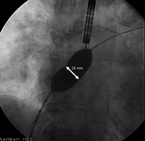
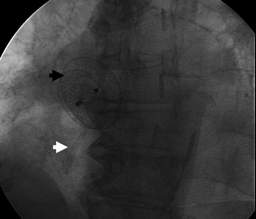
When admitted to our center the patient has presented without signs of heart failure. Her physical examination revealed irregular cardiac rhythm, cardiac heave, and wide fixed splitting of the second heart sound. Her electrocardiogram showed atrial fibrillation with ventricular response of 70–80 beats per minute. The patient was treated with low molecular weight Heparin for five days prior to catheterization while Coumadin was discontinued. The cardiac catheterization was performed under general anesthesia with elective intratracheal intubation. A 6 Fr sheath was inserted in the right femoral artery and two sheathes (6 Fr and 10 Fr) were inserted in the right femoral vein. The relevant data of the hemodynamic study are summarized in . The right coronary catheterization was performed by a 6 Fr diameter Judkins right coronary guiding catheter (JR4) which was introduced via right femoral artery. The right coronary artery was predilated with a 2.0×10 mm Pleon coronary balloon (Biotronik, 812 Avis Dr., Ann Arbor, MI 48108, USA). After predilatation, a 3.0×12 mm Driver bare metal stent (Medtronic Vascular, 3576 Unocal Place Santa Rosa, CA 95403, USA) was successfully implanted. The secundum ASD was then re‐evaluated by TEE. Its diameter was 18–20 mm with appropriate rims. Its stretched diameter as measured by the 24 mm sizing balloon (AGA Medical Corporation, 682 Mendelssohn Avenue, Golden Value, MN 55427, USA) was 24.5 mm (Figure ). We kept the sizing balloon inflated for 5 min with no elevation of pulmonary artery wedge pressure and no increase of mitral valve regurgitation. A 26 mm Amplatzer ASD occluder was successfully delivered and deployed on first attempt through a 10 Fr delivery system (AGA Medical Corporation, 682 Mendelssohn Avenue, Golden Value, MN 55427, USA ) (Figure ). Complete closure, as judged by color Doppler flow, was immediately achieved. The procedure was uneventful. The patient was successfully extubated and no signs of left heart failure were observed. Additional diuretic therapy was temporarily required due to transient augmentation of peripheral edema and markedly increased tricuspid regurgitation. Ten days following the procedure, a TTE has demonstrated an optimal position of the septal occluder with no residual shunt and no obstruction of adjacent obligatory systemic or pulmonary venous flows. The tricuspid regurgitation has returned to its baseline magnitude. The patient has reported a dramatic improvement in quality of life and in functional capacity. She has begun riding a tricycle whereas prior to the procedure she could hardly walk.
Table I. Hemodynamic parameters
Discussion
The association of ASD and CAD may be observed in adult and especially in elderly patients. Although successful results of combined surgical ASD closure and coronary artery bypass grafting (CABG) have been previously reported Citation[1] modern catheter based techniques may be an attractive alternative to surgical treatment of the both conditions. There are few reports describing combined/consequent percutaneous transcatheter treatment of ASD and CAD. E. Onorato et al. Citation[2] described a cohort of 176 adult patients that underwent transcatheter ASD closure. In six of them PTCA was performed due to unstable angina, stable angina or silent ischemia. The PTCA was followed after 35±15 days by ASD closure in 4 patients and in the other two patients the ASD was occluded 40 days and 30 days respectively before the revascularization. Upasni et al. Citation[3] reported successful ASD closure four days after coronary stenting and balloon pulmonary valvuloplasty.
Tomai et al. Citation[4] described a combined procedure, which included percutaneous stenting of the proximal left anterior descending coronary artery (LAD) and proximal left circumflex coronary artery (LCX) followed by transcatheter ASD closure with Amplatzer septal occluder in a 68 year‐old man with the history of myocardial infarction two years prior to the procedure and mildly reduced left ventricular ejection fraction with severe lateral hypokynesia by TTE. The procedure was complicated by acute left ventricular failure due to abrupt overloading of the left ventricle after sudden interruption of the left to right shunt at atrial level. The administration of high dose of diuretics and adrenaline infusion did not prevent a massive pulmonary edema, which needed re‐intubation and mechanical ventilation. The authors' opinion was that it is necessary to defer transcatheter closure of atrial septal defect in patients with associated ischemic heart disease suitable for coronary revascularization.
Our patient is an elderly woman with two‐vessel CAD, preserved left ventricular function and a large secundum ASD. The transcatheter ASD closure immediately after PTCA was not associated with hemodynamic instability. The patient was extubated at the end of the procedure in the catheterization laboratory with no clinical or radiological signs of left heart failure during the postprocedural course. This may be attributed to the preserved left ventricular function and adequate medication.
The precise mechanisms of left heart failure after ASD closure is not entirely clear. While coronary revascularization may improve contractility of viable myocardium, the performance of PTCA prior to ASD closure may prevent the development of left heart failure. Masked left ventricular restriction is another possible mechanism of hemodynamic deterioration after ASD occlusion in elderly patient. The study of Ewert et al. Citation[5] showed that this phenomenon might be predicted by significant elevation of left atrial pressure and restrictive mitral inflow pattern after balloon occlusion of atrial septal defect. Since coronary revascularization may improve diastolic ventricular function Citation[6], Citation[7] we suggest that PTCA should be performed prior to ASD closure and may prevent sudden elevation of left ventricular filling pressure after intra‐atrial shunt occlusion.
This report demonstrates the feasibility of combined percutaneous transcatheter treatment of patients with secundum ASDs and CAD. These procedures may be safe and effective even in elderly patient. Coronary revascularization, which improves systolic and diastolic left ventricular function, may prevent hemodynamic instability after ASD closure. It is emphasized, that meticulous hemodynamic evaluation is important before and during the procedure. Repeated pulmonary capillary wedge or left atrial pressure measurements while the ASD is open and with temporary balloon occlusion of the ASD for at least 10–15 min are mandatory in all adult patients over the age of 60 years. When the wedge/left‐atrial pressure goes up with occlusion of the ASD, the procedure must be abandoned. In these cases adequate preprocedural medical management using diuretics and after load—reducing drugs (e.g. angiotensin converting enzyme (ACE) inhibitors), as well the use of fenestrated atrial septal occluders may allow successful ASD closure Citation[8]. In our department, it is common practice to administer a low doses of diuretics before the transcatheter ASD closure in all adult patients over the age of 60 years. Echocardiographic data of left ventricular systolic and diastolic function, evaluation of myocardial viability by isotopic scan or stress echocardiography and pulmonary capillary wedge pressure and/or left atrial pressure monitoring during the procedure may help to select the suitable patients for the combined procedure, and to optimize therapeutic strategies.
References
- Harjula A., Kupari M., Kyosola K., Ventila M., Hartel G., Maamies T., et al. Early and late results of surgery for atrial septal defect in patients aged over 60 years. J Cardiovasc Surg (Torino) 1988; 29: 134–9
- Onorato E., Pera I., Lanzone A., Ambrosini V., Rubino P., Trabattoni D., et al. Transcatheter treatment of coronary artery disease and atrial septal defect with sequential implantation of coronary stent and Amplatzer septal occluder: preliminary results. Catheter Cardiovasc Interv 2001; 54: 454–8
- Upasani P. T., Lal P., Pandley A. K., Sachdeva S. M., Kanwar S. Interventional therapy for multiple cardiac defects. Indian Heart J 2002; 54: 306–8
- Tomai F., Gaspardone A., Papa M., Polisca P. Acute left ventricular failure after transcatheter closure of a secundum atrial septal defect in a patient with coronary artery disease: a critical reappraisal. Catheter Cardiovasc Interv 2002 Jan; 55((1))97–9
- Ewert P., Berger F., Nagdyman N., Kretschmar O., Dittrich S., Abdul‐Khaliq H., et al. Masked left ventricular restriction in elderly patients with atrial septal defects: a contraindication for closure?. Catheter Cardiovasc Interv 2001; 52: 177–80
- Schannwell C. M., Schneppenheim M., Plehn G., Strauer B. E. Parameters of left ventricular diastolic function 48 hours after coronary angioplasty and stent implantation. J Invasive Cardiol 2003 Jun; 15((6))326–33
- Hedman A., Samad B. A., Larsson T., Zuber E., Nordlander R., Alam M. Improvement in diastolic left ventricular function after coronary artery bypass grafting as assessed by recordings of mitral annular velocity using Doppler tissue imaging. Eur J Echocardiogr 2005; 6: 202–9, Epub 2004 Dec 18
- Holzer R., Cao Q. L., Hijazi Z. M. Closure of a moderately large atrial septal defect with a self‐fabricated fenestrated Amplatzer septal occluder in an 85‐year‐old patient with reduced diastolic elasticity of the left ventricle. Catheter Cardiovasc Interv 2005; 64: 513–8

