A 78-year-old man presented with stable angina, whose coronary risk factors included hypertension and remote tobacco use. Coronary angiography revealed a total occlusion of the proximal left anterior descending artery (LAD) and a stenosis in the right coronary artery (RCA). The patient was pretreated with aspirin and ticlopidine for one month prior to procedure. After administration of 6,000 units of heparin, percutaneous coronary intervention for the LAD lesion was performed. After successful guide wire crossing of the chronic total occlusion (CTO) and rotational atherectomy, three sirolimus-eluting stents (SES, 2.5×18 mm, 3.0×33 mm, 3.5×23 mm, Cypher, Cordis) were implanted in the LAD using 20 atm with optimal angiographic result.
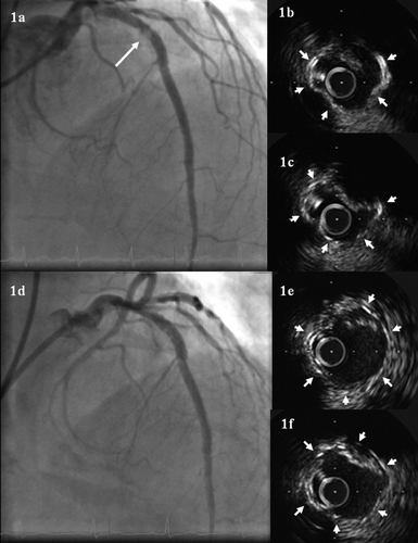
Discharged on dual antiplatelet therapy, the patient returned for staged angioplasty for the RCA 2 weeks later. Coronary angiography revealed a stenosis with haziness within the SES (a). Intravascular ultrasound (IVUS) examination showed a mobile hyperechoic mass (b,c). Following an unsuccessful thrombectomy (Thrombuster, Kaneka Medix corp, Japan), the stenosis was dilated with a balloon, resulting in partial resolution (d,e,f). Angioplasty for the stenosis in his RCA was postponed.
Figure 1. (a) Coronary angiography showing a stenosis with haziness in the proximal left anterior descending artery (LAD) two weeks after sirolimus-eluting stents (SES) implantation. (b) and (c) Intravascular ultrasound imaging of the stented segment at two weeks of follow-up. The maximal plaque prolapse cross sectional area was 2.5 mm2 with a percentage of plaque prolapse burden: 40% of stent area. Stent struts were labeled with arrows. (d) Coronary angiography showing partial resolution of the stenosis with haziness after balloon dilatation. (e) and (f) Intravascular ultrasound imaging of the stented segment after balloon dilatation.
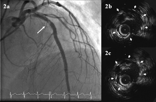
One month later, before staged angioplasty for the RCA, coronary angiography again revealed a similar stenosis with haziness within the SES (a). IVUS examination also showed a similar image (b,c). An additional SES (3.5×13 mm, Cypher, Cordis) was implanted with optimal angiographic/IVUS results and 10 month follow-up coronary angiography showed no restenosis.
Figure 2. (a) Coronary angiography showing a similar stenosis with haziness in the proximal LAD six weeks after SES implantation. (b) and (c) Intravascular ultrasound imaging of the stented segment at six weeks of follow-up.
Previous IVUS studies demonstrated that plaque prolapse was more frequent in chronic total occlusion lesions and long lesions Citation[1], Citation[2]. Although exact explanation remains unknown, severe calcification with chronic total occlusion and/or partial wire passage and stent implantation in subintimal space may be associated with repeated plaque prolapse.
Given the clinical course similar with a previous report (recurrence of thrombosis) Citation[3], possibility of thrombus could not be excluded. With currently available ultrasound technology, however, it is difficult to determine exactly what the observed mobile hyperechoic mass was Citation[4].
Although SES has dramatically reduced restenosis in most lesions, a recent study suggested unsatisfactory long-term results after SES implantation for CTO lesions Citation[5]. In addition to advanced systemic factors, several lesion/procedural factors affect long-term outcomes in the treatment for CTO lesions. Even with DES, re-occlusion sometimes occurs in the treatment for CTO lesions. Due to collateral flow or conditioning, re-occlusion often occurs asymptomatically. Early plaque prolapse and/or thrombus may be an important initiating or contributing factor for re-occlusion. In this case, treated CTO lesion would have led to re-occlusion if follow-up CAG had not been performed. Although optimal treatment remains unclear for such a case, early detection (repeat angiography or other imaging devices such as computed tomography) may improve long-term outcome after SES implantation in the treatment of CTO lesions.
Acknowledgements
The authors thank Heidi N Bonneau, RN, MS, CCA, for her editorial review of the manuscript. Declaration of interest: The authors report no conflicts of interest. The authors alone are responsible for the content and writing of the paper.
References
- Futamatsu H, Sabate M, Angiolillo DJ, Jimenez-Quevedo P, Corros C, Morikawa-Futamatsu K, et al. Characterization of plaque prolapse after drug-eluting stent implantation in diabetic patients: A three-dimensional volumetric intravascular ultrasound outcome study. J Am Coll Cardiol. 2006; 48: 1139–45
- Kim SW, Mintz GS, Ohlmann P, Hassani SE, Fernandez S, Lu L, et al. Frequency and severity of plaque prolapse within Cypher and Taxus stents as determined by sequential intravascular ultrasound analysis. Am J Cardiol 2006; 98: 1206–11
- van Werkum JW, Heestermans AA, de Korte FI, Kelder JC, Suttorp MJ, Rensing BJ, et al. Long-term clinical outcome after a first angiographically confirmed coronary stent thrombosis: An analysis of 431 cases. Circulation 2009; 119: 828–34
- Mintz GS, Nissen SE, Anderson WD, Bailey SR, Erbel R, Fitzgerald PJ, et al. American College of Cardiology clinical expert consensus document on standards for acquisition, measurement and reporting of intravascular ultrasound studies (IVUS). A report of the American College of Cardiology task force on clinical expert consensus documents. J Am Coll Cardiol. 2001; 37: 1478–92
- Garcia-Garcia HM, Daemen J, Kukreja N, Tanimoto S, van Mieghem CA, van der Ent M, et al. Three-year clinical outcomes after coronary stenting of chronic total occlusion using sirolimus-eluting stents: Insights from the rapamycin-eluting stent evaluated at Rotterdam cardiology hospital-(RESEARCH) registry. Catheter Cardiovasc Interv. 2007; 70: 635–9