
ABSTRACT
Sporadic Creutzfeldt-Jakob disease (sCJD) is a fatal progressive neurodegenerative disease. Multimodal approaches, including electroencephalogram, diffusion-weighted imaging (DWI) of brain MRI, and cerebrospinal fluid biomarkers, have been applied to increase the diagnostic accuracy of sCJD. Although previous studies suggested DWI could be the most useful modality for sCJD diagnosis, whether metabolism changes underlying in sCJD are still poorly understood. To the best of our knowledge, there are only one case using the technique of arterial spin labeling (ASL) to detection and follow-up of perfusion changes in CJD. Herein, we described a 71-year-old woman presented with progressive cognitive decline, behavioral and psychological symptoms for two months. The patient died one month later after her admission. As far as we know, this is the first report using the combination of fluorodeoxyglucose positron emission tomography and ASL to explore the metabolism changes in sCJD. Our case exemplifies the difficulty clinicians may face in the diagnosis of sCJD.
Introduction
Prion disease is a rare and fatal neurodegenerative disorder that affects mammals and humans, with a mortality rate of 100%. Sporadic Creutzfeldt-Jakob disease (sCJD) is the most common form of prion disease [Citation1]. It is generally characterized by rapidly progressing dementia (RPD), behavioral and psychiatric disorders, cerebellar ataxia, vision disturbances, akinetic mutism state and myoclonus [Citation2]. To date, the multimodal imaging studies such as diffusion-weighted imaging (DWI) hyperintensity, electroencephalography (EEG), and cerebrospinal fluid (CSF) biomarkers are utilized in conjunction with the clinical picture to try to make the diagnosis of CJD without brain biopsy.
Although previous studies suggested DWI could be the most useful modality for sCJD diagnosis, whether some metabolism changes underlying in sCJD are still poorly understood. Only quite a few studies using the 18F-fluorodeoxyglucose positron emission tomography (FDG-PET) to investigate the metabolism changes in sCJD [Citation3]. To the best of our knowledge, there are only one case using the technique of arterial spin labeling (ASL) to detection and follow-up of perfusion changes in CJD [Citation4]. We herein described a 71-year-old woman presenting with typical clinical features, the findings of MRI, triphasic morphology of EEG and positive 14–3-3 proteins in CSF. As far as we know, this is the first report using the combination of FDG-PET and ASL to explore the metabolism changes in sCJD.
Case
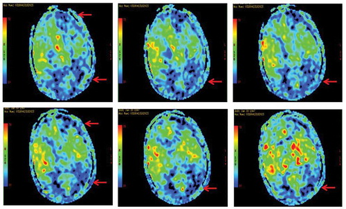
A 71-year-old woman presented with progressive cognitive decline, behavioral and psychological symptoms such as visual hallucinations for two months. Initial neurological examination revealed rapidly progressive dementia, ataxia, tremor and involuntary movements, myoclonus, visual neglect, mutism, startle myoclonus and dystonia, urinary incontinence. The biomarkers of autoimmune encephalitis were unremarkable. The 14–3-3 proteins in CSF were positive. Cranial MRI indicated diffusion restriction revolved in entire cortex more on the right, presenting with hyperintensive signal in the cortex (). The EEG showed periodic discharges over bilateral hemisphere with triphasic morphology (). The findings of FDG-PET () and ASL () both showed extensive hypoperfusion in bilateral cortex, thalamus and cerebellum, especially lateralized to the left hemisphere. The patient died three months after the initial onset of symptoms. The patient had no risk factors for familial, new variant, or iatrogenic CJD. sCJD was the final diagnosis.
Figure 1. MRI (A) findings of bilateral cortical diffusion restriction presenting with hyperintensive signal in the cortex on diffusion weighted imaging. EEG (B) showed slowing and periodic discharges over bilateral hemisphere with triphasic morphology.
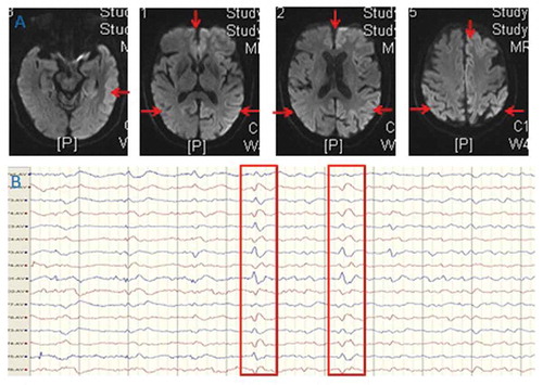
Figure 2. FDG-PET showed hypoperfusion in bilateral cortex, thalamus and cerebellum, especially lateralized to the left hemisphere.
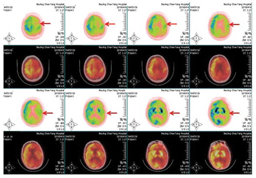
Figure 3. ASL indicated severe extensive hypoperfusion in the cerebral cortex, basal ganglia, especially lateralized to the left hemisphere.
Discussion
sCJD is a prion disease consequent to accumulation of misfolded pathogenic prion protein in the brain. As an untreatable prion encephalopathy, early and accurate diagnosis of sCJD is essential to avoid iatrogenic transmission and to distinguish CJD from other potentially treatable dementia [Citation5]. Common presenting symptoms are RPD, behavioral abnormalities, mood disturbance, sleep disorders, startle myoclonus, extra-pyramidal signs and cerebellar abnormalities. Current clinical diagnostic criteria mostly depend on clinical features and periodic sharp-wave complexes on EEG, DWI hyperintensity, and the presence of 14–3-3 in CSF. Although DWI could be the most useful modality for the diagnosis of sCJD [Citation6,Citation7], however, the gold standard for definitive diagnosis is still considered to be histopathological confirmation. Therefore, the diagnosis of sCJD is often challenging in clinical practice.
Supportive investigations such as EEG, MRI and CSF biomarkers have a relatively low diagnostic sensitivity and specificity in sCJD. Considering to brain metabolism, however, a recent study indicated the [18F]-AV-1451 PET was not suitable for patients with RPD due to sCJD, due to different pathological process [Citation8]. As for the technique of FDG-PET, it has an established role in the diagnostic workup of patients with sCJD [Citation9,Citation10]. We have summarized a table showing the changes in FDG-PET in sCJD patients reported from literature (). To date, there are only one case using ASL to detection and follow-up of perfusion changes in CJD [Citation4]. In this study, ASL showed severe hypoperfusion in the cerebral cortex, basal ganglia and thalami but this was least marked in the thalami [Citation4]. However, the combination of FDG-PET and ASL is quite rare. To the best of our knowledge, our study is the first case using combined FDG-PET and ASL to explore the metabolism changes in patients with sCJD.
Table 1. The changes in FDG-PET in sCJD patients reported from literature.
The strengths of our case were listed as follows. Firstly, our case was typical for sCJD due to very rapid onset of cognitive deficits, myoclonus, ataxia, visual neglect, and tremor. Brain imaging, CSF biomarkers, and neurophysiological findings also revealed typical findings for sCJD as well. Although sCJD and dementia with Lewy bodies (DLB) have overlapping clinical symptoms, however, the given this patient’s clinical presentation was more consistent with CJD than DLB. As a result, our case exemplifies the difficulty clinicians may face in the diagnosis of sCJD. Secondly, in spite that the features of DWI have been pivotal in the diagnostic workup of sCJD and to be the best predictor for sCJD [Citation7,Citation11], however, due to the complementary advantages, the combined techniques of PET and ASL may give us some more important information to detect the metabolism changes underlying in sCJD. Thus, our findings of perfusion changes such as PET and ASL have been proved to be more promising values in clinical practice [Citation12]. As far as we know, this is the first report using the combination of FDG-PET and ASL to explore the metabolism changes in sCJD. Thirdly, our study provides an update on the neuroimaging patterns of sCJD, emphasizing the relevance of MRI and PET/ASL, and highlighting clinical, genetic, and imaging correlations in sCJD. Although CJD is typically associated with diffuse cerebral pathology, our case also conform it could also present with unilateral neurologic symptoms and concordant MRI and EEG findings [Citation13].
Conclusion
Herein, we reported a sCJD with the typical clinical presentations, neuroimaging features, triphasic morphology of EEG, positive 14–3-3 proteins in CSF, also with the evidences of hypometabolism using PET and ASL. One of the limitation was the histopathology was not available in our case. Novel ultrasensitive seeding assays such as the real-time quaking-induced conversion of CSF could provide promising diagnostic information than tests for surrogate markers of sCJD in CSF in future study.
Disclosure statement
No potential conflicts of interest are disclosed
Additional information
Funding
References
- Manix M, Kalakoti P, Henry M, et al. Creutzfeldt-Jakob disease: updated diagnostic criteria, treatment algorithm, and the utility of brain biopsy. Neurosurg Focus. 2015;39(5):E2.
- Iwasaki Y. Creutzfeldt-Jakob disease. Neuropathology. 2017;37(2):174–188.
- Renard D, Vandenberghe R, Collombier L, et al. Glucose metabolism in nine patients with probable sporadic Creutzfeldt-Jakob disease: FDG-PET study using SPM and individual patient analysis. J Neurol. 2013;260(12):3055–3064.
- Chen S, Guan M, Shang JK, et al. Reduced cerebral blood flow in genetic prion disease with PRNP D178N-129M mutation: an arterial spin labeling MRI study. J Clin Neurosci. 2015;22(1):204–206.
- Bizzi A, Peoc’h K. Amended diagnostic protocol increases the early diagnosis of sporadic Creutzfeldt-Jakob disease. Neurology. 2018;91:155–156. [Epub ahead of print].
- Suzuki K, Kawasaki A, Nagashima T, et al. Diffusion-weighted MRI abnormalities antedate the onset of sporadic Creutzfeldt-Jakob disease. Neurology. 2016;87(8):843–845.
- Forner SA, Takada LT, Bettcher BM, et al. Comparing CSF biomarkers and brain MRI in the diagnosis of sporadic Creutzfeldt-Jakob disease. Neurol Clin Pract. 2015;5(2):116–125.
- Day GS, Gordon BA, Perrin RJ, et al. In vivo [(18)F]-AV-1451 tau-PET imaging in sporadic Creutzfeldt-Jakob disease. Neurology. 2018;90(10):e896–e906.
- Duignan J, Healy GM, Hughes NM, et al. FDG-PET diagnoses of sporadic Creutzfeldt-Jakob disease: radiology- Pathology correlation. Qjm. 2018. [Epub ahead of print]. DOI:10.1093/qjmed/hcy071
- Matias-Guiu JA, Guerrero-Marquez C, Cabrera-Martin MN, et al. Amyloid- and FDG-PET in sporadic Creutzfeldt-Jakob disease: correlation with pathological prion protein in neuropathology. Prion. 2017;11(3):205–213.
- Henkel K, Zerr I, Hertel A, et al. Positron emission tomography with [(18)F]FDG in the diagnosis of Creutzfeldt-Jakob disease (CJD). J Neurol. 2002;249(6):699–705.
- Engler H, Lundberg PO, Ekbom K, et al. Multitracer study with positron emission tomography in Creutzfeldt-Jakob disease. Eur J Nucl Med Mol Imag. 2003;30(1):85–95.
- Hamaguchi T, Kitamoto T, Sato T, et al. Clinical diagnosis of MM2-type sporadic Creutzfeldt-Jakob disease. Neurology. 2005;64(4):643–648.
- Okamura N, Shiga Y, Furumoto S, et al. In vivo detection of prion amyloid plaques using [(11)C]BF-227 PET. Eur J Nucl Med Mol Imag. 2010;37(5):934–941.
- Zhang WJ, Westover MB, Keary CJ. Premortem diagnosis of sporadic Creutzfeldt-Jakob disease aided by positron-emission tomography imaging. AJNR Am J Neuroradiol. 2011;32(1):E18.
- Zhang Y, Minoshima S, Vesselle H, et al. A case of Creutzfeldt-Jakob disease mimicking corticobasal degeneration: FDG PET, SPECT, and MRI findings. Clin Nucl Med. 2012;37(7):e173–175.
- Xing XW, Zhang JT, Zhu F, et al. Comparison of diffusion-weighted MRI with 18F-fluorodeoxyglucose-positron emission tomography/CT and electroencephalography in sporadic Creutzfeldt-Jakob disease. J Clin Neurosci. 2012;19(10):1354–1357.
- Kim EJ, Cho SS, Jeong BH, et al. Glucose metabolism in sporadic Creutzfeldt-Jakob disease: a statistical parametric mapping analysis of (18) F-FDG PET. Eur J Neurol. 2012;19(3):488–493.
- Nennesmo I, Kumlien E, et al. Imaging astrocytosis with PET in Creutzfeldt-Jakob disease: case report with histopathological findings. Int J Clin Exp Med. 2012;5(2):201–207.
- Zhao W, Zhang JT, Xing XW, et al. Chinese specific characteristics of sporadic Creutzfeldt-Jakob disease: a retrospective analysis of 57 cases. PLoS One. 2013;8(3):e58442.
- Furukawa F, Ishibashi S, Sanjo N, et al. Serial magnetic resonance imaging changes in sporadic Creutzfeldt-Jakob disease with valine homozygosity at codon 129 of the prion protein gene. JAMA Neurol. 2014;71(9):1186–1187.
- Euskirchen P, Buchert R, Koch A, et al. Sporadic Creutzfeldt-Jakob disease with mesiotemporal hypermetabolism. J Neurol Sci. 2014;345(1–2):278–280.
- Ortega-Cubero S, Pagola I, Luquin MR, et al. Clinical and neuroimaging characteristics of 14 patients with prionopathy: a descriptive study. Neurologia. 2015;30(3):144–152.
- Prieto E, Dominguez-Prado I, Riverol M, et al. Metabolic patterns in prion diseases: an FDG PET voxel-based analysis. Eur J Nucl Med Mol Imag. 2015;42(10):1522–1529.
- Miyazawa N. Creutzfeldt-Jakob disease mimicking alzheimer disease and dementia with lewy bodies-findings of FDG PET with 3-dimensional stereotactic surface projection. Clin Nucl Med. 2017;42(5):e247–e248.
- Renard D, Castelnovo G, Collombier L, et al. FDG-PET in Creutzfeldt-Jakob disease: analysis of clinical-PET correlation. Prion. 2017;11(6):440–453.
- Mente KP, O’Donnell JK, Jones SE, et al. Fluorodeoxyglucose positron emission tomography (FDG-PET) correlation of histopathology and MRI in prion disease. Alzheimer Dis Assoc Disord. 2017;31(1):1–7.
- Fragoso DC, Goncalves Filho AL, Pacheco FT, et al. Imaging of Creutzfeldt-Jakob disease: imaging patterns and their differential diagnosis. Radiographics. 2017;37(1):234–257.
- Yung B, Moeller JJ, Bitar M, et al. Teaching neuroimages: hemispheric-onset Creutzfeldt-Jakob disease with concordant MRI and EEG findings. Neurology. 2010;75(16):e66.