

ABSTRACT
Background/objective
Salivary Mucoepidermoid Carcinoma (MEC) is a heterogeneous malignancy, whose molecular characteristics extend beyond CRTC1/3:MAML2 fusion. This study describes the immunohistochemical expression of CD133, CD44, OCT4, and Mammalian ENA (MENA) in Salivary MEC and their potential relevance to molecular profiling.
Methods
This study tested forty cases of MAML2-rearranged MEC using Immunohistochemistry (IHC) for the expression of CD133, CD44, OCT4, and MENA. Specific antibodies for each marker were utilized (CD133, CD44, OCT4, and MENA). The POU5F1 FISH probe from Empire Genomics (USA) was also employed to detect any alteration corresponding to OCT4. The selection of these adult stem markers as targets in our study is based on their established associations with cancer stem cells and the possible roles in tumorigenesis, metastasis, and treatment resistance in some carcinomas and adenocarcinomas.
Results
The investigation demonstrated a negative expression pattern for CD133, CD44, and OCT4 immunostains in all cases, questioning the involvement in the pathogenesis of salivary MEC. In contrast, MENA showed a diffuse positive expression in all cases. Furthermore, the POU5F1 FISH probe revealed no alterations in the forty samples analyzed.
Conclusion
These findings suggested the noninvolvement of cancer stem cells in the pathogenesis of salivary MEC. The overexpression of MENA suggests its co-expression that may extend beyond stemness or pluripotency.
Introduction
The etiopathogenesis of MEC remains unknown, but emerging evidence suggests the existence of a tumorigenic population of neoplastic cells emanating from pluripotent cells of the excretory-duct derived cells, inducing differentiations into mucin-producing (mucous), highly prolific basaloid cells (intermediate) and epidermoid (squamoid) cells to different extents. Detecting cancer stem cells in mucoepidermoid carcinomas (MECs) has prompted conflicting results from various methodic approaches.Citation1–3 One point of controversy within the findings generated based on the cancer stem cell hypothesis is whether these “allegedly” neoplastic cells originate from actual tissue-specific stem cells with a slow rate of division and longevity within the tissue. Another concern involves the accumulation of mutations in in vitro studies, or the utilization of immunodeficient mice, leading to a lack of a human microenvironment and subsequent differences in growth and tumor initiation, especially that the studied cases were not tested molecularly.Citation1 The molecular labeling of MAML2-rearranged MECs versus non-MECs seems to be of paramount diagnostic and prognostic significance, with possible therapeutic implications.Citation4 Moreover, it remains uncertain whether stem cells are definitively established as the origin of cancer especially in molecularly labeled carcinomas. Regarding MECs that demonstrate a canonical fusion of CRTC1/3::MAML2 that activates transcriptions mediated by cAMP response element-binding, the role of cancer stem cells in oncogenesis is underexplored due to a lack of suitable research models (such as cell lines and xenograft modelsCitation5 and the unavailability of molecular human clones that facilitate the identification of cell sub-populations with unique tumorigenic potential, given the newly proposed (super)enhancer hypothesis.Citation6–9
The robustness of cancer stem cell studies is limited by the spatial configuration of intratumoral maximal stemness or pluripotency, potential confusion with other confounders such as stemness, epithelial-mesenchymal transition (EMT), and tumor invasion, and the heterogeneity in the expression of stemness-related markers.Citation10–12 The heterogeneous distribution of stemness within the tumor can make it challenging to obtain a representative sample for analysis. Changes in stemness might be wrongly attributed to EMT or tumor invasion, leading to inaccurate conclusions about the functional significance of cancer stem cells.Citation13–15
Cancer stem cell markers frequently studied in carcinomatous lesions include OCT4,Citation10,Citation12,Citation16,Citation17 SOX2,Citation18–22 Nanong,Citation21,Citation23,Citation24 CD133,Citation25–28 MENACitation29 and SALL4.Citation30,Citation31 We are exploring the molecular detection of POU5F1 and its corresponding IHC marker OCT4, along with other pertinent markers (CD133, CD44, and MENA) to achieve a more comprehensive analysis. POU5F1 involvement may activate transcriptional regulation of pluripotent stem cells by sumoylation, glycosylation, phosphrylation or polyubiquitination.
Method
Case Selection
Forty MAML2-rearranged parotid MECs and ten normal salivary tissue harvested during excision of mucoceles were initially enrolled. The included cases were randomly selected from the registry starting from 2019 to 2023. The included cases were graded according to the AFIP grading system. Clinical information for all cases was obtained from electronic medical records from which the cases were retrieved. The rationale for selecting specific targets such as CD133, CD44, OCT4, and MENA in our study stems from their known associations with cancer stem cells (CSCs) and their potential roles in tumorigenesis, metastasis, and treatment resistance.Citation32,Citation33 These markers have been implicated in CSC self-renewal, tumor initiation, metastasis, and resistance to therapy.Citation11,Citation34 OCT4 is a transcription factor crucial for maintaining stemness in embryonic stem cells, and its expression has been linked to CSC properties in several cancer types. MENA has been associated with cancer cell invasion and metastasis, and its overexpression correlates with poor prognosis in multiple cancers.Citation29
Immunohistochemistry
To prepare the tissue samples, 4 µm-thick sections underwent deparaffinization in xylene followed by gradual rehydration using alcohol. Epitope retrieval was performed by immersing the samples in a Tris-EDTA buffered solution with a pH of 8.0 (Target Retrieval Solution; Dako, Glostrup, Denmark) in a water bath at 97°C. For antigen detection, anti-CD133 (dilution of 1:100, recombinant anti-CD133 antibody, Abcam) was utilized. The antibody, acquired from Lab Vision Corporation, USA, was specifically designed for localizing antigens in formalin-fixed paraffin-embedded tissue sections. Additionally, anti-OCT-4 polyclonal antibody (SC-8629; 1:50 dilution; Santa Cruz Biotechnology), anti-CD44 (Monoclonal, MA5–1389, ThermoFisher), and anti-MENA (monoclonal, clone dilution 1:500, Dako, Agilent Technologies, Santa Clara, CA, USA) were employed. All specimens were counterstained with hematoxylin.
The immunohistochemical process involved applying Biotinylated Goat Anti-Polyvalent and incubating for 10 minutes at room temperature, followed by a 5-minute PBS wash. Streptavidin Peroxidase was then applied and incubated for 10 minutes at room temperature, with another PBS wash for 5 minutes. The development of the colored reaction product using (3,3ʹ-Diaminobenzidine tetrahydrochloride) DAB as a chromogen included adding 1 drop (40 ul) DAB plus chromogen to 2 ml of DAB plus substrate, swirling to mix, applying to the tissue, and incubating for 5–15 minutes. Slides were counterstained with Mayer’s hematoxylin bath and covered using permanent mounting media (Canada balsam). Positive immunoreactive deposits manifested as horse-reddish to brown reactions at the site of the target antigen. For quality control, OCT4 was tested on prostate tissue sections, CD133 on human pancreatic carcinoma, CD44 on human esophageal carcinoma, and MENA on human lung cancer, all serving as positive controls according to the respective manufacturer’s instructions. Positive and negative controls were processed in parallel.
Fluorescence in situ Hybridization
For fluorescence in situ hybridization (FISH) analysis on formalin-fixed paraffin-embedded sections, a commercially available POU5F1 dual-color break-apart probe (Empire Genomics, USA) was employed. The FISH analysis, conducted using fluorescence microscopy, classified cells with two fused signals as normal, indicating the presence of one orange and one green fluorochrome that were not displaced from each other. Cells displaying rearrangements in the POU5F1 gene exhibited orange and green signals situated at a distance from each other. To ensure the accuracy of the analysis, tissues from a confirmed salivary clear-cell myoepithelial carcinoma containing the POU5F1 rearrangement served as a positive control, while normal salivary tissue was utilized as a negative control. Both positive and negative controls were processed simultaneously for comparison and validation.
Statistical Analysis
The grading was conducted according to AFIP by two pathologists (EA and WB). Grading according to AFIP criteriaCitation35 involved assessing several parameters, including the presence of a cystic component occupying less than 20%, perineural invasion, necrosis, the number of mitoses equal to or greater than four per ten high-power fields (HPF), and the presence of anaplasia. Each parameter was assigned a certain number of points: +2 points were assigned if the parameter was present, while no points were added if it was absent for the cystic component occupying less than 20%, perineural invasion, and necrosis; +3 points were assigned for the presence of four or more mitoses per ten HPF; and + 4 points were assigned for anaplasia. Following the scoring of these parameters, the grading of the lesions was computed. Based on the AFIP scoring system, lesions were categorized into different grades. A total score ranging from 0 to 4 indicated a low-grade lesion, a score of 5 or 6 indicated an intermediate-grade lesion, and a score of 7 to 14 indicated a high-grade lesion. The subjective grading was cross-checked using the automated grader. Calculation of descriptive statistics was performed using SPSS. The Kruskal-Wallis test compared the expressions of CD133, CD44, OCT4 and MENA across the studied groups representing different histological variants. Log-rang and Cox proportional hazards regression analyses were used to analyze the association between CD133, CD44, OCT4 and MENA expression levels and survival outcomes.
Results
Clinicopathological Findings
Retrospective studies showed conflicting links between clinical pictures (e.g., tumor stage, lymphovascular/bony invasion, recurrence and metastatic behavior) with histological features. Therefore, we included as many clinicopathologically diverse MECs to attain representative effect size (Set at α= 0.05, corresponding to 90%, and an effect size of 0.844). The distribution of the studied 40 cases based on gender demonstrated a slightly higher prevalence in females, with 55% of the cases, compared to 45% in males. This sex distribution was nearly evenly proportional. The mean age of approximately 42.4 years indicates that MECs affect individuals in their early to mid-adulthood. The age range spanning from 12 to 62 years highlights the broad spectrum of ages at which MECs can manifest. The distribution of diagnoses showcases the prevalence of low-grade MEC, constituting the majority at 52.5%. Intermediate-grade MEC represents 37.5% of the cases, while high-grade MEC accounts for 10%. The clinical TNM staging and athological TNM grading were directly proportional. The majority of cases (70%) exhibit a localized tumor without regional lymph node involvement or distant metastasis (cT2N0M0). However, variations in staging, including cases with lymph node involvement and distant metastasis, heightens the clinical heterogeneity within the cohort. The variability in age and gender distribution highlighted the wide-ranging impact of MECs across different populations. The majority of cases shared a common clinical TNM stage of cT2N0M0, indicative of a localized tumor without regional lymph node involvement or distant metastasis. This staging aligned with the typical presentation of MEC as a locally confined malignancy. Notably, case 21 deviated from this trend, demonstrating a more advanced stage (cT2N2M1). shows the clinical data relating to the 40 patients of MEC enrolled in the study.
Table 1. Clinicopathological profile of the 40 parotid cases.
The prevalence of low-grade MEC according to the AFIP grading was ineluctably high. Lymph node involvement was evident in cases 2, 8, and 21 ().
Table 2. Histologic profile of the 40 parotid cases and their grading according to the AFIP system.
Immunohistochemistry
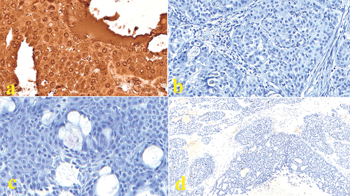
We found that the MENA was overexpressed in the MECs. Nevertheless, all normal salivary glands tested negative for the anti-Mena antibody. The 40 cases showed negative immunostaining for CD133, CD44, and OCT4 (). Univariate analysis with the Kaplan-Meier method demonstrated that disease-free survival was significantly reduced in high-grade MECs compared to low-grade MECs. However, all cases showed strong cytoplasmic MENA expression, irrespective of grading and staging. It should be noted that statistical analysis was not informative.
Figure 1. Micrograph showing (a) Immunoreactive deposits appeared as reddish-brown reactions where the target antigen was located (MENA) in conventional low-grade MEC (40×); (b) Negative immunostaining for CD133 in low-grade oncocytic MEC (40×); (c) Negative immunostaining for CD44 in low-grade oncocytic MEC (40×). (d) Negative immunostaining for OCT4 in high-grade solid MEC (10×).
Molecular Findings
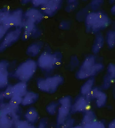
The molecular findings indicate that there were no alterations detected in any case when using the POU5F1 dual-color break-apart probe (). In this context, a dual-color break-apart probe is typically employed in fluorescence in situ hybridization to identify structural alterations or rearrangements in a specific gene, in this case, POU5F1.
Figure 2. Photomicrograph showing retained signals for POU5F1 FISH probe.
Statistical Analysis
The Kaplan-Meier survival plots demonstrated a significant reduction in 5-year DFS in high-grade cases, those with nodal metastasis, and advanced TNM clinical stages (III + IV). Due to the consistent and uniform expression of all markers across cases (either consistently positive or consistently negative), Cox regression analysis did not reveal significant differences between groups.
Discussion
Analyzing cancer stem cells solely based on the expression of specific IHC markers represents an oversimplification that introduces potential confounding factors by co-targeting irrelevant co-expressions.Citation36 This approach also overlooks subsets of cancer stem cells with distinct marker profiles, preventing accurate attributions to specific pathways. Even though benign salivary neoplasms, including 10 pleomorphic adenomas and 10 Warthin’s tumors, exhibited negative immunostaining for MENA, positive expression was observed in a few cases of salivary duct carcinomas, carcinomas ex pleomorphic adenoma, acinic cell carcinomas, squamous cell carcinomas, and high-grade mucoepidermoid carcinomas. Further investigation and analysis would be needed to understand the underlying reasons for the observed positive expression in those particular cases.Citation37 In our study, regardless of grade phenotype (low-, intermediate, or high-grade), all MECs demonstrated diffuse positive cytoplasmic staining for MENA. However, the nuclear expression in MECs was too weak to be considered as having high intensity compared to the nonspecific background, indicating immunonegativity. CD133, CD44, OCT4 showed neither nuclear, cytoplasmic nor membranous expression in the studied 40 parotid MECs.
Positivity in immunostaining must be attributed to the expressing cellular population. It was alleged that aternal cells produce intermediate cells that are regarded as the progenitor of epidermoid cells, mucous secreting cells and clear cells. However, this was not sufficient to explain the diversity of cellular differentiation in MECs (e.g. differentiation into oncocytes, sebocytes, acin, spindle cells or polygonal cells) that show genetic alternations on FISH assay for MAML2 and CRTC1/3 probes. Therefore, attributing immunopositive staining for each neoplatic cell is mandatory. The diffuse membranous and cytoplasmic expressions for β -catenin is considered nonspecific. Only nuclear immunostaining expression of β-catenin counts, as in the positive control. This is important for many salivary gland neoplasms that commonly show nonspecific diffuse membranous immunostaining pattern (including the recently described palisading adenocarcinoma). In Hodgkin’s lymphoma, the background (non-Reed-Sternberg cells) is reactive for both CD20 and CD3. Fascin, and CDMay 15, also be nonspecifically positive. Not all weak immunopositivity counts. For instance, HER2/neu weak positive of less than 10% is considered negative in breast carcinomas (Supplementary Figures).
Although this finding seemed perplexing initially, given the negativity of normal salivary gland tissue and the absence of positivity for other stem cell markers in neoplastic components, there are possible explanations. Dysregulated phosphorylation of emerin in MECs could disrupt normal cellular functions and contribute to tumorigenic signaling pathways. Changes in nuclear architecture and chromatin repositioning could influence the accessibility of genes involved in tumorigenesis. Altered chromatin organization may contribute to the dysregulation of key genes related to cell proliferation, differentiation, and apoptosis.Citation38 The ability of human homologue of Enabled, Mena (encoded by ENAH and also known as hMena), an actin regulatory protein belonging to the Ena/VASP family, to modulate transcription via direct signaling across the nuclear membrane implies a direct involvement in the regulation of gene expression. This could lead to the activation or suppression of specific pathways related to cancer development in MECs. Dysregulation in these processes might result in increased cytoplasmic levels of Mena, influencing cell motility, migration, and invasive potential, which are commonly associated with cytoplasmic overexpression in cancer cells. It also affects the membrane localization of MENA, influencing cellular adhesion, signaling, or membrane-associated processes that are relevant to cancer progression. But the main guarantor is the nuclear expression, encoding the transcriptional machinery and contributing to the altered gene expression patterns observed in pathogenesis.
OCT4 is mainly expressed in unfertilized oocytes, zygotesand primordial germ cells. Its silencing aids in differentiation, cell engineering, and oncogenesis, while its expression ertains to stem cell characteristization. Methylation Studies have shown that OCT4 is regulated by DNA methylation of CpG of the promoter and exon in the human trophoblast cells. Unlike previous studies on CD133, CD44, OCT4 in salivary carcinomasCitation12,Citation39–42 and POU5F1,Citation43,Citation44 the consistent immunonegativity observed for these markers, and the lack of molecular alterations in POU5F1 in MAML2-rearranged MEC challenge the prevailing assumptions regarding the role of cancer stemness in elucidating its high-grade transformation.
CD44, a cell surface adhesion receptor overexpressed in numerous cancers, facilitates metastasis by adhering to the cell surface, interacting with extracellular matrix ligands, and its cleavage, shedding, and elevated soluble levels in patients’ serum serve as indicators of tumor burden and metastasis in colon and gastric cancers.Citation45–47 CD133, an adult stem cell marker, is expressed to varying degrees in both normal and cancerous tissues, indicating its involvement in cellular dynamics and potential implications for disease progression.Citation48,Citation49
The absence of alterations in the POU5F1 gene may have specific implications depending on the context of the study. For example, in cancer research, alterations in certain genes might be associated with tumor development or progression. In the absence of alterations, it could imply that POU5F1 may not play any role in the molecular characteristics or progression of the studied Salivary MEC cases. This should elicit open-end questions about alternative explanations. While the absence of stemness markers and molecular alterations questions the traditional paradigm, it also raises concerns on the accuracy of diagnosing the previously diagnosed MECs given that all stem-marker-based studied on MECsCitation1,Citation2,Citation50 did not report the molecular status of MAML2. It was suggested that TWIST-regulated lncRNA-Hh induces the expression of OCT4 by promoting the Hedeghog pathway and maintains the tumorigenicity and self-renewal ability of breast CSCs. Moreover, The involvement of PI3K/Akt, Wnt/β-catenin signaling, Hippo signaling pathways were proposed theoretically without unequivocal evidence. However, non-involvement of OCT4 in MECs could be attributed to the mediation of MAML2-regulating LINC00473.Citation51
Exploring genomic and clinical disparities among extra-salivary MECs (e.g., pancreatic and thymic) compared to those of the parotid gland could shed light on the influence of topography-specific oncogenetic pathways on their behavior. However, accessibility to pancreatic and thymic MEC samples was not attainable at our institutes. Furthermore, incorporating miRNA analysis or transcriptomics into future studies may unveil additional regulatory mechanisms at the molecular level, enriching our understanding of MEC biology. Unfortunately, our samples did not pass quality control for NGS testing. While our study was hypothesis-based, acknowledging the small sample size, it is worth noting that some authorsCitation52,Citation53 have derived new taxonomies from similar sample sizes, suggesting the potential for insightful findings. However, we acknowledge this limitation, considering that the minimum effect size for MEC should be 125. Additionally, our study did not include many high-grade MECs because we do not believe they truly exist. Most reported high-grade MECs turned out to be metastases from squamous cell carcinoma, negative for MAML2 rearrangement on molecular assay and lacking conspicuous mucocytes on morphological assessment or histochemical staining (Periodic Acid-Schiff staining/Mucicarmine).Citation54,Citation55
Conclusion
The role of MENA in MEC appears to involve co-expression without direct relevance to gene regulation or influence on cancer-related pathways. While MENA’s potential ability to modulate transcription hints at a role in gene regulation, its aberrant cytoplasmic expression may not be specific, as it does not align with MENA’s typical nuclear molecular pathway. This highlights the importance of considering both the site and intensity of marker expression when assessing its cytomorphologic staining. Furthermore, the consistent lack of immunoreactivity for CD133, CD44, and OCT4, as well as the absence of POU5F1 alterations, observed in high-grade MECs, challenges prevailing assumptions regarding cancer stemness, particularly in MEC. Stemness may however be involved in other salivary cancers originating from intercalated ducts.
Institutional Review Board Approval
Obtained (IRB Number: AUAREC202300006–07).
Statement on Informed Consent
Archival cases; not applicable.
Supplemental Material
Download Zip (26.5 MB)Disclosure Statement
No potential conflict of interest was reported by the author(s).
Supplementary Data
Supplemental data for this article can be accessed online at https://doi.org/10.1080/19424396.2024.2358554
Additional information
Notes on contributors
Ebtissam Alerraqi
Ebtissam Alerraqi is a head and neck pathologist, currently pursuing post-doc research on salivary gland epigenomics/transcriptomics. She is also an associate editor at a number of prestigious journals, including LWW’s IJS Oncology. Alerraqi welcomes to help pathologists worldwide confirm the diagnosis of the their challenging cases by testing corresponding FFPE samples molecularly at her lab.
Wafaey Badawy
Wafaey Badawy, MBBCh, MSc, MD, is a distinguished consultant histopathologist and the head of the Pathology Department at Military Industries Hospital (AKMICH) in Alkharj, Kingdom of Saudi Arabia. He holds a Master’s and an MD degrees in Pathology from Cairo University and is certified by the European Board of Pathology.
References
- Adams A, Warner K, Pearson AT, et al. ALDH/CD44 identifies uniquely tumorigenic cancer stem cells in salivary gland mucoepidermoid carcinomas. Oncotarget. 2015;6(29):26633–8. doi: 10.18632/oncotarget.5782.
- Sadeghi H, Saffar H, Taheri P, et al. Prognostic significance of cancer stem cell markers in patients with salivary gland carcinomas. Appl Immunohistochem Mol Morphol. 2022;30(4):284–290. doi: 10.1097/PAI.0000000000001006.
- Silva LC, Borgato GB, Wagner VP, et al. Repurposing NFκB and HDAC inhibitors to individually target cancer stem cells and non-cancer stem cells from mucoepidermoid carcinomas. Am J Cancer Res. 2023;13(4):1547–1559. http://www.ncbi.nlm.nih.gov/pubmed/37168350%0Ahttp://www.pubmedcentral.nih.gov/articlerender.fcgi?artid=PMC10164805.
- Skálová A, Bradová M, Michal M, et al. Molecular pathology in diagnosis and prognostication of head and neck tumors. Virchows Arch. 2024;484(2):215–231. doi: 10.1007/s00428-023-03731-2.
- Zhang L, Li L, Wang Y, et al. MC3 mucoepidermoid carcinoma cell line enriched cancer stem-like cells following chemotherapy. Oncol Lett. 2014;7(5):1569–1575. doi: 10.3892/ol.2014.1902.
- Jiang Y, Jiang YY, Xie JJ, et al. Co-activation of super-enhancer-driven CCAT1 by TP63 and SOX2 promotes squamous cancer progression. Nat Commun. 2018;9(1):9. doi: 10.1038/s41467-018-06081-9.
- Lee HK, Willi M, Liu C, et al. Cell-specific and shared regulatory elements control a multigene locus active in mammary and salivary glands. Nat Commun. 2023;14(1):14. doi: 10.1038/s41467-023-40712-0.
- Othman BK, Steiner P, Leivo I, et al. Rearrangement of KMT2A characterizes a subset of pediatric parotid mucoepidermoid carcinomas arising metachronous to Acute Lymphoblastic Leukemia. Fetal Pediatr Pathol. 2023;42(5):796–807. doi: 10.1080/15513815.2023.2241903.
- Drier Y, Cotton MJ, Williamson KE, et al. An oncogenic MYB feedback loop drives alternate cell fates in adenoid cystic carcinoma. Nat Genet. 2016;48(3):265–272. doi: 10.1038/ng.3502.
- Schoenhals M, Kassambara A, De Vos J, Hose D, Moreaux J, Klein B. Embryonic stem cell markers expression in cancers. Biochem Biophys Res Comm. 2009;383(2):157–162. doi: 10.1016/j.bbrc.2009.02.156.
- Han J, Fujisawa T, Husain SR, et al. Identification and characterization of cancer stem cells in human head and neck squamous cell carcinoma. BMC Cancer. 2014;14(1):14. doi: 10.1186/1471-2407-14-173.
- Ramachandran I, Ganapathy V, Gillies E, et al. Wnt inhibitory factor 1 suppresses cancer stemness and induces cellular senescence. Cell Death Dis. 2014;5(5):e1246–e1246. doi: 10.1038/cddis.2014.219.
- Engels B, Rowley DA, Schreiber H. Targeting stroma to treat cancers. Semin Cancer Biol. 2012;22(1):41–49. doi: 10.1016/j.semcancer.2011.12.008.
- Kong D, Banerjee S, Ahmad A, et al. Epithelial to mesenchymal transition is mechanistically linked with stem cell signatures in prostate cancer cells. PLOS ONE. 2010;5(8):5. doi: 10.1371/journal.pone.0012445.
- Weygant N, Qu D, May R, et al. DCLK1 is a broadly dysregulated target against epithelial-mesenchymal transition, focal adhesion, and stemness in clear cell renal carcinoma. Oncotarget. 2015;6(4):2193–2205. doi: 10.18632/oncotarget.3059.
- Lim YC, Oh SY, Cha YY, et al. Cancer stem cell traits in squamospheres derived from primary head and neck squamous cell carcinomas. Oral Oncol. 2011;47(2):83–91. doi:10.1016/j.oraloncology.2010.11.011.
- Featherston T, Yu HH, Dunne JC, et al. Cancer stem cells in moderately differentiated buccal mucosal squamous cell carcinoma express components of the Renin–Angiotensin system. Front Surg. 2016;3:3. doi: 10.3389/fsurg.2016.00052.
- Tseng CH, Lu PH, Wang YP, et al. Enrichment of SOX2-positive cells in BRAF V600E mutated and recurrent ameloblastoma. J Pers Med. 2022;12(1):77. doi: 10.3390/jpm12010077.
- Emmerson E, May AJ, Berthoin L, et al. Salivary glands regenerate after radiation injury through SOX2‐mediated secretory cell replacement. EMBO Mol Med. 2018;10(3):e8051. doi: 10.15252/emmm.201708051.
- Vijayakumar G, Narwal A, Kamboj M, et al. Association of SOX2, OCT4 and WNT5A expression in oral epithelial dysplasia and oral squamous cell carcinoma: an immunohistochemical study. Head Neck Pathol. 2020;14(3):749–757. doi: 10.1007/s12105-019-01114-1.
- Virant-Klun I, Kenda-Suster N, Smrkolj S. Small putative NANOG, SOX2, and SSEA-4-positive stem cells resembling very small embryonic-like stem cells in sections of ovarian tissue in patients with ovarian cancer. J Ovarian Res. 2016;9(1):1–15. doi: 10.1186/s13048-016-0221-3.
- Vaddi PK, Stamnes MA, Cao H, et al. Elimination of SOX2/OCT4-associated prostate cancer stem cells blocks tumor development and enhances therapeutic response. Cancers (Basel). 2019;11(9):11. doi: 10.3390/cancers11091331.
- Swain N, Thakur M, Pathak J, et al. SOX2, OCT4 and NANOG: The core embryonic stem cell pluripotency regulators in oral carcinogenesis. J Oral Maxillofac Pathol. 2020;24(2):368. doi: 10.4103/jomfp.jomfp_22_20.
- Lequerica-Fernández P, Suárez-Canto J, Rodriguez-Santamarta T, et al. Prognostic relevance of cd4+, cd8+ and foxp3+ tils in oral squamous cell carcinoma and correlations with pd-l1 and cancer stem cell markers. Biomedicines. 2021;9(6):653. doi: 10.3390/biomedicines9060653.
- Wang SS, Gao XL, Liu X, et al. CD133+ cancer stem-like cells promote migration and invasion of salivary adenoid cystic carcinoma by inducing vasculogenic mimicry formation. Oncotarget. 2016;7(20):29051–29062. doi: 10.18632/oncotarget.8665.
- Saigusa S, Tanaka K, Toiyama Y, et al. Correlation of CD133, OCT4, and SOX2 in rectal cancer and their association with distant recurrence after chemoradiotherapy. Ann Surg Oncol. 2009;16(12):3488–3498. doi: 10.1245/s10434-009-0617-z.
- Shevchenko V, Arnotskaya N, Korneyko M, et al. Proteins of the Wnt signaling pathway as targets for the regulation of CD133 + cancer stem cells in glioblastoma. Oncol Rep. 2019;41. doi: 10.3892/or.2019.7043.
- Herpel E, Jensen K, Muley T, et al. The cancer stem cell antigens CD133, BCRP1/ABCG2 and CD117/c-KIT are not associated with prognosis in resected early-stage non-small cell lung cancer. Anticancer Res. 2011;31(12):4491–4500. https://s2.0-84855181703&partnerID=40&md5=ab03e9af90545dd33e79b794d23b7f0a.
- Hu K, Huang P, Luo H, et al. Mammalian-enabled (MENA) protein enhances oncogenic potential and cancer stem cell-like phenotype in hepatocellular carcinoma cells. FEBS Open Bio. 2017;7(8):1144–1153. doi: 10.1002/2211-5463.12254.
- Forghanifard MM, Ardalan Khales S, Javdani-Mallak A, et al. Stemness state regulators SALL4 and SOX2 are involved in progression and invasiveness of esophageal squamous cell carcinoma. Med Oncol. 2014;31(4):31. doi: 10.1007/s12032-014-0922-7.
- Kulkarni S, Alampally H, Guddattu V, et al. Expression of Fascin and SALL4 in odontogenic cysts and tumors: an immunohistochemical appraisal. F1000res. 2022;11:1578. doi: 10.12688/f1000research.126091.1.
- Chiou SH, Wang ML, Chou YT, et al. Coexpression of Oct4 and nanog enhances malignancy in lung adenocarcinoma by inducing cancer stem cell–like properties and epithelial–mesenchymal transdifferentiation. Cancer Res. 2010;70(24):10433–10444. doi: 10.1158/0008-5472.CAN-10-2638.
- Panaccione A, Zhang Y, Mi Y, et al. Chromosomal abnormalities and molecular landscape of metastasizing mucinous salivary adenocarcinoma. Oral Oncol. 2017;66:38–45. doi: 10.1016/j.oraloncology.2016.12.011.
- Yin X, Zhang BH, Zheng SS, et al. Coexpression of gene Oct4 and Nanog initiates stem cell characteristics in hepatocellular carcinoma and promotes epithelial-mesenchymal transition through activation of Stat3/Snail signaling. J Hematol Oncol. 2015;8(1):8. doi: 10.1186/s13045-015-0119-3.
- Guzzo M, Andreola S, Sirizzotti G, et al. Mucoepidermoid carcinoma of the salivary glands: Clinicopathologic review of 108 patients treated at the national cancer institute of Milan. Ann Surg Oncol. 2002;9(7):688–695. doi: 10.1007/BF02574486.
- Xie J, Lin LS, Huang XY, et al. The NOTCH1-HEY1 pathway regulates self-renewal and epithelial-mesenchymal transition of salivary adenoid cystic carcinoma cells. Int J Biol Sci. 2020;16(4):598–610. doi: 10.7150/ijbs.36407.
- Gurzu S, Krause M, Ember I, et al. Mena, a new available marker in tumors of salivary glands? Eur J Histochem. 2012;56(1):8. doi: 10.4081/ejh.2012.e8.
- Li Mow Chee F, Beernaert B, Griffith BGC, et al. Mena regulates nesprin-2 to control actin–nuclear lamina associations, trans-nuclear membrane signalling and gene expression. Nat Commun. 2023;14(1):14. doi: 10.1038/s41467-023-37021-x.
- Xu W, Wang Y, Qi X, et al. Prognostic factors of palatal mucoepidermoid carcinoma: A retrospective analysis based on a double-center study. Sci Rep. 2017;7(1):7. doi: 10.1038/srep43907.
- Nanduri LSY, Lombaert IMA, Van Der Zwaag M, et al. Salisphere derived c-Kit+ cell transplantation restores tissue homeostasis in irradiated salivary gland. Radiother Oncol. 2013;108(3):458–463. doi: 10.1016/j.radonc.2013.05.020.
- Li W, Tamamura R, Wang B, et al. Expressions of ABCG2, CD133, and podoplanin in salivary adenoid cystic carcinoma. Biomed Res Int. 2014;2014:1–11. doi: 10.1155/2014/132349.
- de Oliveira Moura JMB, Gonzaga AKG, Queiroz SIML, et al. Immunohistochemical expression of OCT4 and CD44 in major and minor salivary gland neoplasms. Braz Oral Res. 2021;35:e073. doi: 10.1590/1807-3107bor-2021.vol35.0073.
- Möller E, Stenman G, Mandahl N, et al. POU5F1, encoding a key regulator of stem cell pluripotency, is fused to EWSR1 in hidradenoma of the skin and mucoepidermoid carcinoma of the salivary glands. J Pathol. 2008;215(1):78–86. doi: 10.1002/path.2327.
- Antonescu CR, Katabi N, Zhang L, et al. EWSR1-ATF1 fusion is a novel and consistent finding in hyalinizing clear-cell carcinoma of salivary gland. Genes Chromosomes Cancer. 2011;50(7):559–570. doi: 10.1002/gcc.20881.
- Senbanjo LT, Chellaiah MA. CD44: A multifunctional cell surface adhesion receptor is a regulator of progression and metastasis of cancer cells. Front Cell Dev Biol. 2017;5:5. doi: 10.3389/fcell.2017.00018.
- Chaudhury S, Panda S, Mohanty N, et al. Can Immunoexpression of cancer stem cell markers prognosticate tongue squamous cell carcinoma? A systematic review and meta-analysis. J Clin Med. 2023;12(8):2753. doi: 10.3390/jcm12082753.
- Lee HJ, Choi YS, Kim SJ, et al. CD44 and CD133 as cancer stem cell markers for gastric cancer. J Gastric Cancer. 2010;10(3):99–105. doi: 10.5230/jgc.2010.10.3.99.
- Wu Y, Wu PY. CD133 as a marker for cancer stem cells: Progresses and concerns. Stem Cells Dev. 2009;18(8):1127–1134. doi: 10.1089/scd.2008.0338.
- Shareef MM, Wasfy ESR, Abu Safia HS, Marey KI. Stem cell markers nestin and CD133 are markers of aggressive behavior in astrocytic tumors. Egypt J Pathol. 2015;35(1):47–52. doi: 10.1097/01.xej.0000465418.44975.00.
- Marleen S, Rachmadi L, Handjari DR, et al. CD44 expression as a potential favorable marker for prognosis in mucoepidermoid carcinoma of salivary gland. Bali Med J. 2022;11(1):106–111. doi: 10.15562/bmj.v11i1.2793.
- Chen Z, Lin S, Li J, et al. CRTC1-MAML2 fusion-induced lncRNA LINC00473 expression maintains the growth and survival of human mucoepidermoid carcinoma cells. Oncogene. 2018;37(14):1885–1895. doi: 10.1038/s41388-017-0104-0.
- Katabi N, Ghossein R, Ali S, et al. Prognostic features in mucoepidermoid carcinoma of major salivary glands with emphasis on tumour histologic grading. Histopathology. 2014;65(6):793–804. doi: 10.1111/his.12488.
- Elham F, Mandour E. Towards a synergistic grading system for mucoepidermoid carcinoma. Adv Clin Exp Dent. 2023;4(2):3–14. doi: 10.21608/ACED.2023.232002.1022.
- El-Nagdy SY, Shetty P, Abdullahab B, et al. YAP1 rearrangement in parotid sclerosing mucoepidermoid carcinoma. J Calif Dent Assoc. 2023;52(1):2283225. doi: 10.1080/19424396.2023.2283225.
- Abdullah B, Al Qeshty OAM, El-Nagdy SY, et al. Heterogeneity in PD-L1 expression in MAML2-rearranged mucoepidermoid carcinoma. Pathol Res Pract. 2024;253:253. doi: 10.1016/j.prp.2023.155005.