
ABSTRACT
Colon cancer is a malignant tumor that seriously affects human health. Recently, studies revealed that the expression of MTBP enhanced the proliferation and metastasis of many types of cancer cells. And the data also showed that MTBP has the potential to regulate the expression of ZEB2. However, it is unclear whether MTBP can affect the proliferation, migration and invasion of colon cancer cells by modulating the expression of ZEB2. In this study, we established the MTBP overexpression and knockdown colon cancer cells with the transfection. Next, CCK-8 and transwell assays were carried out to determine the changes of the proliferation and invasion of colon cancer cells, respectively. After that, we overexpressed the ZEB2 in these MTBP knockdown colon cancer cells. Finally, the invasion and migration of these cells were detected with the same methods. We revealed that overexpression of MTBP enhanced the proliferation and invasion of colon cancer cells. Moreover, suppression of MTBP repressed the proliferation, migration and invasion of colon cancer cells. Furthermore, MTBP promoted the expression of ZEB2. The overexpression of ZEB2 abolished the MTBP knockdown induced inhibition of the migration and invasion of colon cancer cells. These results implied that MTBP enhanced the proliferation, migration and invasion of colon cancer cells by activating the expression of ZEB2.
Introduction
Colon cancer is one of the three leading causes of tumor-related deaths in the world, with 1.02 million new cases and 530,000 deaths each year (Dienstmann et al. Citation2017). In recent years, due to the continuous improvement of surgical procedures and the update of chemotherapy, the treatment effect of colon cancer has been significantly improved, and the pain of patients has been significantly alleviated (Snaebjornsson et al. Citation2017). Despite the enhanced efficacy on the treatment of colon cancer, the mortality and prognosis of colon cancer patients have not been significantly improved. There was study have shown that metastasis was the leading cause of death in colon cancer patients, and more than one-third of colon cancer patients will eventually suffered from the metastasis of colon cancer, which brought great difficulties to clinical treatment and seriously affected the quality of life of patients (Gray et al. Citation2017).
The E-box zinc finger binding protein family of zinc finger structure transcription factors is a key transcription factor of embryonic development process, including E-box zinc finger binding protein 1 (ZEB1) and E-box zinc finger binding protein 2 (ZEB2) (Wang et al. Citation2019). ZEB2 is mainly located in the nucleus. Furthermore, ZEB2 is also one of the transcription factors which activat the epithelial–mesenchymal transition (EMT) process (Li et al. Citation2019). Higher levels of ZEB2 induced the EMT of cells by decreasing the levels of E-cadherin thereby enhancing the metastasis of these cells (Si et al. Citation2015). In addition, some studies have confirmed that the expression of ZEB2 was elevated in many malignant tumor tissues (Sun et al. Citation2020; Wu et al. Citation2020). High levels of ZEB2 enhanced the metastatic ability of breast cancer, prostate cancer cells and enhanced the drugs resistance of tumor cell (Si et al. Citation2015; Hanrahan et al. Citation2017). In colon cancer cells, the decreased levels of ZEB2 could suppress the EMT process and the metastasis of cancer cells (Zhu et al. Citation2019).
Furthermore, MDM2 binding protein (MTBP) was initially identified as a protein that interacted with the cancer related protein MDM2 through yeast two-hybrid screening (Boyd et al. Citation2000). And the higher levels of MDM2 induced decreased levels of P53 in multiple types of cancer cells (Zanjirband and Rahgozar Citation2019). In addition, the expression of MTBP could also drive the development of many types of cancers. MTBP promoted the invasion and metastasis of hepatocellular carcinoma cells by enhancing the MDM2 induced degradation of E-cadherin (Lu et al. Citation2015). MTBP promoted the migration and invasion of lung cancer cells by activating the ZEB2 induced EMT of these cells (Pan et al. Citation2018). However, whether the expression of MTBP could regulate the migration and invasion of colon cancer cells by modulating the levels of ZEB2 in these cells is unclear. We detected the effect of MTBP on the invasion of colon cancer cells in this study. And the results of our research also offered the new target and strategy for the treatment of colon cancer patients.
Material and methods
Cell culture and treatment
Human normal intestinal epithelial cells (HIEC) and colon cancer cell lines (HT29, SW480, SW620 and HCT116) were purchased from the ATCC (Manassas, VA, USA). All these cells were cultured with the RPMI-1640 medium (Hyclone, USA) supplemented with 10% fetal bovine serum (Gibco, USA). These cells were cultured in the 37 °C humid atmosphere with 5% CO2. Lentivirus (Genechem, Shanghai, China) was used for the establishment of the overexpression and knockdown MTBP SW620 cells. Overexpression ZEB2 lentivirus (Genechem, Shanghai, China) was also used for the overexpression of ZEB2 in MTBP knockdown colon cancer cells. Polybrene (Genecham, Shanghai, China) was used for the promotion of the transfection efficacy. All the operations during the transfection were complied with the instruction.
Collection of clinical samples
60 pairs of colon cancer samples and tissue samples adjacent to cancer were collected from patients. All the patients agreed for the research. This experiment was also approved by the ethics review committee of Cancer Hospital of Xinjiang Medical University.
CCK-8 assays
All the cells were made into cell suspension. Then these cells were plated into five 96 well plates (2 × 10³ cells per well). After the adhesion of these cells, culture medium was used for the dilution of the CCK-8 and added into the 96 well plates. Then these cells were incubated under 37°C for 1 h. Finally, the absorbance of these cells was detected with the spectrophotometer (Thermo Fisher Scientific, USA). And the absorbance of the other four 96 well plates were detected after 24, 48, 72 and 96 h.
Transwell assays
Before the experiments, all the cells were cultured with the serum free medium for 12 h. Then these cells were plated into the upper layer of the 8 µm boyden chamber (Corning, USA). For the detection of the invasion of these cells, the Matrigel (Corning, USA) were diluted with the serum free medium and added into the upper room of boyden chamber. For the detection of the migration of these cells, there was no Matrigel in the upper room of the boyden chamber. The complete medium was added into the lower room of the boyden chamber. Then, the boyden chamber was placed into the incubator for 24 h. Then 70% ethanol was used to fix the cells passing through the 8 µm holes. Next, crystal violet (Thermo Fisher Scientific, USA) was used for the staining of these cells. Finally, the cells on the membranes were observed with the inverted microscope (Olympus, Japan).
qRT-PCR
Total RNA was isolated with the Trizol (Thermo Fisher Scientific, USA) method. The RNA was reverse transcribed into cDNA with the commercial kits (Takara, Japan). And these cDNA was amplified with the ABI 7500 system (Thermo Fisher Scientific, USA). The results were analyzed with 2−ΔΔCT methods. The primers used in this research were MTBP forward primer 5′-ACTGAGAATCAGCCGGACTT-3′ reverse primer 5′-C TGCACTGCAAAGAACCACT-3′ ZEB2 forward primer 5′-GATGAAATAAGGGAGGGTGG-3′ reverse primer 5′-CCTCAAAATCTGATGTGCAA-3′ GAPDH forward primer 5′-GCACCGTCAAGGC TGAGAAC-3′ reverse primer 5′-TGGTGAAGACGCCAG TGGAT-3′.
Western blotting
RIPA buffer (Beyotime, China) was used for the collection of protein samples. BCA (Beyotime, China) was used for the determination of the concentration of these protein samples. Next, SDS-PAGE gel (Beyotime, China) was used for the separation of these proteins. And the PVDF membranes (Millipore, USA) were used for the adsorption of these proteins. Next, these membranes were blocked with the BSA (Beyotime, China) for 2 h. After that, the primary antibodies were incubated with these membranes at 4°C overnight. The primary antibodies applied in this were MTBP (Abcam, ab115529), ZEB2 (Abcam, ab138222), CD44 (Abcam, ab51037), CD133 (Abcam, ab222782) and β-actin (Abcam, ab8227). All these primary antibodies were diluted with the BSA (Beyotime, China) with the ratio (1:1000). These membranes were incubated with the second antibodies for 2 h in the second day. The second antibodies were also diluted with the BSA with the ratio (1:2000). Finally, immunoreactive signals were determined with the Pierce Western Blotting Substrate (Millipore, USA). The bands were quantified with the ImageJ software (National Institutes of Health, USA).
Statistical analysis
Graphpad Prism 7.0 (GraphPad Software Inc., USA) was used for the analysis of these data in this research. All the experiments of this study were repeated for three times. The data in this paper was displayed with mean ± SD. The comparison between diverse groups was performed with the student’s t test. The difference was considered as statistically significant when the values of p was less than 0.05.
Results
The expression of MTBP was promoted in the colon cancer tissues and cells
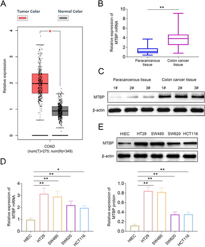
By querying the database (TCGA, https://www.cancer.gov/), we found that the levels of MTBP in colon cancer tissues was obviously higher than that in normal tissues ((A)). Next, we collected the paracancerous tissues and colon cancer tissues from the hospital. And the results ((B,C)) of qRT-PCR and western blotting showed that the levels of MTBP in colon cancer tissues were higher than that in paracancerous tissues. We also detected the expression of MTBP in normal intestinal epithelial cells (HIEC) and colon cancer cells (HT29, SW480, SW620 and HCT116). The results ((D,E)) also showed that the mRNA and protein levels of MTBP in HIEC cells were lower than that in colon cancer cells.
Figure 1. The expression of MTBP was promoted in the colon cancer tissues. (A) The expression of MTBP in colon cancer tissues and normal tissues was obtained from the TCGA database. (B, C) The expression of MTBP in colon cancer tissues and normal tissues was detected with the qRT-PCR and western blotting, respectively. (D, E) The expression of MTBP in normal intestinal epithelial cells and colon cancer cells was determined with qRT-PCR and western blotting. *p < 0.05, **p < 0.01.
MTBP promoted the proliferation, migration and invasion of colon cancer cells
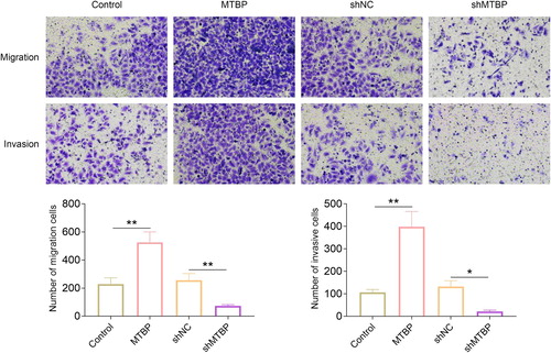
In this part, we established the MTBP overexpression and knockdown SW620 cells. Then, western blotting was used for the detection of the expression of MTBP in these cells. Results ((A)) showed that the levels of MTBP were inhibited in knockdown group and enhanced in overexpression group. The results ((B)) of CCK-8 also showed that the proliferation of the knockdown cells was suppressed compared to the cells of sh-NC groups and overexpression of MTBP promoted the proliferation of SW620 cells. Next, the changes of migration and invasion of SW620 cells were detected with the transwell assays. As shown in , the number of SW620 cells crossing the bottom membrane was promoted after the overexpression of MTBP and the knockdown of MTBP induced the decreased number of cells crossing the bottom membrane. These results indicated that the MTBP enhanced the migration and invasion of SW620 cells.
Figure 2. MTBP enhanced the proliferation of colon cancer cells. (A) The expression of MTBP in MTBP overexpression or knockdown SW620 cells was detected with the western blotting. (B) The proliferation of MTBP overexpression or knockdown SW620 cells was determined with the CCK-8. **p < 0.01.
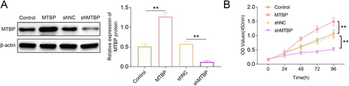
Figure 3. MTBP promoted the migration and invasion of colon cancer cells. Transwell assays were performed to detect the migration and invasion of MTBP overexpression or knockdown SW620 cells. *p < 0.05, **p < 0.01.
MTBP enhanced the stemness of colon cancer cells
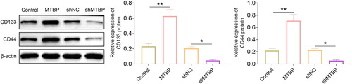
The stem cell characteristics of cancer cells means the strong proliferation and metastasis ability of these cells. And the expression of CD133 and CD44 was the characteristic of cancer stem cells (Aghajani et al. Citation2019). In this study, we found that the expression of CD133 and CD44 was promoted by the overexpression of MTBP. Meanwhile, the levels of MTBP were decreased by the suppression of MTBP ().
Figure 4. MTBP induced the expression of CD133 and CD44 in colon cancer cells. Western blotting was used for the detection of the expression of CD133 and CD44 in MTBP overexpression or knockdown SW620 cells. *p < 0.05, **p < 0.01.
MTBP promoted the proliferation, migration and invasion of colon cancer cells by activating the expression of ZEB2
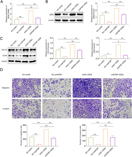
There was study revealed that the expression of MTBP could promote the proliferation and metastasis of lung cancer cells by activating the expression of ZEB2 (Pan et al. Citation2018). In our study, we found that the overexpression of MTBP also promoted the expression of ZEB2 in SW620 cells. However, the inhibition of MTBP suppressed the expression of ZEB2 ((A,B)). Next, we overexpressed the ZEB2 in MTBP knockdown SW620 cells and corresponding negative control cells. The results ((A,B)) showed that the expression of ZEB2 in the overexpression cells (shMTBP + ZEB2) was higher than that in the negative control group (NC + shMTBP). And the expression of CD133 and CD44 was also determined with the western blotting in these groups. As shown in (C), the levels of CD133 and CD44 were increased after the overexpression of ZEB2 in these cells. Furthermore, the results ((D)) of transwell assays showed that overexpression of ZEB2 abolished the inhibitory effects of knockdown MTBP on the migration and invasion of SW620 cells.
Figure 5. MTBP promoted the expression of ZEB2 in colon cancer cells. (A, B) The protein and mRNA levels of ZEB2 in MTBP overexpression or knockdown SW620 cells was determined with the western blotting. * p < 0.05, **p < 0.01.
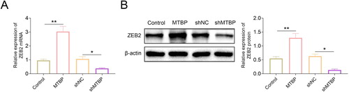
Figure 6. Overexpression of ZEB2 rescued the migration and invasion of colon cancer cells which was suppressed with the knockdown of MTBP. (A, B) The mRNA and protein levels of ZEB2 in ZEB2 overexpression SW620 cells was detected with the western blotting. (C) The expression of CD133 and CD44 in ZEB2 overexpression SW620 cells was detected with the western blotting. (D) Transwell assays were used for the detection of the migration and invasion of ZEB2 overexpression SW620 cells. * p < 0.05, **p < 0.01.
Discussion
Colon cancer is a common malignant tumor of the digestive tract in clinical practice. In addition, the incidence and mortality of this malignant tumor are both high (Walter et al. Citation2019). In recent years, with the changes of dietary structure, the incidence of colon cancer in China is increasing year by year and the patient population is also gradually getting younger (Zhang et al. Citation2015). The pathogenesis of the colon cancer has not yet been fully elucidated. However, most studies currently believed that the occurrence and development of colon cancer are the result of the combined effects of environmental and genetic factors (Wang et al. Citation2020). Furthermore, intestinal homeostasis, diet, alcohol and physical exercise are also critical factors which affect the occurrence of colon cancer (AlMutairi et al. Citation2019). The current treatment of colon cancer is still based on surgery, and chemotherapy and radiotherapy are used to assist colon cancer treatment. However, the blood metastasis, peritoneal metastasis and lymph node metastasis of colon cancer cells after surgery could cause the recurrence of colon cancer (Martínez-Meza et al. Citation2019). The spread of cancer cells caused by blood metastasis was a crucial factor which affected the survival rate of colon cancer patients (Ntavatzikos et al. Citation2019). Therefore, we urgently need to develop a new therapy to inhibit the metastasis and spread of colon cancer cells. And the suppression of the metastasis of colon cancer cells could also improve the survival rate of colon cancer patients.
Furthermore, the expression of MTBP also promoted the development of multiple types of cancer (Grieb et al. Citation2014; Wang et al. Citation2017). In the initial study, MTBP was identified as a tumor suppressor (Boyd et al. Citation2000). Subsequent study has confirmed that the biological function of MTBP is independent of MDM2, and the increased the levels of MTBP could promote the tumor progression by activating the expression of Myc (Odvody et al. Citation2010). In addition, the expression of MTBP also promoted the metastasis of hepatoma carcinoma cells by inducing the MDM2 mediated decreased levels of E-cadherin (Lu et al. Citation2015). The levels of MTBP were increased in triple negative breast cancer cells and promoted the development of breast cancer (Grieb et al. Citation2014). In our study, we found that the expression of MTBP in SW620 cells was moderate and therefore SW620 cells was selected for the establishment of MTBP overexpression or knockdown colon cancer cells. Moreover, we also revealed that overexpression of MTBP promoted the proliferation, migration and invasion of colon cancer cells. And these results also indicated that the expression of MTBP could induce the development of colon cancer.
In addition, the stem cell characteristics of cancer cells could also promote cells to obtain strong proliferation and metastasis ability (Lytle et al. Citation2018). The expression of the CD133 and CD44 were the marks of the stem cells (Singh et al. Citation2019). There was study revealed that the expression of MTBP promoted the proliferation of glioblastomas cells by activating the stem cell characteristic of these cells (Song et al. Citation2019). In our study, we also found that the expression of MTBP promoted the expression of CD133 and CD44 in colon cancer cells. This result also implied that the expression of MTBP could promote the development of colon cancer by activating the stem cell characteristic of colon cancer cells.
On the other hand, ZEB2 is also the protein which promoted the development of multiple types of cancer cells (De Coninck et al. Citation2019). Furthermore, there was study suggested that miR-498 repressed the proliferation and metastasis of cancer cells by inhibiting the expression of ZEB2 (Zhang et al. Citation2019). LncRNA-CTS could also enhance the proliferation and metastasis of cervical cancer cells by activating the expression of EMT (Feng et al. Citation2019). Moreover, the expression of miR-3653 suppressed the metastasis and EMT of colon cancer cells by inhibiting the expression of ZEB2 (Zhu et al. Citation2019). In this study, we found that the expression of MTBP has the potential to regulate the expression of ZEB2 by querying the database. The results of experiments also showed that the MTBP promoted the expression of ZEB2. Overexpression of ZEB2 abolished the inhibitory effect of MTBP knockdown on the migration and invasion of colon cancer cells. These results indicated that the expression of MTBP promoted the proliferation, migration, and invasion of colon cancer cells by enhancing the expression of ZEB2.
Above all, we clarified that the expression of MTBP promoted the proliferation, migration, and invasion of colon cancer cells by activating the ZEB2. The results of our research also provided the new target and therapy strategy for the clinic treatment of colon cancer patients.
Ethics approval
Ethical approval was obtained from the Ethics Committee of Cancer Hospital of Xinjiang Medical University.
Statement of informed consent
Written informed consent was obtained from a legally authorized representative(s) for anonymized patient information to be published in this article.
Availability of data and materials
All data generated or analyzed during this study are included in this published article.
Authors’ contributions
Paerhati shayimu and Aikeremu yusufu designed the study, supervised the data collection, Aizimaiti rehemutula analyzed the data, interpreted the data, Darebai redati, Rexida jiapaer and Rousidan tuerdi prepare the manuscript for publication and reviewed the draft of the manuscript. All authors have read and approved the manuscript.
Disclosure statement
No potential conflict of interest was reported by the author(s).
Additional information
Funding
References
- Aghajani M, Mansoori B, Mohammadi A, Asadzadeh Z, Baradaran B. 2019. New emerging roles of CD133 in cancer stem cell: signaling pathway and miRNA regulation. J Cell Physiol. 234(12):21642–21661.
- AlMutairi M, Parine NR, Shaik JP, Aldhaian S, Azzam NA, Aljebreen AM, Alharbi O, Almadi MA, Al-Balbeesi AO, et al. 2019. Association between polymorphisms in PRNCR1 and risk of colorectal cancer in the Saudi population. PloS One. 14(9):e0220931.
- Boyd MT, Vlatkovic N, Haines DS. 2000. A novel cellular protein (MTBP) binds to MDM2 and induces a G1 arrest that is suppressed by MDM2. J Biol Chem. 275(41):31883–31890.
- De Coninck S, Berx G, Taghon T, Van Vlierberghe P, Goossens S. 2019. ZEB2 in T-cells and T-ALL. Adv Biol Regul. 74:100639.
- Dienstmann R, Mason MJ, Sinicrope FA, Phipps AI, Tejpar S, Nesbakken A, Danielsen SA, Sveen A, Buchanan DD, et al. 2017. Prediction of overall survival in stage II and III colon cancer beyond TNM system: a retrospective, pooled biomarker study. Ann Oncol Off J Eur Soc Med Oncol. 28(5):1023–1031.
- Feng S, Liu W, Bai X, Pan W, Jia Z, Zhang S, Zhu Y, Tan W. 2019. LncRNA-CTS promotes metastasis and epithelial-to-mesenchymal transition through regulating miR-505/ZEB2 axis in cervical cancer. Cancer Lett. 465:105–117.
- Gray RT, Cantwell MM, Coleman HG, Loughrey MB, Bankhead P, McQuaid S, O'Neill RF, Arthur K, Bingham V, et al. 2017. Evaluation of PTGS2 expression, PIK3CA mutation, aspirin Use and colon cancer survival in a population-based cohort study. Clin Transl Gastroenterol. 8(4):e91.
- Grieb BC, Chen X, Eischen CM. 2014. MTBP is overexpressed in triple-negative breast cancer and contributes to its growth and survival. Mol Cancer Res MCR. 12(9):1216–1224.
- Hanrahan K, O'Neill A, Prencipe M, Bugler J, Murphy L, Fabre A, Puhr M, Culig Z, Murphy K, et al. 2017. The role of epithelial-mesenchymal transition drivers ZEB1 and ZEB2 in mediating docetaxel-resistant prostate cancer. Mol Oncol. 11(3):251–265.
- Li N, Babaei-Jadidi R, Lorenzi F, Spencer-Dene B, Clarke P, Domingo E, Tulchinsky E, Vries RGJ, Kerr D, et al. 2019. An FBXW7-ZEB2 axis links EMT and tumour microenvironment to promote colorectal cancer stem cells and chemoresistance. Oncogenesis. 8(3):13.
- Lu S, Zhou W, Wei H, He L, Li L. 2015. MTBP promotes the Invasion and metastasis of hepatocellular carcinoma by enhancing the MDM2-mediated degradation of E-cadherin. Dig Dis Sci. 60(12):3681–3690.
- Lytle NK, Barber AG, Reya T. 2018. Stem cell fate in cancer growth, progression and therapy resistance. Nat Rev Cancer. 18(11):669–680.
- Martínez-Meza S, Díaz J, Sandoval-Bórquez A, Valenzuela-Valderrama M, Díaz-Valdivia N, Rojas-Celis V, Contreras P, Huilcaman R, Ocaranza MP, et al. 2019. AT2 receptor mediated activation of the tyrosine phosphatase PTP1B blocks caveolin-1 enhanced migration, invasion and metastasis of cancer cells. Cancers (Basel). 11:9.
- Ntavatzikos A, Spathis A, Patapis P, Machairas N, Vourli G, Peros G, Papadopoulos I, Panayiotides I, Koumarianou A. 2019. TYMS/KRAS/BRAF molecular profiling predicts survival following adjuvant chemotherapy in colorectal cancer. World J Gastrointest Oncol. 11(7):551–566.
- Odvody J, Vincent T, Arrate MP, Grieb B, Wang S, Garriga J, Lozano G, Iwakuma T, Haines DS, et al. 2010. A deficiency in Mdm2 binding protein inhibits Myc-induced B-cell proliferation and lymphomagenesis. Oncogene. 29(22):3287–3296.
- Pan B, Han H, Wu L, Xiong Y, Zhang J, Dong B, Yang Y, Chen J. 2018. MTBP promotes migration and invasion by regulation of ZEB2-mediated epithelial-mesenchymal transition in lung cancer cells. Onco Targets Ther. 11:6741–6756.
- Si W, Huang W, Zheng Y, Yang Y, Liu X, Shan L, Zhou X, Wang Y, Su D, et al. 2015. Dysfunction of the reciprocal feedback loop between GATA3- and ZEB2-nucleated repression programs contributes to breast cancer metastasis. Cancer Cell. 27(6):822–836.
- Singh SR, Aggarwal P, Hou SX. 2019. Cancer stem cells and stem cell tumors in drosophila. Adv Exp Med Biol. 1167:175–190.
- Snaebjornsson P, Jonasson L, Olafsdottir EJ, van Grieken NCT, Moller PH, Theodors A, Jonsson T, Meijer GA, Jonasson JG. 2017. Why is colon cancer survival improving by time? A nationwide survival analysis spanning 35 years. Int J Cancer. 141(3):531–539.
- Song Y, Zhang L, Jiang Y, Hu T, Zhang D, Qiao Q, Wang R, Wang M, Han S. 2019. MTBP regulates cell survival and therapeutic sensitivity in TP53 wildtype glioblastomas. Theranostics. 9(20):6019–6030.
- Sun S, Yang X, Qin X, Zhao Y. 2020. TCF4 promotes colorectal cancer drug resistance and stemness via regulating ZEB1/ZEB2 expression. Protoplasma. 257(3):921–930.
- Walter V, Boakye D, Weberpals J, Jansen L, Haefeli WE, Martens UM, Knebel P, Chang-Claude J, Hoffmeister M, et al. 2019. Decreasing Use of chemotherapy in older patients with stage III colon cancer irrespective of comorbidities. J Natl Comprehens Cancer Network JNCCN. 17(9):1089–1099.
- Wang J, Cai H, Liu Q, Xia Y, Xing L, Zuo Q, Zhang Y, Chen C, Xu K, et al. 2020. Cinobufacini inhibits colon cancer Invasion and Metastasis via suppressing Wnt/β-catenin Signaling pathway and EMT. Am J Chin Med. 48(3):703–718.
- Wang W, Chen Z, Jin J, Long Z, Liu X, Cai H, Zhou Y, Huang H, Wang Y. 2017. MDM2 binding protein as a predictor of metastasis and a novel prognostic biomarker in patients with gastric cancer. Oncol Lett. 14(6):6409–6416.
- Wang F, Zhu W, Yang R, Xie W, Wang D. 2019. LncRNA ZEB2-AS1 contributes to the tumorigenesis of gastric cancer via activating the Wnt/β-catenin pathway. Mol Cell Biochem. 456(1-2):73–83.
- Wu X, Zhu H, Xie Y, Gu X, Zhang L, Huang L. 2020. Knockdown of ZEB2-AS1 inhibits cell invasion and induces apoptosis in colorectal cancer. J B.U.ON. Off J Balkan Union Oncol. 25(1):194–201.
- Zanjirband M, Rahgozar S. 2019. Targeting p53-MDM2 interaction using small molecule inhibitors and the challenges needed to be addressed. Curr Drug Targets. 20(11):1091–1111.
- Zhang Y, Shi J, Huang H, Ren J, Li N, Dai M. 2015. Burden of colorectal cancer in China. Zhonghua liu Xing Bing xue za zhi=Zhonghua Liuxingbingxue Zazhi. 36(7):709–714.
- Zhang X, Xu X, Ge G, Zang X, Shao M, Zou S, Zhang Y, Mao Z, Zhang J, et al. 2019. Mir-498 inhibits the growth and metastasis of liver cancer by targeting ZEB2. Oncol Rep. 41(3):1638–1648.
- Zhu W, Luo X, Fu H, Liu L, Sun P, Wang Z. 2019. MiR-3653 inhibits the metastasis and epithelial-mesenchymal transition of colon cancer by targeting Zeb2. Pathol Res Pract. 215(10):152577.