
 ?Mathematical formulae have been encoded as MathML and are displayed in this HTML version using MathJax in order to improve their display. Uncheck the box to turn MathJax off. This feature requires Javascript. Click on a formula to zoom.
ABSTRACT
The American Association of Endodontists (AAE) released a case difficulty assessment form to help general dentists and students treat cases within their expertise or refer advanced cases to reduce the risk of iatrogenic errors. The purpose of this study was to determine the incidence of iatrogenic complications after the use of the case difficulty assessment form. Arandom sample of 1000 cases that received root canal treatment in undergraduate clinics during the academicyear (2016–2017) was selected. Case difficulty assessment was made for each case before treatment onset. Once the case was approved for treatment in the undergraduate clinics, the endodontic treatments were performed following the standard procedure the endodontic department of the Dentistry School of King Abdulaziz University mandates. Digital radiographs were obtained during routine root canal treatment and were evaluated by four observers to detect any iatrogenic errors, after which the data were analyzed statistically. Iatrogenic errors were correlated significantly with case difficulty (p= 0.003), and were detected in 22.1% of all teeth treated in the undergraduate clinics. Underfilling accounted for the highestpercent of errors detected (8.4%), followed by ledge formation (4.2%). Molar teeth had the highest frequency of errors, and mesio-buccal roots of maxillary molars showed the highest percentage of errors. The AAE developed asignificantly useful tool to determine the difficulty of each case treated in undergraduate clinics, and following their recommendations will minimize the risk of iatrogenic errors.
1. Introduction
One of the important role of endodontic treatment is to eliminate primary bacterial infection, prevent reinfection, and allow periapical tissues to heal [Citation1]. The outcome of such treatment is determined largely by meticulous techniques of preparing the canal system that may result in a success rate as high as 94% [Citation2,Citation3].
Poor preparation techniques and low quality root fillings are significant factors in the healing of periapical tissue [Citation4–Citation6]. Iatrogenic errors during the mechanical preparation phase, such as perforations, ledges, and instrument fractures, compromise the treatment [Citation7]. Thus, endodontic iatrogenic errors should be kept to a minimum, as they affect the quality of the treatment and may jeopardize the outcome subsequently.
Several studies have shown that undergraduate students perform treatments with low technical quality because of insufficient experience [Citation8]. Yet, the occurrence of procedural errors cannot be avoided even with close clinical supervision [Citation9,Citation10]. In addition, patients who require endodontic therapy in undergraduate clinics present with different levels of difficulties, such as severely curved, calcified, and C-shaped canals.
The American Association of Endodontists (AAE) released a case difficulty assessment form to classify endodontic cases as minimally, moderately, or highly difficult (http://www.aae.org/caseassessment/) that provides a practical method to make case selection more efficient and consistent. This form helps practitioners plan endodontic treatment and provides a more useful method with which to make referral decisions. The assessment form is divided into four categories of considerations: patient, diagnostic, treatment, and additional considerations. Guidelines are given for each level of difficulty to help determine whether the case is appropriate for general dentists/students or needs to be referred to a specialized endodontist.
The purpose of this study was to determine the incidence of iatrogenic complications: corono-cervical, furcation, strip, lateral root, and apical perforations, ledge formation, fractured instruments, overfilling, and underfilling after the use of the case difficulty assessment form in all cases treated in the undergraduate clinics in the Dentistry School at King Abdulaziz University, Jeddah, Saudi Arabia.
2. Materials and methods
This study commenced after obtaining ethical approval from the Research Ethics Committee of King Abdulaziz University (Registration No. 075-05-18). The study has been conducted in full accordance with the World Medical Association Declaration of Helsinki.
Sample size was calculated based on the major outcome (iatrogenic errors). The following equation was used: [Citation11,Citation12]
Z1-α/2 = Standard normal variate (at 5% type 1error (P < 0.05) it is 1.96 and at 1% type 1 error (P < 0.01) it is 2.58). As in majority of studies P values are considered significant below 0.05, hence 1.96 is used in formula.
p = Expected proportion in population based on previous studies or pilot studies.
d = Absolute error or precision – Has to be decided by researcher
The sample size calculation revealed that if there is truly correlation between the use of case difficulty assessment and the occurrence of iatrogenic errors, then at least 200 cases with iatrogenic errors are required to produce a statistical significant result with probability (power) = 0.95. The Type I error probability associated with the test of this null hypothesis is a = .05.
In this cross-sectional retrospective study, a random sample of 1000 patients who received root canal treatment in the undergraduate clinics during academic year 2016–2017 was selected. All cases were from patients between 18–80 years of age referred within the academic year to the Dentistry School. Undergraduate 5th and 6th year students were able to treat endodontic cases with single, double, or multi-rooted teeth that have simple root canals. Advanced, complex cases that require strong experience and advanced techniques, such as teeth with severely curved roots, double curvature, calcified canals, or C-shaped canals were referred to the post-graduate clinics.
During the first visit, clinical and radiographic evaluations were performed. After clinical and radiographic examinations and assessments, a case difficulty assessment was required before treatment begins. If it is highly difficult, the case was referred automatically to post-graduate clinics. Once cases were approved for treatment in the undergraduate clinic, endodontic treatments were performed followed the standard protocol of treatment of the endodontic department in King Abdulazizi University. After rubber dam application to ensure proper isolation, straight-line access was achieved using Gates-Glidden drills number 2–4 (Premier Dental, Norristown, PA, USA). Working lengths were determined using digital radiographs and apex locators (Root ZX, J. Morita Inc., Irvine, California, USA). All canals were instrumented with the step-back technique using either Nickel Titanium (NiTi) K-files (DENTSPLY, Tulsa, Oklahoma, USA) with 0.02 taper or ProTaper Next (PTN; DENTSPLY Maillefer, Ballaigues, Switzerland). Root canals were irrigated continuously with 5.25% sodium hypochlorite solution. Root fillings were carried out with gutta percha (Spident Co. Ltd., Incheon, Republic of Korea) and Tubli-Seal™ Zinc Oxide Eugenol Root Canal Sealer (Kerr, Salerno, Italy) using the cold lateral compaction technique. The teeth were restored temporarily and postoperative radiographs were taken.
Iatrogenic errors were recorded from the inter-operative or immediate postoperative digital radiographs of each case, as well as any progress notes that mentioned one of the procedural errors. Digital radiographs were evaluated with Kodak Dental Imaging Software version 7 for Windows (Carestream Health, Inc., Rochester, New York, USA), which provides the option to magnify, change contrast, etc. Each root canal was investigated separately in multi-rooted teeth, i.e. molars and premolars.
Four observers (two dental interns and two endodontic consultants) evaluated all radiographs. All observers were calibrated initially [Citation13]. The method of viewing the radiographs was standardized and an evaluation form was designed to record the information gathered from the radiographs. Then the examiners agreement was measured by Cohen kappa test [Citation14,Citation15]. using one hundred radiographs for assessment.
Iatrogenic errors were defined using strict criteria. Ledge formation was identified when the filling material appeared to be 1 mm or shorter than the working length, or when the root filling deviated from the original canal path. Furcation perforation was recorded if any obturating material extruded from the furcation in multi-rooted teeth. Strip perforation was recognized when filling material was seen in the lateral (inner) wall of curved roots. Root perforation was reported when filling material extruded from any other area of a root other than the furcational area or the convex wall of curved roots. Fractured instrument was determined when a fractured instrument was observed inside a root canal or its tip extended into the periapical area either clinically or radiographically.
Once the investigation of radiographs was completed, all associated data were gathered and analyzed statistically.
3. Statistical analysis
Descriptive statistics were performed for each type of iatrogenic error. The Chi-squared test was performed to determine whether case difficulty affected the occurrence of endodontic iatrogenic errors and influenced the likelihood of endodontic mishaps. Data were analyzed using SPSS v. 20.0 (SPSS Inc., Chicago, USA). The p value was set at 0.05.
4. Results
This retrospective cross-sectional study measured the incidence of iatrogenic errors committed by undergraduate dental students at King Abdulaziz University after the use of the AAE case difficulty assessment form. The k-values obtained for the inter-examiner reliability was 0.80, which indicate strong agreement. A total of 1000 cases was evaluated for iatrogenic errors. Three hundred fifty cases were treated by 5th year students and 650 cases by 6th year students. Of that number, only 221 (22.1%) were found to have errors; 72 cases (7%) by 5th year students and 149 cases (15%) by 6th year students.
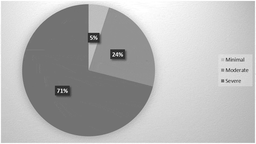
More than half of the cases were categorized as minimally difficult (54%) compared with the moderately difficult category (27%) and highly difficult category (19%). shows the percentage of cases of each difficulty associated with iatrogenic errors; 11 cases (5%) in the minimally difficult level were found to have errors. The remaining 53 cases (24%) in the moderately difficult category and 157 cases (71%) in the highly difficult category demonstrated errors.
Figure 1. The percentage of iatrogenic errors in the high, moderate, and minimal difficulty categories
shows the percentage of all types of iatrogenic errors. Corono-cervical perforation was detected in one 6th year student case, 0.1% of the total number of cases and 0.5% of all cases with iatrogenic errors. Furcation perforation was found in two 6th year student cases, 0.2% of the total number of cases and 1% of all cases with iatrogenic errors. Strip perforation was seen in three 6th year student cases, 0.3% of the total number of cases and 1.2% of all cases with iatrogenic errors. Lateral root perforation was found in seven cases, three treated by 5th year and four by 6th year students, 0.7% of the total number of cases and 3% of all cases with iatrogenic errors. Apical perforation was detected in 12 cases, two on the part of 5th year and 10 on the part of 6th year students, 1.2% of the total number of cases and 5.3% of all cases with iatrogenic errors. Thus, all types of perforations accounted only for 25 cases (2.5% of the total number of cases and 11% of the iatrogenic errors cases).
Figure 2. Percentage of iatrogenic errors in the total number of teeth treated (N = 1000) in each academic year
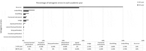
Other types of errors, such as ledge formation, were detected in 42 cases, 15 of 5th year and 27 of 6th year students’ cases, 4.2% of the total cases and 19% of the iatrogenic error cases. Fractured instruments were found in 30 cases, 13 of 5th year and 17 of 6th year students’ cases, 3% of the total cases and 13.5% of the iatrogenic errors. Overfilling was detected in 40 cases, 12 of 5th year and 28 of 6th year students, 4% of the total cases and 18% of iatrogenic errors. Underfilling accounted for the greatest number of iatrogenic errors detected, and was reported in 84 cases, 27 of 5th year and 57 of 6th year students, 8.4% of the total cases and 38% of iatrogenic errors cases.
The data on the relation between the presence of iatrogenic errors and tooth type in the dental arch are presented in . Of all teeth with procedural errors, maxillary and mandibular molars demonstrated the highest frequency of errors (161 cases, 73%) followed by premolars in both arches (42 cases, 18.5%) and anterior teeth (19 cases, 8.5%).
Table 1. The relation between the presence of iatrogenic errors and tooth type; N (%)
Furthermore, the mesiobuccal roots of maxillary molars showed the highest percentage of errors (58 cases: 26% of the total sample with iatrogenic errors), followed by the buccal roots of maxillary premolars (47 cases: 21% of cases with iatrogenic errors) and the mesial roots of mandibular molars (43 cases: 19.5% of cases with iatrogenic errors) as shown in .
Table 2. The relation between the presence of iatrogenic errors and root type; N (%)
Iatrogenic errors were associated significantly with case difficulty (p = 0.003), as shown in . However, there was no significant correlation between academic year and case difficulty (p = 0.828) or between academic year and types of iatrogenic errors (p = 0.635). In addition, there was no significant relation between root type and iatrogenic errors (p = 0.119) while the association between tooth type and iatrogenic errors was significant (p = 0.020).
Table 3. The relation between the presence of iatrogenic errors and case difficulty; N (%)
5. Discussion
The purpose of this study was to determine the incidence of iatrogenic complications, including the presence of ledges, perforations, fractured instruments, overfilling, and underfilling in cases treated by undergraduate dental students at King Abdulaziz University after the case difficulty assessment form was introduced.
Two dental interns and two endodontic consultants evaluated five periapical radiographs per case. All radiographs were taken during routine root canal treatment. A periapical radiograph is the ideal method used to determine the quality of root canal treatments performed in undergraduate or postgraduate teaching institutions [Citation9,Citation16–Citation19]. Although conventional periapical radiographs are limited because they provide a two-dimensional image of a three-dimensional object [Citation9], Alver et al [Citation20]. found no significant difference in different imaging techniques’ ability to detect different iatrogenic errors.
According to the AAE case difficulty assessment form [Citation21], the conditions listed should be considered potential risk factors that may complicate treatment. Minimally difficult cases exhibited factors listed only in the Minimal difficulty category, and a competent practitioner with limited experience should be able to achieve a predictable treatment outcome. Moderately difficult cases are complex and exhibit one or more factors listed in the Moderate difficulty category, and achieving a predictable treatment outcome will be challenging for a competent, experienced practitioner. A case is considered highly difficult if it includes at least one factor in the high difficulty category and multiple factors in the moderate or minimal difficulty categories. This category is considered to be challenging even for experienced practitioners.
In our study, the total percentage of iatrogenic errors was approximately 22%. Although this percentage is somewhat high compared to Kulic et al [Citation22], who reported only 7 teeth with iatrogenic errors and Lynch and Burke [Citation8], who did not detect any iatrogenic errors, it is considerably lower compared to Yousuf, Khan and Mehdi [Citation23], who identified iatrogenic errors in 32.8% of the teeth, and Haji-Hassani et al [Citation24]. in 66% of the teeth. The most common iatrogenic errors encountered in our study were underfilling (8.4%), followed by ledge formation (4.2%), overfilling (4%), and fractured instrument (3%). Balto et al [Citation25]. reported that the most common iatrogenic errors found in their study were ledges (14%), followed by apical transportations (7%) and apical perforations (7%). This high percentage of iatrogenic errors could be attributable to the failure to follow the AAE guidelines strictly with respect to referring difficult cases. At the end of the academic year, when a certain number of cases must be completed before graduation, students take the risk of treating difficult cases that may increase the probability of iatrogenic errors.
Underfilling usually occurs if incomplete mechanical instrumentation or ledge formation of the root canal occurs during the procedure. Incomplete instrumentation results from incorrect measurement of the working length or canal blockage with debris attributable to inadequate irrigation and recapitulation of canal patency [Citation7]. In our study, underfilling accounted for the highest percentage of iatrogenic errors, and was found in 8.4% of the total number of cases treated by undergraduate students. Mandibular molars had the highest incidence of underfilling, 18% of all cases with iatrogenic errors. The fact that the anatomy of such teeth has curved roots that hold more than one canal in a root may explain this finding [Citation4].
With respect to ledge formation, the rate reported in this study was 4.2% of the total number of cases treated in undergraduate clinics. This percentage was lower than most other studies have reported, including Greene and Krell, 46% [Citation26], Kapalas and Lambrianidis, 52% [Citation27], Eleftheriadis and Lambrianidis, 39% [Citation16], Khabbaz et al., 54.8% [Citation28], Mozayeni et al., 26% [Citation29], and Balto et al., 14% [Citation25]. However, it was close to Smadi et al., 5.2% [Citation30], Vukadinov et al., 2.8% [Citation9], and Eskandarloo et al., 2.8% [Citation31]. Anterior and premolar teeth exhibited ledge formation less frequently than did molars. Molars have a higher prevalence of narrow and curved canals that make root canal treatment more challenging. The low percentage of ledge formation in our study may be attributable to the use of the Gates-Glidden to maintain straight line access, which helps decrease the occurrence of ledges, as previous studies have reported [Citation32,Citation33]. In addition, the use of NiTi files and step-down or passive step-back methods have been proposed to reduce ledge formation [Citation34]. The analysis of canal curvature related to the presence of ledges indicated that there was a significant difference between the percentages reported in moderately curved and straight canals. Canal curvature was the principal factor related to the occurrence of ledges in canals with moderate curvature [Citation16,Citation18].
Overfilling was detected in 4% of all cases treated in undergraduate dental clinics. This may be attributable to inadequate length determination or over-instrumentation. Molars have the shortest roots compared to other teeth, which makes them more susceptible to this type of error [Citation23]. Over-instrumentation damages the apical constriction, which makes it difficult to obtain an adequate apical seal and confine obturation materials within the root canal system. Over-instrumentation in the undergraduate clinic may be attributable to the lack of attention to reference points.
Fractured instruments were found in only 3% of the total number of cases. Our study is consistent with that of Haug et al [Citation35], who reported that the incidence of instrument separation was 2.3%, and of Iqbal et al [Citation36], who reported 1.9% for fractured instruments. Moreover, Rafeek et al [Citation37]. identified fractured instruments in 1.5% of the root canals.
All types of perforations accounted only for 2.5% of the total number of cases treated in the undergraduate clinics. This percentage was significantly lower than that Eskandarloo et al. which was 17.6% of the cases [Citation38]. However, it was comparable to Eleftheriadis and Lambrianidis [Citation16], Yavari et al [Citation39], and Smadi et al.’s [Citation30] results, in which perforations ranged from 1.9% to 2.7%. This low percentage of perforation could be attributable to two reasons: the use of case difficulty assessment form is required for each case, and any difficult case was referred to postgraduate students. Thus, there were minimal complicated teeth in our sample.
The percentage of iatrogenic errors in this study was evaluated according to tooth type and a significant difference was found among them. Upper and lower molars demonstrated the highest incidence of iatrogenic errors. This result is consistent with those in other studies that reported a correlation between the frequency of iatrogenic errors and the tooth type evaluated [Citation25,Citation28,Citation29]. Moreover, several other studies have indicated that most of the procedural errors occurred in molars and teeth that possessed severely curved canals [Citation16,Citation17,Citation25,Citation40,Citation41]. The AAE considers first molar teeth moderately difficult and they require treatment by skilled dental students under the supervision of highly-qualified endodontic instructors, or should be referred to a graduate student or endodontist. Therefore, it is recommended to avoid having 5th year students treat molar teeth until they master single or double straight root canals, after which they can treat molar teeth in their next year.
This study had some limitations including its retrospective design. In addition, this study was conducted at one dental school and its results may not be generalizable to students at other schools. On a positive note, all assessments for this study took place throughout the same year, which would minimize errors due to different levels of skills. Future study with a prospective design and multiple schools is required to minimize bias and confounding variables.
6. Conclusion
This study showed that endodontic iatrogenic errors are associated significantly with the difficulty of the case. The AAE has provided an important and valuable tool, the case difficulty assessment form, which can help the students and general dentists evaluate the level of difficulty associated with each case critically. The awareness and realization of the practitioners’ limits makes case selection more efficient and minimizes the risk of endodontic iatrogenic errors.
Acknowledgments
I thank Eng. Ahmed Bamousa heartily for his guidance and suggestions during this study, particularly in designing the graphs. This research received no financial support.
Disclosure statement
No potential conflict of interest was reported by the authors.
References
- Sjogren U, Figdor D, Persson S, et al. Influence of infection at the time of root filling on the outcome of endodontic treatment of teeth with apical periodontitis. Int Endod J. 1997;30(5):297–7.
- Imura N, Pinheiro ET, Gomes BP, et al. The outcome of endodontic treatment: a retrospective study of 2000 cases performed by a specialist. J Endod. 2007;33(11):1278–1282.
- Lazarski MP, Walker WA 3rd, Flores CM, et al. Epidemiological evaluation of the outcomes of nonsurgical root canal treatment in a large cohort of insured dental patients. J Endod. 2001;27(12):791–796.
- Smith CS, Setchell DJ, Harty FJ. Factors influencing the success of conventional root canal therapy–a five-year retrospective study. Int Endod J. 1993;26(6):321–333.
- Sjogren U, Hagglund B, Sundqvist G, et al. Factors affecting the long-term results of endodontic treatment. J Endod. 1990;16(10):498–504.
- Dugas NN, Lawrence HP, Teplitsky PE, et al. Periapical health and treatment quality assessment of root-filled teeth in two Canadian populations. Int Endod J. 2003;36(3):181–192.
- Lin LM, Rosenberg PA, Lin J. Do procedural errors cause endodontic treatment failure? J Am Dent Assoc. 2005;136(2):187–193. quiz 231
- Lynch CD, Burke FM. Quality of root canal fillings performed by undergraduate dental students on single-rooted teeth. Eur J Dent Educ. 2006;10(2):67–72.
- Vukadinov T, Blazic L, Kantardzic I, et al. Technical quality of root fillings performed by undergraduate students: a radiographic study. ScientificWorldJournal. 2014;2014:751274.
- Zambon da Silva P, Carlos Ribeiro F, Machado Barroso Xavier J, et al. Radiographic evaluation of root canal treatment performed by undergraduate students, part I; iatrogenic errors. Iran Endod J. 2018;13(1):30–36.
- Lwanga K, Lemeshow S. Sample size determination in health studies: A practical manual. Geneva, Switzerland: World Health Organization. 1991;25.
- Charan J, Biswas T. How to calculate sample size for different study designs in medical research? Indian J Psychol Med. 2013;35(2):121–126.
- Reit C. The influence of observer calibration on radiographic periapical diagnosis. Int Endod J. 1987;20(2):75–81.
- Valachovic RW, Douglass CW, Berkey CS, et al. Examiner reliability in dental radiography. J Dent Res. 1986;65(3):432–436.
- Hunt RJ. Percent agreement, Pearson’s correlation, and kappa as measures of inter-examiner reliability. J Dent Res. 1986;65(2):128–130.
- Eleftheriadis GI, Lambrianidis TP. Technical quality of root canal treatment and detection of iatrogenic errors in an undergraduate dental clinic. Int Endod J. 2005;38(10):725–734.
- Er O, Sagsen B, Maden M, et al. Radiographic technical quality of root fillings performed by dental students in Turkey. Int Endod J. 2006;39(11):867–872.
- Dadresanfar B, Mohammadzadeh Akhlaghi N, Vatanpour M, et al. Technical quality of root canal treatment performed by undergraduate dental students. Iran Endod J. 2008;3(3):73–78.
- Schneider SW. A comparison of canal preparations in straight and curved root canals. Oral Surg Oral Med Oral Pathol. 1971;32(2):271–275.
- Alves RAA, Souza JB, Alencar AHG, et al. Detection of procedural errors with stainless steel and NiTi instruments by undergraduate students using conventional radiograph and cone beam computed tomography. Iran Endod J. 2013;8(4):160.
- American Association of Endodontists. Available from: http://www.aae.org/caseassessment/. 2010.
- Kulić L, Nogo-Živanović D, Krunic J, et al. Radiological assessment of the quality of root canal fillings in teeth endodontically treated at students’ practical sessions. Serb Dent J. 2011; 58.
- Yousuf W, Khan M, Mehdi H. Endodontic procedural errors: frequency, type of error, and the most frequently treated tooth. Int J Dent. 2015;2015:673914.
- Haji-Hassani N, Bakhshi M, Shahabi S. Frequency of iatrogenic errors through root canal treatment procedure in 1335 charts of dental patients. J Int Oral Health. 2015;7(Suppl 1):14–17.
- Balto H, Al Khalifah S, Al Mugairin S, et al. Technical quality of root fillings performed by undergraduate students in Saudi Arabia. Int Endod J. 2010;43(4):292–300.
- Greene KJ, Krell KV. Clinical factors associated with ledged canals in maxillary and mandibular molars. Oral Surg Oral Med Oral Pathol. 1990;70(4):490–497.
- Kapalas A, Lambrianidis T. Factors associated with root canal ledging during instrumentation. Endod Dent Traumatol. 2000;16(5):229–231.
- Khabbaz MG, Protogerou E, Douka E. Radiographic quality of root fillings performed by undergraduate students. Int Endod J. 2010;43(6):499–508.
- Mozayeni MA, Asnaashari M, Modaresi SJ. Clinical and radiographic evaluation of procedural accidents and errors during root canal therapy. Iran Endod J. 2006;1(3):97–100.
- Smadi L, Hammad M, El-Ma’aita A. Evaluation of the quality of root canal treatments performed by dental undergraduates: is there a need to review preclinical endodontic courses. Am J Educ Res. 2015;3(12):1554–1558.
- Hendi SS, Karkehabadi H, Eskandarloo A. Iatrogenic errors during root canal instrumentation performed by dental students. Iran Endod J. 2018;13(1):126–131.
- Barbieri N, Leonardi DP, Baechtold MS, et al. Influence of cervical preflaring on apical transportation in curved root canals instrumented by reciprocating file systems. BMC Oral Health. 2015;15:149.
- Sousa K, Andrade-Junior CV, Silva JM, et al. Comparison of the effects of tripleGates and gates-glidden burs on cervical dentin thickness and root canal area by using cone beam computed tomography. J appl oral sci. 2015;23(2):164–168.
- Walia HM, Brantley WA, Gerstein H. An initial investigation of the bending and torsional properties of Nitinol root canal files. J Endod. 1988;14(7):346–351.
- Haug SR, Solfjeld AF, Ranheim LE, et al. Impact of case difficulty on endodontic mishaps in an undergraduate student clinic. J Endod. 2018;44(7):1088–1095.
- Iqbal MK, Kohli MR, Kim JS. A retrospective clinical study of incidence of root canal instrument separation in an endodontics graduate program: a PennEndo database study. J Endod. 2006;32(11):1048–1052.
- Rafeek RN, Smith WA, Mankee MS, et al. Radiographic evaluation of the technical quality of root canal fillings performed by dental students. Aust Endod J. 2012;38(2):64–69.
- Eskandarloo A, Karkehabadi H, Hoseini Hashemi SZ, et al. Radiographic quality of root canal obturation performed by fifth year students of Hamadan dental school. Iran Endod J. 2017;12(2):236–241.
- Yavari H, Samiei M, Shahi S, et al. Radiographic evaluation of root canal fillings accomplished by undergraduate dental students. Iran Endod J. 2015;10(2):127–130.
- Barrieshi-Nusair KM, Al-Omari MA, Al-Hiyasat AS. Radiographic technical quality of root canal treatment performed by dental students at the dental teaching center in Jordan. J Dent. 2004;32(4):301–307.
- Alsulaimani RS, Al-Manei KK, Alsubait SA, et al. Effects of clinical training and case difficulty on the radiographic quality of root canal fillings performed by dental students in Saudi Arabia. Iran Endod J. 2015;10:268–273.