ABSTRACT
Methanol bears semblance to ethanol in smell and taste, thus, individuals who indulge in alcohol may fall back on it in societies where alcohol consumption is illegal or difficult to come by despite the life-threatening neurologic sequelae of methanol toxicity. Stroke is an uncommon outcome of methanol poisoning. We presented two cases of methanol-induced infarctive and hemorrhagic stroke in biological brothers who were simultaneously involved in an illicit ingestion of methanol. One of them developed infarctive stroke while the other had infarctive stroke with hemorrhagic transformation. We have highlighted the differences and similarity in the course of their illnesses.
1. Introduction
Methanol is an organic constituent of several commercially available industrial solvents and a component of adulterated alcoholic beverages. It is consumed either intentionally through abuse or attempted suicide or inadvertently through misuse or accident [Citation1,Citation2]. It has also been described in victims of fraudulent adulteration of alcoholic drinks [Citation3].
Methanol is oxidized to formaldehyde in the liver and ultimately formic acid resulting in severe metabolic acidosis. Formic acid is more than six times as toxic when compared to methanol. It is a known cause of generalized body weakness, nausea, vomiting, headache, abdominal pain, breathlessness, convulsion, and on rare occasions, neurological sequelae such as convulsion and coma. Nevertheless, cerebral infarcts and cerebral hemorrhages are considered rare complications of acute methanol poisoning. Occurrence of intracerebral hemorrhage has been attributed to the application of systemic anticoagulation during hemodialysis by some authors [Citation4,Citation5]. Combination of infarctive stroke, hemorrhagic stroke, and blindness is somewhat rare and only few cases of stroke and blindness have been reported [Citation6].
In spite of the life-threatening nature of methanol poisoning, it is poorly recognized by health care givers and it is often not considered in the differential diagnosis of stroke in developing countries [Citation6].
We report two cases of stroke that occurred simultaneously in biological brothers following illicit ingestion of methanol.
2. Cases
2.1. Case 1
RM, a 47-year-old mason was referred to the emergency department with a two-day history of nausea, vomiting, headache, abdominal pain, confusion and loss of consciousness following illicit use of a substance containing methanol. The symptoms occurred about twelve hours after consumption of the substance with his younger brother both of whom are foreigners resident in Saudi Arabia. Additionally, he had history of low urine output which started a day after the onset of the illness. He had no history of cardiovascular risk factors.
The salient findings on examination were: a middle-age man, unconsciousness (GCS = 3/15), hypotension, quadriplegia, minimally reactive midriasis bilaterally, and bilateral papilledema.
He was admitted with a presumed diagnosis of methanol intoxication complicated by stroke and acute renal injury. He was intubated and transferred to the intensive care unit (ICU).
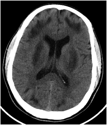
His arterial blood gases showed severe metabolic acidosis. Findings of the other laboratory investigations included normal chest X-ray, high methanol blood level leukocytosis, hyperglycemia, normal PT, normal PTTK, normal INR, elevated serum lactate, elevated creatinine and urea level with high anion gap and high osmolal gap. His blood and urine culture yielded no organism. ECG showed tachycardia and the initial brain CT was normal (30 minutes after admission). Intravenous fluid, proton pump inhibitor and bicarbonate were administered and the patient had two sessions of hemodialysis. Follow-up CT on day 4 showed low-density confluent lesions in superficial white matter, frontal, occipital lobes and putamen consistent with acute toxic edema and infarction (). His EEG showed generalized polymorphic delta waves suggestive of encephalopathy. After the foregoing treatment, the patient regained consciousness six days into admission. However, he was blind in both eyes and quadriplegic. He died about 2 months after his presentation in our facility.
2.2. Case 2
MMM, a 37-year-old mason who is a younger brother of case 1 was brought to the emergency department with his elder brother with a two-day history of nausea, vomiting, headache, fever, abdominal pain, confusion and altered consciousness after use of illicit alcoholic beverage. His urine output was adequate. He had no history of cardiovascular risk factors.
The salient findings on his examination were: coma (GCS = 3/15), acidotic breathing pattern, pyrexia (temperature = 39°C) tachycardia (P.R = 108 beats/min), minimally reactive midriasis bilaterally, and papilledema.
He was admitted with a presumed diagnosis of methanol intoxication complicated by stroke, intubated, mechanically ventilated and transferred to the intensive care unit (ICU).
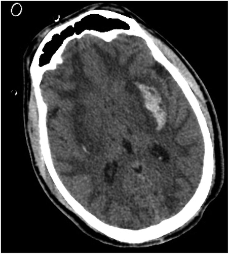
Arterial blood gases showed severe metabolic acidosis (pH, 6.8; pCO2 27; Bicarbonate, 7.6). He had normal chest X-ray, high methanol blood level, normal WBC normal RBS, normal PT, normal PTTK, normal INR, elevated serum lactate, creatinine kinase = 6.1 mmol, normal creatinine, and normal urea level with high anion gap and high osmolal gap (48 mEq/L). His complete blood count and liver function tests at presentation were within normal limits. The initial sepsis work-up was normal, ECG showed tachycardia and the initial brain CT (60 minutes after admission) revealed low density confluent lesions in superficial white matter, frontal, occipital lobes and putamen consistent with acute toxic edema and infarction as well as bilateral putaminal hemorrhagic changes (left ≫right) (). His EEG showed generalized polymorphic delta waves suggestive of encephalopathy. Blood culture, 3 weeks into his admission, yielded multiresistance Klebsiella.
Figure 2. Case 2 brain CT showing hypodense confluent lesions in superficial white matter, frontal, occipital lobes and putamen consistent with acute toxic edema and infarction as well as bilateral putaminal hemorrhagic changes (left ≫right).

Intravenous fluid, proton pump inhibitor, lactulose and bicarbonate were administered, nasogastric tube was inserted and the patient had two sessions of hemodialysis. Following the treatment, his GCS only increased to 10/15 four weeks on admission. He was alive at the time of this report.
3. Discussion
Methanol is a clear, colorless highly toxic solvent with a smell and taste similar to that of ethanol. It is commonly used in anti-freeze solutions, varnishes, paint, and fuel [Citation7]. It is perceived as an alternative to ethanol in societies where sales of ethanol or its consumption is unquestionably forbidden and illegal. Thus, illicit use of methanol, as exemplified by these two cases, is more likely to occur in such society.
The mode of manifestation of methanol poisoning may vary from one patient to another, so is the susceptibility to methanol intoxication. In agreement with the mode of presentation in the index cases, the most common clinical presentations include nausea, vomiting, headache, abdominal pain, dizziness, and general body weakness which are often non-specific.
In the two cases presented, the symptoms started within twelve hours of ingestion of methanol. Classically, a latent period of 12 to 24 hours often follows methanol consumption [Citation8,Citation9]. The interval between the ingestion and the appearance of symptoms, which correlated with the volume of methanol ingested [Citation10], has been partly attributed to the time it takes for the formation of formaldehyde and formic acid which are the toxic metabolites from methanol and partly ascribed to the time lag between methanol ingestion and clinical manifestation [Citation7,Citation11].
Less commonly, however, patients with methanol toxicity may present with many complications like high anion gap, metabolic acidosis, acute kidney injury, visual disturbances, and neurologic deficit [Citation7,Citation11] as seen in the two cases presented. Generally, cases of methanol toxicity with brain lesions are more likely to be in those that are severely poisoned and are more likely to have acidosis on admission compared with those without CNS complications [Citation11].
Advancement in neuroimaging techniques has enabled a better understanding of the symptomatology of methanol poisoning. Cerebral infarction (necrosis) with extensive edema particularly in the basal ganglia (putamen) was observed in the brain CT of the first case while the brain CT of his younger brother showed bilateral putaminal hemorrhage in addition to infarction despite taking the same agent at the same time [Citation6].
The most characteristic brain CT and brain MRI findings in methanol poisoning are bilateral putaminal infarction or necrosis with varying occurrence of hemorrhage in the same area [Citation8]. According to Li et al., the possible mechanism for infarction is methanol-induced cerebral vasospasm resulting from a marked elevation of intra-cytosolic calcium in cerebrovascular smooth muscle cells [Citation12]. The reason for cerebral infarction in these selective regions is still obscured. However, cogent hypotheses proposed include peculiar venous drainage pattern of the basal ganglia, relative accumulation of methanol and its metabolites in high concentrations in basal ganglia region [Citation13], and high metabolic demand of the putamen by virtue of its anatomical location within the boundary zones of vascular perfusion [Citation14]. Nonetheless, anti-coagulation during hemodialysis may also contribute to hemorrhagic transformation of infarcted areas of the brain [Citation8] but in the second case, the putaminal hemorrhage existed before he had hemodialysis and as such could not have been a consequence of hemodialysis. Contrary to the report of Zakharov et al. who accounted that the intracerebral hemorrhage in methanol toxicity evidently appear in the later stage of the disease, sometimes after 10 to14 days of hospitalization [Citation11], the second case in this report was actually admitted with bilateral putaminal hemorrhage.
Aside from the areas of lesions in the index cases, other parts of the cerebrum and cerebellum have rarely been reported [Citation15]. It is also worthy of note that, this finding is by no means peculiar to methanol toxicity only as it can also be found in a number of other conditions such as Wilson disease and Leigh disease [Citation14].
Diagnosis of methanol poisoning is based on the presence of severe metabolic acidosis with high anion or osmolal gap and elevated serum methanol levels. Methanol poisoning is a medical emergency and its management includes prevention of conversion of methanol into formic acid by administering ethanol because of its affinity for alcohol dehydrogenase enzyme. Other resuscitative and therapeutic measures for methanol toxicity include gastric lavage, administration of bicarbonate to combat the life-threatening acidosis and hemodialysis. [Citation15]
The current report emphasizes the need to lower the threshold of suspicion for methanol as a risk factor for coma and stroke. The low index of suspicion is key, particularly in young and middle-aged patients, as early diagnosis and prompt commencement of treatment could go a long way to prevent or limit neurological complications and hence, prognostically rewarding.
4. Conclusion
We reported ischemic and hemorrhagic stroke that occurred in two biological brothers after illicit ingestion of methanol with special emphasis on similarities and differences in their clinico-radiologic presentation. Although, cerebral infarction and cerebral hemorrhage are rare complications of methanol intoxication, they should be given consideration in the management of such cases. Similarly, health care workers should be mindful of the possibility of methanol ingestion in young or middle-age individuals with stroke in societies where alcohol consumption is illegal.
Disclosure statement
No potential conflict of interest was reported by the authors.
References
- Bucaretchi F, De Capitani EM, Madureira PR, et al. Suicide attempt using pure methanol with hospitalization of the patient soon after ingestion: case report. Sao Paulo Med J. 2009;127:108–110.
- Zakharov S, Navratil T, Pelclova D. Non-fatal suicidal self-poisonings in children and adolescents over a 5-year period (2007–2011). Basic Clin Pharmacol Toxicol. 2013;112:425–430.
- Kuteifan K, Oesterlé H, Tajahmady T, et al. Necrosis and haemorrhage of the putamen in methanol poisoning shown on MRI. Neuroradiology. 1998;40:158–160.
- Askar A, Al-Suwaida A. Methanol intoxication with brain hemorrhage: catastrophic outcome of late presentation. Saudi J Kidney Dis Transplant Off Publ Saudi Cent Organ Transplant Saudi Arab. 2007;18:117–122.
- Giudicissi Filho M, Holanda CV, Nader NA, et al. Bilateral putaminal hemorrhage related to methanol poisoning: a complication of hemodialysis? Case report. Arq Neuropsiquiatr. 1995;53:485–487.
- Kim H-J, Na J-Y, Lee Y-J, et al. An autopsy case of methanol induced intracranial hemorrhage. Int J Clin Exp Pathol. 2015;8:13643–13646.
- Gupta N, Sonambekar AA, Daksh SK, et al. A rare presentation of methanol toxicity. Ann Indian Acad Neurol. 2013;16:249–251.
- Blanco M, Casado R, Vázquez F, et al. CT and MR imaging findings in methanol intoxication. AJNR Am J Neuroradiol. 2006;27:452–454.
- Rubinstein D, Escott E, Kelly JP. Methanol intoxication with putaminal and white matter necrosis: MR and CT findings. AJNR Am J Neuroradiol. 1995;16:1492–1494.
- Halavaara J, Valanne L, Setälä K. Neuroimaging supports the clinical diagnosis of methanol poisoning. Neuroradiology. 2002;44:924–928.
- Zakharov S, Kotikova K, Vaneckova M, et al. Acute methanol poisoning: prevalence and predisposing factors of haemorrhagic and non-haemorrhagic brain lesions. Basic Clin Pharmacol Toxicol. 2016;119:228–238.
- Li W, Zheng T, Wang J, et al. Methanol elevates cytosolic calcium ions in cultured canine cerebral vascular smooth muscle cells: possible relation to CNS toxicity. Alcohol Fayettev N. 1999;18:221–224.
- Koopmans RA, Li DK, Paty DW. Basal ganglia lesions in methanol poisoning: MR appearance. J Comput Assist Tomogr. 1988;12:168–169.
- Hsu HH, Chen CY, Chen FH, et al. Optic atrophy and cerebral infarcts caused by methanol intoxication: MRI. Neuroradiology. 1997;39:192–194.
- Jain N, Himanshu D, Verma SP, et al. Methanol poisoning: characteristic MRI findings. Ann Saudi Med. 2013;33:68–69.

