
ABSTRACT
Kwashiorkor syndrome is a form of severe protein-energy malnutrition characterized by protein deficiency and bilateral extremity swelling. Worldwide, most affected regions include Southeast Asia, South Africa and Central America; it is rare in developed countries such as the USA. We report a case of profound kwashiorkor in a 38-year-old male with an underlying psychiatric disorder and restricted diet who presented with extensive abdominal distention and systemic findings indicative of protein malnutrition.
1. Introduction
Kwashiorkor syndrome, also known as ‘edematous malnutrition,’ was classified as a public health crisis by the World Health Organization (WHO) in the 1950s; it is a nutritional disorder associated with inadequate protein intake and peripheral edema [Citation1].
2. Case report
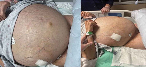
A 38-year-old Caucasian male with a history of schizophrenia and noncompliance with medications presented to the emergency department with abdominal swelling and altered mental status. Per the patient’s mother, he self-diagnosed himself with a gluten allergy and had been avoiding foods with gluten, sugar, and salt for the past three months. For the past 5 years, he followed an exclusively plant-based diet. Following a recent loss of a family member, the patient became reclusive; he rarely left his room and had limited oral intake with small amounts of raw vegetables every two to three days for several weeks. It was unclear when the abdominal swelling had started, nor the duration of his confusion, as the patient would cover himself with his sheets and stay in bed a majority of the time. The patient resided in New York his entire life with no travel outside of the USA; he had no known hepatic or gastrointestinal (GI) disorders. His mother denied any use of alcohol or recreational drugs by the patient. The patient’s medication list was limited to risperidone 1 mg every twelve hours. Review of systems could not be obtained given the patient’s mental status.
Vital signs revealed an oral temperature of 98.0°F, BP 142/105 mmHg, heart rate 139 bpm, respiratory rate 30/min, saturating at 89% on room air, and a BMI of 20.1 kg/m2. Physical exam revealed altered mental status; cachexia with sunken orbits, poor oral hygiene, extensive abdominal distention, hypoactive bowel sounds, diffuse tenderness to palpation, and a positive fluid wave. There was no evidence of gastrointestinal bleeding. 3+ pitting edema of the bilateral lower extremities was noted. Initial laboratory studies were significant for hemoglobin 10.0 g/dL, creatinine 1.5 mg/dL, lactic acid 4.4 mmol/L, HCO3 15 mEq/L, ammonia 65 µmol/L and albumin 1.8 g/dL; liver function tests were within normal limits, INR was 1.2. Subsequent iron studies revealed iron of 25 ug/dL, with a total iron binding capacity of 133 ug/dL, a ferritin of 75 ng/mL, and a percent saturation of 18.8%. The vitamin B12 level was 1447 pg/mL, and folate was 4.44 ng/mL. A CT of the chest, abdomen, and pelvis without contrast showed extensive abdominal and pelvic ascites along with bilateral infiltrates and consolidation of the right lower lung lobe. An abdominal ultrasound revealed a large amount of ascites and a lower extremity venous ultrasound demonstrated deep vein thromboses (DVT) in the common femoral veins bilaterally. A computer tomography angiogram (CTA) was negative for acute pulmonary embolism.
The patient was admitted to the medical intensive care unit for acute hypoxic respiratory failure secondary to ascites and community acquired pneumonia. The patient was started on continuous heparin infusion with an initial bolus and antibiotics with piperacillin-tazobactam 3.375 grams (g) every six hours and intravenous vancomycin 1 g every twelve hours; he was also started on intravenous albumin 25 g every six hours for 4 doses. On the next day, the patient underwent an ultrasound-guided paracentesis with 5.5 liters of ascitic fluid removed and a 6 Fr drainage catheter was placed, anticipating the need for further drainage. Workup for chronic liver disease including ceruloplasmin, alpha antitrypsin level and a hepatitis panel were unrevealing. Analysis of the ascitic fluid revealed a serum-ascites albumin gradient (SAAG) greater than 1.1, total ascitic protein of less than 2.5 g/dL, and ascitic fluid lactic dehydrogenase (LDH) level of less than 225 Sigma units (SU), findings suggestive of a transudative pathology (). A transthoracic echocardiogram showed normal heart function without significant valvular disease or any right heart strain. The patient was started on enteral feeds with close monitoring for signs of refeeding syndrome. On hospital day eight, intravenous heparin was transitioned to direct acting oral anticoagulation in the form of apixaban for DVT treatment. However, the patient was noted to have a decline of his hemoglobin to 6.4 g/dl and he received a transfusion of one unit of packed red blood cells. A fecal occult blood test was positive. Anticoagulation was held and an inferior vena cava filter was placed by a vascular surgeon. The patient underwent an upper endoscopy that showed severe portal hypertensive gastropathy. During his hospitalization, the patient had persistent re-accumulation of abdominal ascites for which he underwent repeat paracentesis. A total of 28.5 L of fluid was removed over the course of the hospitalization. The case was discussed with the patient and his family and a liver biopsy was recommended; however, the patient refused. After medical optimization, the patient was transferred to the medical floor and was eventually discharged home.
3. Discussion
Kwashiorkor is almost exclusively seen among the pediatric population in developing countries where there is limited food supply and is rarely seen in the developed countries such as the USA [Citation1]. According to the WHO, as of 2019, 6.9% of children under the age of 5 worldwide, and 0.4% in the USA suffer from a form of malnutrition called wasting, when a child is too thin for his or her height due to the failure to gain weight [Citation2]. Kwashiorkor is a severe form of protein energy malnutrition believed to be caused by protein deficiency. Albumin, a protein, acts to increase oncotic pressure and holds fluids within the vasculature; in kwashiorkor syndrome, profound hypoalbuminemia due to protein deficiency causes an imbalance between the oncotic and hydrostatic pressures across the capillary blood vessel walls, which ultimately results in intravascular fluids escaping into the third space, causing abdominal distension, ascites, and peripheral edema; other signs include hepatomegaly and marked muscle atrophy [Citation3].
The diagnosis of kwashiorkor is made based on the constellation of clinical presentation, a detailed dietary history of low protein intake, physical exam, low body mass index, and laboratory evidence of low blood protein levels and electrolyte disturbances [Citation4]. If paracentesis is performed, abdominal ascitic fluid analysis usually shows a transudative pattern. Differentiating kwashiorkor from another type of malnutrition, marasmus, can be supported by etiology as well as physical examination. Marasmus is due to low calorie intake whereas kwashiorkor is due to low protein consumption. Patients with kwashiorkor are more likely to have lower extremity edema and a protuberant abdomen, as in the patient that presented to our hospital; patients with marasmus appear wasted, cachectic, and sarcopenic [Citation5]. Kwashiorkor is more likely in this patient given his age, clinical presentation with abdominal swelling and edema, and the history of poor diet comprised solely of vegetables with no significant protein intake. The normal liver enzyme levels, INR of 1.2, and lack of alcohol use made the differential diagnosis of a decompensated liver disease with secondary thrombosis less likely. Other complications to be considered for this patient include medication induced liver disease, however, risperidone, which was the only medication this patient was taking, very rarely causes liver disease, and it would not explain other clinical finings we seen in this patient.
Another complicating factor in this patient’s hospital course to consider is the anemia that was present at baseline and the bilateral DVT that were found on lower extremity ultrasound. This can be associated with the low protein state, specifically albumin, as hypoalbuminemia has been associated with hypercoagulable states and this incurs risks of renal vein thrombosis and DVTs [Citation6]. It is difficult to determine whether the steady fall in hemoglobin from the admission level of 10.0 g/dL to 6.4 g/dL was due to bleeding after initiation of heparin for the DVT or to fluid redistribution with improved albumin levels and subsequent hemodilution, which is supported by the evidence of a concomitant decrease in platelets, leukocytes, and hemoglobin as the albumin rises.
The mainstay of treatment and prevention of kwashiorkor is initiation of a protein-rich diet [Citation7]. However, these patients are at risk of developing refeeding syndrome when treatments are instituted, which is characterized by metabolic and electrolyte changes. The WHO has developed guidelines and a three-phase management approach for managing severe malnutrition, in which phase 1 involves initial resuscitation and stabilization, phase 2 introduces nutritional rehabilitation, and phase 3 involves follow up for prevention of recurrence [Citation8–10].
Kwashiorkor is a treatable condition with appropriate therapy and follow up. Global initiatives are in progress to lower the prevalence of this devastating disease, which predominantly affects the underprivileged and the underserved. An interprofessional approach must be taken for this condition. Although it is rare in the USA, it can happen as a result of severe neglect, malnutrition, very restricted diet, or underlying psychiatric disorders such as in the patient in the above case [Citation10]. It is important to know that this is a chronic and complex calorie deprivation disorder that affects almost every organ in the body. Awareness of this condition with prompt diagnosis is essential to prevent treatment delays.
Informed consent
Informed patient consent was obtained for this case report.
Authors’ contributions
S. Arcieri, S. Cheung, and A. Belkin performed chart review, literature review, and preparation of the manuscript.
A. Pillai and R. Gupta provided revision and final approval of the manuscript.
Disclosure statement
No potential conflict of interest was reported by the author(s).
References
- Palm CV, Frølich JS, Snogdal LS, et al. Kwashiorkor: an unexpected complication to anorexia nervosa. BMJ Case Rep. 2016;2016:bcr2016215638.
- World Health Organization. UNICEF/WHO/The World bank group joint child malnutrition estimates: levels and trends in child malnutrition: key findings of the 2020 edition; 2020
- Sarah L. Lappin. Proc Natl Acad Sci U S A. 2019 Jan 15;116(3):723-731. ... Copyright © 2021, StatPearls Publishing LLC.
- Dean R, Schwartz R. The serum chemistry in uncomplicated kwashiorkor. Br J Nutr. 1953;7(1–2):131–147.
- Bhutta ZA, Berkley JA, Bandsma RH, et al. Severe childhood malnutrition. Nat Rev Dis Primers. 2017;3(1):1–18.
- Gigante A, Barbano B, Sardo L, et al. Hypercoagulability and nephrotic syndrome. Curr Vasc Pharmacol. 2014;12(3):512–517.
- Dean RF. Treatment and prevention of kwashiorkor. Bull World Health Organ. 1953;9(6):767–783.
- World Health Organization. Management of severe malnutrition: a manual for physicians and other senior health workers. Geneva (Switzerland): World Health Organization; 1999.
- Ahmed S, Ejaz K, Mehnaz A, et al. WHO feeding guidelines for inpatient management of malnourished children. J Coll Physicians Surg Pak. 2014 Jul;24(7):493–497.
- WHO. Management of severe malnutrition: a manual for physicians and other senior health workers. Geneva: World Health Organization; 1999.