ABSTRACT
Background: Pulmonary hypertension (PH) is a progressive disorder of the pulmonary circulation, associated with diverse medical conditions. Exercise limitation is the most prominent symptom in PH. Exercise capacity, commonly assessed through a six-minute walk test (6MWT), correlates with both functional status and survival in PH. Few studies have analysed the relation between respiratory function and exercise limitation. Therefore, we investigated the relationship between resting pulmonary function, exercise capacity, and exertional desaturation, assessed through the 6MWT, in unselected PH patients.
Methods: Fifty consecutive patients with PH diagnosis, referred for pulmonary function testing (lung volume, spirometry, and diffusing capacity for carbon monoxide (DLCO)) and 6MWT, were recruited at Molinette University Hospital, Turin.
Results: The majority of the patients (54%) had PH due to left heart disease. Airway obstruction (FEV1/VC-ratio < 0.7) was found in 46% of the patients and they performed significantly worse in the 6MWT than unobstructed patients (307 m vs. 377 m). Patients with PH due to left heart disease also performed significantly poorer 6MWT when airway obstruction was present (305 m vs. 389 m). Twenty-two patients (44%) presented exertional desaturation upon 6MWT. Lower DLCO divided by the alveolar volume (DLCO/VA), FEV1/VC-ratios and resting PaO2-values were significantly correlated with exertional desaturation after adjustments for age, sex, BMI, and smoking habits. DLCO/VA was the main determinant of exertional desaturation in a stepwise regression model.
Conclusions: Spirometric parameters of airway obstruction were related to walk distance and exercise-induced desaturation in PH patients. This suggests a place for spirometry in clinical monitoring of PH patients.
Introduction
Pulmonary hypertension (PH) is a diverse group of entities affecting the pulmonary circulation and is defined by a resting mean pulmonary arterial pressure over 25 mmHg, confirmed by right heart catheterization [Citation1,Citation2]. A WHO classification divides PH into five major subtypes based on pathological, pathophysiological, and therapeutic characteristics: (1) pulmonary arterial hypertension (PAH); (2) PH due to left heart disease; (3) PH due to interstitial lung disease and/or hypoxia (PH-ILD); (4) chronic thromboembolic PH (CTEPH); and (5) PH with unclear and/or multifactorial mechanisms [Citation1].
While the abnormalities of the cardiovascular system in PH are well described, it is unclear to what extent the respiratory system is affected [Citation3]. The abnormal pulmonary vessels could affect the function of the adjacent airways and lung parenchyma and contribute to symptoms. Contradictory results exist regarding the presence of airway obstruction in PAH, with studies reporting no differences [Citation4,Citation5], or a lower ratio of forced expiratory volume in 1 sec to forced vital capacity (FEV1/VC) compared with controls [Citation6,Citation7]. Restrictive pulmonary function pattern can be found in up to 50% of PAH patients and is also found in patients with PH due to left heart disease [Citation8]. The most consistent lung function limitation in PH is an abnormal gas transfer assessed through diffusing capacity for carbon monoxide (DLCO) [Citation3,Citation4,Citation9,Citation10], which is found in the large majority of PH patients. The alveolo-capillary membrane function can also be assessed by adjusting the diffusing capacity for the ‘accessible’ alveolar volume (DLCO/VA) [Citation11]. However, similar value of DLCO and DLCO/VA was found as predictor for pulmonary gas exchange [Citation12]. Since PH can have diverse underlying causes, the reduced DLCO may be attributed to different essential mechanisms such as ventilation-perfusion mismatch, thickening of the alveolar capillary membrane due to endothelial cell proliferation, reductions in pulmonary capillary blood volume, low cardiac output, hypoxic vasoconstriction, and right heart dysfunction [Citation13]. Even though reduced DLCO is a common finding in PH, it is not included in the risk stratification protocols of PH patients [Citation14], and the clinical significance of DLCO impairment in PH is less obvious [Citation15,Citation16]. Furthermore, the relation of the functional limitation, assessed through six-minute walk distance (6MWD), and extensive lung function, with measurements of spirometry, lung volumes, and DLCO, have not been thoroughly characterized in patients with PH.
Exertional dyspnoea and exercise intolerance are the main clinical findings in PH. These have been attributed to decreased cardiac output, under-perfusion of the alveoli caused by remodelled small pulmonary arteries, hyperventilation, and respiratory and peripheral muscle dysfunction [Citation17–Citation19]. Exercise capacity correlates with survival and functional status in PH [Citation20,Citation21]. As a result, exercise capacity, commonly assessed through the six-minute walk test (6MWT), has been a mandatory, if not a primary, outcome measure in the majority of the recent clinical trials in PH [Citation22]. Moreover, the 6MWT also evaluates exercise-induced oxygen desaturation, known to occur often in patients with pulmonary vascular disease [Citation23]. However, few studies have attempted to explore the relationship between pulmonary function and exertional desaturation in PH patients [Citation24].
The aim of the present study was to investigate the relationship of resting pulmonary function including DLCO with exercise capacity and exertional desaturation, assessed through the 6MWT, in patients suffering from PH.
Materials and methods
Participants
A total of 50 consecutive patients visiting, during the period 2012–2013, the Pulmonary Function Testing Unit of Molinette University Hospital, Turin, Italy, upon referral from the cardiology consultant for pulmonary evaluation, which included pulmonary function test and six-minute walk test, were included in the study. These patients were previously diagnosed with pulmonary hypertension, based upon hemodynamic data attained by right heart catheterization (mean pulmonary arterial pressure above 25 mm Hg), and were subsequently divided into PH subgroups according to guidelines [Citation25]. Information on comorbidities listed in was extracted from the medical journals. Heart disease was defined as ischemic disease/valvular disease/grow-up congenital heart disease/cardiomyopathy/left ventricular diastolic dysfunction. Among PH group I patients, two had ongoing treatment with bosentan and two had ongoing treatment with sildenafil.
Table 1. Patient characteristics
Pulmonary function tests
Spirometry was performed using a computerized water-sealed Stead-Wells spirometer (Biomedin, Padua, Italy). Lung volumes were obtained through using the Baires System (Biomedin, Padua, Italy) and Helium dilution technique. All lung function testing was performed following the standards outlined by ATS/ERS [Citation26,Citation27]. Reference values were used in accordance with standardized lung function testing [Citation28] and lung function parameters were expressed as both absolute values and % of predicted values.
Pre-bronchodilatory spirometry was used for all subjects with the exception of patients with previously diagnosed COPD (n = 10), for whom post-bronchodilatory values were used.
Gas transfer for carbon monoxide (DLCO) was measured with the single-breath technique, using the Baires System (Biomedin, Padua, Italy) with a gas mixture of 0.3% CO, 10% helium, and balance air.
The lung function parameters used in the present analyses were forced expiratory volume in 1 sec (FEV1), peak expiratory flow (PEF), forced expiratory flow at 25–75% of vital capacity (FEF25–75) and Vital Capacity (VC) from spirometry, total lung capacity (TLC), residual volume (RV), RV/TLC-ratio, DLCO, and DLCO divided by the alveolar volume (DLCO/VA).
Six-minute walk test
Six-minute walk tests were conducted in accordance with guidelines [Citation29]. Briefly, walk distance was measured after the subjects had walked as far as possible in 6 min in a 30 m corridor at sea level. Peripheral capillary oxygen saturation (SpO2) and heart rate were measured before and immediately after the 6MWT using a pulse oximeter with a finger sensor. Reference values according to Chetta et al. [Citation30] were used and 6MWD was expressed as both absolute value and % of predicted value.
Acquisition of baseline SpO2 was obtained when patients were relaxed and in the sitting position. Exercise-induced oxygen desaturation was defined in accordance with the Royal College of Physicians’ guidelines [Citation31] as a minimum of 5% reduction between arterial oxygen saturation measured through pulse oximetry pre- and post-test.
Arterial blood gases (ABG)
Partial arterial pressure of carbon dioxide (PaCO2) and partial arterial pressure of oxygen (PaO2) were measured using the GEM 4000 Premiere analyser (Instrumentation Laboratory, Lexington, USA).
Statistical analyses
Statistical analyses were performed using a computer software program (STATA 12.1, StataCorp, College Station, TX, USA). Mean ± standard deviation (SD) was used to present the descriptive statistics.
Simple linear regression was used to analyse the relation between different single lung function parameters and 6MWD. These relations were tested for consistency in a multiple linear regression model that included sex, age, BMI, and smoking habits, in addition to the lung function parameters. Finally, a stepwise regression model including arterial blood gases, resting SpO2, all lung function parameters which showed statistically significant correlation to 6MWD, sex, age, BMI, and smoking habits was used to determine the most important predictors of 6MWD.
Unpaired t-tests were used to compare 6MWD in patients with normal and decreased lung function, respectively, when such grouping was done. Decreased lung function was defined as a value below 80% predicted, with the exception of the FEV1/VC-ratio, for which it was defined as an absolute value below 0.7 (and for specific subanalyses as a value below the lower limit of normal (LLN)).
A p value <0.05 was considered as statistically significant.
Ethics
The trial was reviewed and approved by the Interaziendale Ethical Review Board in Turin (reference number: 370/378/70/2011, approved 28 September 2011). All patients gave written informed consent to the study.
Results
Population characteristics
Patient characteristics for the whole group at inclusion are presented in . The majority of the patients were categorized as PH groups 1 and 2 according to the WHO classification and four patients (8%) were diagnosed to have developed PH due to underlying lung diseases. Most patients were either ex- or current smokers. The majority (69%) of the investigated subjects suffered from heart disease. One-fifth of the subjects had a medical record of COPD diagnosis.
Lung function parameters are presented in . Reduced DLCO was found in 46 patients (92%) and 5 patients (10%) showed reduced TLC. A total of 23 patients presented signs of airway obstruction defined as FEV1/VC < 0.70, while 15 patients showed FEV1/VC < LLN. 27 patients had decreased FEV1 and 26 patients had decreased PEF, defined as a value below 80% of the predicted normal value. Two patients had reduced oxygen saturation at rest (SpO2 < 90%).
Table 2. Indices of lung function, respiratory parameters and 6MWD
Lung function indices and 6MWD in all subjects
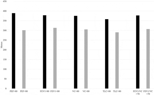
In general, decreased lung function indices (<80% predicted) were associated with shorter 6MWD, as shown in . Twenty-four of the patients (48%) displayed airway obstruction, defined as FEV1/VC < 0.7, and patients with FEV1/VC < 0.7 had a significantly shorter 6MWD than patients without airway obstruction (307 m vs. 377 m, p = 0.014) (). These results were consistent when airway obstruction was defined as value below LLN and 6MWD was significantly lower in patients with FEV1/VC < LLN than patients with FEV1/VC ≥ LLN (299 m vs. 362 m, p = 0.046).
Figure 1. Decreased lung function parameters in relation to 6MWD

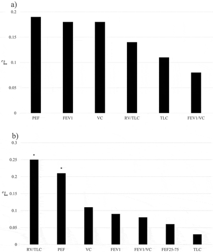
The strongest correlation with 6MWD was found for PEF, followed by VC and FEV1 ()). DLCO, DLCO/VA, RV, SpO2 and arterial gases at rest did not show any association with 6MWD. 6MWD was significantly correlated to FEV1 (r2 = 0.15, p = 0.01), VC (r2 = 0.14, p = 0.02), PEF (r2 = 0.18, p = 0.007) and FEV1/VC (r2 = 0.16, p = 0.01) after patients previously diagnosed with COPD (n = 10) were excluded from the analyses.
Figure 2. Predictive value (expressed as r2) for 6MWD of single lung function measurements for all patients (a) and for PH due to left heart disease (b)

Stepwise regression analysis in all PH subjects, in a model where all lung function parameters (as absolute values), arterial gases, gender, age, smoking habits and BMI were included, yielded PEF as the sole determinant of 6MWD (p = 0.002). A similar stepwise regression analysis, when lung functions were expressed as %predicted instead, revealed PEF (%predicted) as the sole determinant of 6MWD (p = 0.003). PEF remained the variable most strongly associated with 6MWD also after adjusting for gender, age, smoking habits, and BMI ().
Table 3. Correlations between 6MWD and different lung function or physiological parameters
Lung function indices and 6MWD in PH due to left heart disease
Looking specifically at the largest group of PH patients, PH group 2 (n = 27), the strongest relation to 6MWD was found with RV/TLC (r2 = 0.25), followed by PEF (r2 = 0.21), p < 0.05 for both ()). No significant correlations of 6MWD with other lung function parameters were found. Stepwise regression analysis in PH group 2, in a model where all lung function parameters (as absolute values), arterial gases, gender, age, smoking habits, and BMI were included, yielded RV/TLC as the sole determinant of 6MWD (p = 0.01).
The majority of the PH group 2 patients showed FEV1/VC < 0.7 (56% of the subjects). The majority of the patients in PH group 2 also displayed low FEV1 (63% of the subjects) and low PEF (52% of the subjects), both defined as a value below 80% of predicted normal value. The finding of decreased 6MWD related to airway obstruction was also consistent for PH group 2, as patients with FEV1/VC < 0.7 showed a significantly shorter 6MWD (305 m vs. 389 m, p = 0.03).
Lung function and exertional desaturation during 6MWT in all subjects
Twenty-two patients (44%) presented exertional desaturation following the 6MWT. Lower levels of DLCO/VA, resting SpO2, FEV1/VC and PaO2 were significantly associated with exercise-induced oxygen desaturation (). None of the lung volumes obtained through the helium dilution technique showed a significant correlation with exertional desaturation. Lower DLCO/VA (OR 0.02, CI 0.001–0.4, p = 0.01), FEV1/VC-ratios (OR 0.92, CI 0.84–0.997, p = 0.04) and resting PaO2-values (OR 0.95, CI 0.89–0.999, p = 0.04) remained significantly correlated with desaturation also in a logistic regression model adjusted for age, sex, BMI, and smoking habits. After exclusion of COPD patients from above analyses, lower DLCO/VA (OR 0.01, CI 0.0004–0.33, p = 0.009), FEV1/VC-ratios (OR 0.90, CI 0.81–0.99, p = 0.04) and resting SpO2-values (OR 0.67, CI 0.46–0.98, p = 0.04) remained significantly correlated with desaturation in the adjusted logistic regression model.
Table 4. Lung function parameters in relation to desaturation during 6MWT
Stepwise regression analysis including all lung function parameters and arterial gases, gender, age, smoking habits, and BMI revealed DLCO/VA as the main determinant of oxygen desaturation during the 6MWT (p = 0.01). Including only relative values in the stepwise regression analysis, expressed as % predicted, DLCO/VA (%predicted) was shown to be the main determinant of exertional desaturation (p = 0.003).
Lung function and exertional desaturation during 6MWD in PH due to left heart disease
Investigating only PH group 2, 44% of the patients (n = 12) displayed exertional desaturation. Lower levels of DLCO/VA (p = 0.03) and resting SpO2 (p = 0.047) were significantly associated with exercise-induced oxygen desaturation in a single logistic regression model. The relation to lower levels of DLCO/VA was consistent after adjusting for age, sex, BMI, and smoking habits (p = 0.04).
Discussion
The major finding of the present study was that airway obstruction was strongly related to exercise capacity in this group of unselected patients with PH. Furthermore, this finding was consistent in a sub-analysis of patients with PH secondary to left heart disease, the largest group included in the present material. Diffusing capacity for carbon monoxide was not related to exercise capacity, but related to oxygen desaturation during exercise. TLC measurements did not provide any additional information in relation to the studied outcomes, while RV/TLC-ratio was related to exercise capacity.
Airway obstruction was related to a shorter six-minute walk distance. This relation was consistent for different expiratory lung function indices obtained through dynamic spirometry (i.e. PEF, FEV1 and FEV1/VC) and after adjustment for patient characteristics.
Previous studies have shown significant inspiratory and expiratory muscle weakness in idiopathic pulmonary arterial hypertension [Citation19]. This could in fact mirror the finding in our study, where PEF appeared to be the variable most strongly associated with 6MWD.
Airway obstruction in PH has previously been studied and 20–40% of the patients with PH group 1 have shown airway obstruction based on a forced expiratory volume in 1 sec to forced vital capacity ratio of less than 70% [Citation32,Citation33]. Surprisingly, the association between airway obstruction and exercise capacity could also be seen in patients with PH secondary to left heart failure, patients who are more often regarded to be characterized by restrictive lung function [Citation34]. Moreover, in patients with PH group 2, a relation with air trapping, assessed through RV/TLC-ratio, was found which might reflect lung congestion causing air trapping through peri-bronchial cuffs or lung stiffening [Citation35]. One could argue the fact that we speculate regarding air trapping as a potential explanation when we use the RV- and TLC-values at rest, as it is suggested that increasing lung hyperinflation during exercise (dynamic hyperinflation) is more closely related to exercise intolerance and clinical dyspnoea than airflow limitation and static hyperinflation [Citation36]. We hypothesized in advance that DLCO would have a large impact on exertional desaturation in patients suffering from PH [Citation37]. However, in the present material, an unexpected significant association was found between FEV1/VC and exercise-induced desaturation.
No relation between diffusing capacity for carbon monoxide and walk distance was found in the present material. The diffusing capacity relates inversely to the mean pulmonary arterial pressure and this is in line with results from echocardiography studies, which found no relation between pulmonary arterial pressure and 6MWD [Citation20]. We hypothesize that the patients were characterized as pulmonary hypertensive, e.g. reduced diffusion capacity, makes airway obstruction more influential in the outcome of 6MWT due to a tendency of uniformity for DLCO. This is in fact in line with previous findings from our group in which we could demonstrate that in patients suffering from COPD, e.g. airway obstruction, DLCO is more closely linked to reduced walk distance [Citation38]. On the other hand, DLCO/VA was revealed as the main determinant of exercise-induced desaturation, which is in line with reported interdependence between pulmonary diffusion and oxygen desaturation during exercise in patients with diffuse systemic sclerosis and interstitial lung disease [Citation39].
Total lung capacity measurements obtained through the hilum dilution technique did not offer supplementary information. This is in accordance with previous studies where lung volume measurements offered minimal additional information [Citation40–Citation42]. In a study by Armstrong et al. [Citation40], the authors were unable to show any relationship between peak exercise capacity and lung volumes in patients suffering from interstitial pulmonary disease with or without pulmonary hypertension.
The strength of the study is the availability of extensive lung function characterization. However, the study has its limitations as it was a single centre investigation with a relatively low total number of study subjects. This limited the possibilities of performing sub-analyses with regard to type of PH, with the exception of PH group 2, the largest group investigated, where we confirmed the main finding of the present study regarding the relation between airway obstruction and exercise capacity. It could be argued that the term airway obstruction was used quite broadly, and lower PEF or FEV1 might be due to other underlying pathologies, such as decreased respiratory muscle strength or a pulmonary restrictive component. However, the findings were confirmed when airway obstruction was defined as FEV1/VC-ratio (less than 0.7) and a restrictive pattern was seen in only a minority (10%) of the subjects. Another point of argue could be that not all study patients performed lung function testing similarly and mandatory post-bronchodilatory spirometry was carried out only in patients with previously known COPD diagnosis (n = 10). However, we believe that this approach mirrors the functional capacity in COPD patients in a more suitable manner. As patients with COPD are treated with bronchodilator medication, post-bronchodilatory lung function would display their baseline status before 6MWT more accurately, as the patients in other disease categories do not use bronchodilators. Moreover, our main findings were consistent even after exclusion of COPD patients. Unfortunately, we lack more detailed data on smoking history and therefore could not account for this in the regression models. Another limitation of the present study is that no assessment of peripheral muscle function was done, for example muscle strength, as peripheral muscle dysfunction is common in PH and relates with reduced exercise capacity [Citation43]. Another major limitation of the study is the lack of current data on pulmonary arterial pressure as PH-diagnosis was made at a previous and variable time compared with the investigation of lung function and 6MWD. Although PH is a clinical syndrome with different underlying etiology, the key diagnostic feature is an elevated pulmonary artery pressure [Citation44]. Nevertheless, functional parameters such as NYHA class and 6-min walk distance have a prognostic significance superior to most standard resting haemodynamic parameters despite underlying causes of disease [Citation20,Citation45]. However, whilst it is widely accepted that exercise capacity in patients with pulmonary hypertension is limited by cardiac output, the impact of pulmonary function has not been fully investigated.
Conclusion
Airway obstruction was an important predictor of walk distance and exercise-induced desaturation in this unselected material of PH patients with a very low prevalence of PH secondary to lung disease and this finding was consistent after exclusion of subjects with COPD. As 6MWD is an important predictor of mortality in PH patients, it is important to better understand the limitations of 6MWD in individual PH patients, as obstruction is probably independent of PH in most of these patients. The obstructive component that could be identified this way might be a treatable target and therefore it would be clinically useful to map this pulmonary function component in PH patients.
Disclosure statement
No potential conflict of interest was reported by the authors.
Additional information
Notes on contributors
Amir Farkhooy
Amir Farkhooy MD, PhD is program director and medical director of internal medicine in Bollnäs hospital / Sweden. Dr. Farkhooy is a consultant in internal and respiratory medicine and clinical physiology. His main research area is the relationship between lung function and exercise capacity.
Michaela Bellocchia
Michela Bellocchia MD, is currently active staff of the Respiratory Diseases Division at the Cardiothoracic Department in AOU Città della Salute e della Scienza, Molinette Hospital, Turin University, Italy. She coauthored several manuscripts published on international journals, about asthma, COPD and pulmonary hypertension.
Hans Hedenström
Hans Hedenström MD, PhD is associated professor at Uppsala University, Sweden. Dr. Hedenström is a consultant in clinical physiology. His main research areas are exercise capacity and pulmonary function testing.
Daniela Libertucci
Daniela Libertucci MD, PhD is currently active staff of the Respiratory Diseases Division at the Cardiothoracic Department in AOU Città della Salute e della Scienza, Molinette Hospital, Turin University, Italy. She has authored more than 30 publications and her main research area is within pulmonary hypertension and lung transplantation.
Caterina Bucca
Catarina Bucca MD, PhD is professor in Respiratory Medicine at University of Turin, Italy. She has authored more than 200 publications and her main research area is within asthma, chronic obstructive pulmonary disease
Christer Janson
Christer Janson MD, PhD is professor in Respiratory Medicine at Uppsala University, Sweden. He has authored more than 350 publications in the field of obstructive pulmonary disease and has large experience in epidemiological research in respiratory diseases.
Paolo Solidoro
Paolo Solidoro MD, PhD is assistant professor at University of Turin, Italy. Dr. Soildoro is Pulmonology referral in Thoracic Oncologic program . He has authored more than 100 publications and his main research area is within lung transplantation, pulmonary hypertension and advanced life support.
Andrei Malinovschi
Andrei Malinovshi MD, PhD is Professor in Clinical Physiology at Uppsala University, Sweden. He has authored more than 100 publications and his main research area is within pulmonary function testing and biomarkers in respiratory diseases.
References
- Task Force for Diagnosis and Treatment of Pulmonary Hypertension of European Society of Cardiology (ESC), European Respiratory Society (ERS), International Society of Heart and Lung Transplantation (ISHLT), Galiè N, Hoeper MM, Humbert M, et al. Guidelines for the diagnosis and treatment of pulmonary hypertension. Eur Respir J. 2009;34(6):1219–9.
- Rubin LJ. Primary pulmonary hypertension. N Engl J Med. 1997;336(2):111–117.
- Low AT, Medford AR, Millar AB, et al. Lung function in pulmonary hypertension. Respir Med. 2015;109(10):1244–1249.
- Sun XG, Hansen JE, Oudiz RJ, et al. Pulmonary function in primary pulmonary hypertension. J Am Coll Cardiol. 2003;41(6):1028–1035.
- Rich S, Dantzker DR, Ayres SM, et al. Primary pulmonary hypertension. A national prospective study. Ann Intern Med. 1987;107(2):216–223.
- Jing ZC, Xu XQ, Badesch DB, et al. Pulmonary function testing in patients with pulmonary arterial hypertension. Respir Med. 2009;103(8):1136–1142.
- Meyer FJ, Ewert R, Hoeper MM, et al. Peripheral airway obstruction in primary pulmonary hypertension. Thorax. 2002;57(6):473–476.
- Gehlbach BK, Geppert E. The pulmonary manifestations of left heart failure. Chest. 2004;125(2):669–682.
- Agostoni P, Magini A, Andreini D, et al. Spironolactone improves lung diffusion in chronic heart failure. Eur Heart J. 2005;26(2):159–164.
- Wrobel JP, Thompson BR, Williams TJ. Mechanisms of pulmonary hypertension in chronic obstructive pulmonary disease: a pathophysiologic review. J Heart Lung Transplant. 2012;31(6):557–564.
- Hughes JM, Pride NB. Examination of the carbon monoxide diffusing capacity (DL(CO)) in relation to its KCO and VA components. Am J Respir Crit Care Med. 2012;186(2):132–139.
- Kaminsky DA, Whitman T, Callas PW. DLCO versus DLCO/VA as predictors of pulmonary gas exchange. Respir Med. 2007;101(5):989–994.
- Fritz JS, Smith KA. The pulmonary hypertension consult: clinical and coding considerations. Chest. 2016. DOI:10.1016/j.chest.2016.05.010
- Benza RL, Gomberg-Maitland M, Miller DP, et al. The REVEAL registry risk score calculator in patients newly diagnosed with pulmonary arterial hypertension. Chest. 2012;141(2):354–362.
- Kawut SM, Horn EM, Berekashvili KK, et al. New predictors of outcome in idiopathic pulmonary arterial hypertension. Am J Cardiol. 2005;95(2):199–203.
- van der Lee I, Zanen P, Grutters JC, et al. van den Bosch JM. Diffusing capacity for nitric oxide and carbon monoxide in patients with diffuse parenchymal lung disease and pulmonary arterial hypertension. Chest. 2006;129(2):378–383.
- Arena R, Guazzi M, Myers J, et al. Cardiopulmonary exercise testing in the assessment of pulmonary hypertension. Expert Rev Respir Med. 2011;5(2):281–293.
- Babu AS, Myers J, Arena R, et al. Evaluating exercise capacity in patients with pulmonary arterial hypertension. Expert Rev Cardiovasc Ther. 2013;11(6):729–737.
- Meyer FJ, Lossnitzer D, Kristen AV, et al. Respiratory muscle dysfunction in idiopathic pulmonary arterial hypertension. Eur Respir J. 2005;25(1):125–130.
- Miyamoto S, Nagaya N, Satoh T, et al. Clinical correlates and prognostic significance of six-minute walk test in patients with primary pulmonary hypertension. Comparison with cardiopulmonary exercise testing. Am J Respir Crit Care Med. 2000;161(2 Pt 1):487–492.
- Wensel R, Opitz CF, Anker SD, et al. Assessment of survival in patients with primary pulmonary hypertension: importance of cardiopulmonary exercise testing. Circulation. 2002;106(3):319–324.
- Galie N, Manes A, Negro L, et al. A meta-analysis of randomized controlled trials in pulmonary arterial hypertension. Eur Heart J. 2009;30(4):394–403.
- Sun XG, Hansen JE, Oudiz RJ, et al. Exercise pathophysiology in patients with primary pulmonary hypertension. Circulation. 2001;104(4):429–435.
- Fox BD, Langleben D, Hirsch A, et al. Step climbing capacity in patients with pulmonary hypertension. Clin Res Cardiol. 2013;102(1):51–61.
- Galie N, Humbert M, Vachiery JL, et al. ESC/ERS Guidelines for the Diagnosis and Treatment of Pulmonary Hypertension: The Joint Task Force for the Diagnosis and Treatment of Pulmonary Hypertension of the European Society of Cardiology (ESC) and the European Respiratory Society (ERS): endorsed by: Association for European Paediatric and Congenital Cardiology (AEPC), International Society for Heart and Lung Transplantation (ISHLT). Eur Respir J. 2015;46(4):903–975.
- Miller MR, Hankinson J, Brusasco V, et al. Standardisation of spirometry. Eur Respir J. 2005;26(2):319–338.
- Wanger J, Clausen JL, Coates A, et al. Standardisation of the measurement of lung volumes. Eur Respir J. 2005;26(3):511–522.
- Standardized lung function testing. Report working party. Bull Eur Physiopathol Respir. 1983;19(Suppl 5):1–95.
- Laboratories ATSCoPSfCPF. ATS statement: guidelines for the six-minute walk test. Am J Respir Crit Care Med. 2002;166(1):111–117.
- Chetta A, Zanini A, Pisi G, et al. Reference values for the 6-min walk test in healthy subjects 20–50 years old. Respir Med. 2006;100(9):1573–1578.
- Wedzicha JA. Domiciliary oxygen therapy services: clinical guidelines and advice for prescribers. Summary of a report of the Royal College of Physicians. J R Coll Physicians Lond. 1999;33(5):445–447.
- D’Alonzo GE, Bower JS, Dantzker DR. Differentiation of patients with primary and thromboembolic pulmonary hypertension. Chest. 1984;85(4):457–461.
- Burke CM, Glanville AR, Morris AJ, et al. Pulmonary function in advanced pulmonary hypertension. Thorax. 1987;42(2):131–135.
- Wasserman K, Zhang YY, Gitt A, et al. Lung function and exercise gas exchange in chronic heart failure. Circulation. 1997;96(7):2221–2227.
- Breitling S, Ravindran K, Goldenberg NM, et al. The pathophysiology of pulmonary hypertension in left heart disease. Am J Physiol Lung Cell Mol Physiol. 2015;309(9):L924–41.
- Soffler MI, Hayes MM, Schwartzstein RM. Respiratory sensations in dynamic hyperinflation: physiological and clinical applications. Respir Care. 2017;62(9):1212–1223.
- Trip P, Nossent EJ, de Man FS, et al. Severely reduced diffusion capacity in idiopathic pulmonary arterial hypertension: patient characteristics and treatment responses. Eur Respir J. 2013;42(6):1575–1585.
- Farkhooy A, Janson C, Arnardottir RH, et al. Impaired carbon monoxide diffusing capacity is the strongest lung function predictor of decline in 12 minute-walking distance in COPD; a 5-year follow-up study. COPD. 2015;12(3):240–248.
- Rizzi M, Sarzi-Puttini P, Airoldi A, et al. Performance capacity evaluated using the 6-minute walk test: 5-year results in patients with diffuse systemic sclerosis and initial interstitial lung disease. Clin Exp Rheumatol. 2015;33(4 Suppl 91):S142–7.
- Armstrong HF, Schulze PC, Bacchetta M, et al. Impact of pulmonary hypertension on exercise performance in patients with interstitial lung disease undergoing evaluation for lung transplantation. Respirology. 2014;19(5):675–682.
- Glaser S, Noga O, Koch B, et al. Impact of pulmonary hypertension on gas exchange and exercise capacity in patients with pulmonary fibrosis. Respir Med. 2009;103(2):317–324.
- Hosenpud JD, Stibolt TA, Atwal K, et al. Abnormal pulmonary function specifically related to congestive heart failure: comparison of patients before and after cardiac transplantation. Am J Med. 1990;88(5):493–496.
- Malenfant S, Potus F, Mainguy V, et al. Impaired skeletal muscle oxygenation and exercise tolerance in pulmonary hypertension. Med Sci Sports Exerc. 2015;47(11):2273–2282.
- Simonneau G, Gatzoulis MA, Adatia I, et al. Updated clinical classification of pulmonary hypertension. J Am Coll Cardiol. 2013;62(25 Suppl):D34–41.
- Condliffe R, Kiely DG, Gibbs JS, et al. Prognostic and aetiological factors in chronic thromboembolic pulmonary hypertension. Eur Respir J. 2009;33(2):332–338.
