
ABSTRACT
Objective
To assess whether laryngotracheal lesions were more prevalent among tracheostomized post- coronavirus disease 2019 (COVID-19) adult patients compared with a matched historical control group of non-COVID-19 patients.
Methods
An analytical, observational, and cross-sectional study was conducted in a weaning and rehabilitation center. Our study was conducted on tracheostomized adult patients who underwent a fiberoptic bronchoscopy at admission. Non-COVID-19 patients of the historical control group were selected through propensity score (PS) matching. Each post-COVID-19 case was matched in a 1:1 ratio to a non-COVID-19 case. The PS was created based on age, days of endotracheal intubation before tracheostomy, days of artificial airway before bronchoscopy, and days of mechanical ventilation in intensive care unit
Results
Forty-nine matched patients were assessed in each group. The prevalence of airway lesions in post-COVID-19 patients and non-COVID-19 patients of the historical control group was 53.1% and 73.5%, respectively (p = 0.036). In the post-COVID-19 group, 12.2% had severe lesions, and in the historical control group of non-COVID-19 patients, 20.4%. The most prevalent lesion in both groups was granuloma, and lesions were most frequently localized in the subglottic region.
Conclusions
Our findings show a lower prevalence of laryngotracheal lesions in matched tracheostomized post-COVID-19 patients compared to a historical control group of non-COVID.
The most prevalent lesion in both groups was mild (luminal obstruction < 50%) subglottic granuloma.
Introduction
Advances in intensive care have enabled more patients to survive a critical illness, leading to a growing population with long-term care needs. This subgroup of patients commonly requires a tracheostomy because of either prolonged mechanical ventilation or inability to protect the airway. Estenssoro et al. reported that in Argentina, 24.3% of coronavirus disease 2019 (COVID-19) patients hospitalized in the intensive care unit (ICU) underwent a tracheostomy [Citation1], a procedure that facilitates weaning from invasive mechanical ventilation (IMV). During the pandemic, however, the risk of virus aerosolization caused modifications to regular weaning protocols, increasing the number of critically ill patients in need of prolonged intubation and late tracheostomies and predisposing to a higher number of laryngotracheal lesions [Citation2].
Multiple mechanisms to treat COVID-19 patients may further contribute to their susceptibility to laryngotracheal lesions [Citation3]. Some mechanisms include prone positioning for refractory hypoxemia; prothrombotic and antifibrinolytic state of the patient affecting the laryngotracheal microcirculation – predisposing to ischemia and necrosis—; high epithelial viral replication and high dose of corticosteroid use – predisposing to weakness—; low levels of oxygenation that increase the risks of hypoxia and subsequent damage to the tracheal mucosa—; and existing comorbidities [Citation4]. Other factors associated with airway lesions include age, systemic infections, diabetes mellitus, size of the endotracheal and tracheostomy tube, duration of endotracheal intubation (ETI), and increased cuff pressure [Citation5].
Laryngotracheal lesions have been described based on severity as progressive if present one year following extubation or decannulation, which could be a recurrent reason for consultation after discharge [Citation6]. Progressive lesions may require surgical intervention; thus, flexible bronchoscopy is central to diagnosing laryngotracheal lesions and defining the necessary conditions to decannulate. Decannulation is essential as it represents a protective factor against in-hospital mortality: 80% of decannulated patients are discharged compared with 15.8% of non-decannulated patients [Citation7].
The primary objective of this study was to assess, through flexible fiberoptic bronchoscopy, whether laryngotracheal lesions were more prevalent among tracheostomized post-COVID-19 adult patients compared with a matched historical control group of non-COVID-19 patients in a weaning and rehabilitation center. The secondary objective was to describe laryngotracheal lesions among tracheostomized post-COVID-19 adult patients.
Methods
An analytical, observational, and cross-sectional study was conducted in a specialized weaning center (SWC) for the care of post-ICU patients. Our study included adult patients admitted to ICU with polymerase chain reaction confirmed COVID-19 and a tracheostomy cannula who underwent bronchoscopy at SWC admission.
All bronchoscopic procedures were performed within the first week admission in our center with the patient awake under local analgesia with the Ambu® aScope™ 3 Slim flexible videoscope and assessed by a pulmonologist with over 30-year experience. The flexible tube was inserted through the nose and held above the vocal cords, and once the tracheostomy cannula was withdrawn, the tube was then advanced into the tracheobronchial tree.
Bronchoscopic findings were categorized as either normal or pathological, depending on the absence or presence of lesions, respectively. Lesions were classified based on severity as mild (luminal obstruction < 50%) or severe (luminal obstruction > 50%). They were also classified based on localization as glottic, subglottic, at the level of the stoma, tracheal, or bronchial. Lesions were finally classified based on type as granuloma, stenosis, or excessive central airway collapse [Citation8].
Patients without COVID-19 in the historical control group were selected from a database registry that started recording in 2005. However, only patients admitted from 2016 to 2020 were used to avoid possible differences in the care and treatment that patients receive. Patients without COVID-19 from the historical control group were selected by propensity score (PS) matching. Each post-COVID-19 case was matched in a 1:1 ratio to a non-COVID-19 case. The PS was created based on potential predictors of laryngotracheal lesions, such as age, days of ETI before tracheostomy, days of artificial airway before bronchoscopy, and days of mechanical ventilation in ICU. The study protocol was reviewed and approved by the Research Board of Clínica Basilea.
Statistical analysis
Continuous variables with normal distribution were presented as mean and standard deviation (SD). Non-normally distributed variables were presented as median and interquartile range 25–75 (IQR). Categorical variables were presented as number and percentage (n; %). The distribution of numeric variables was assessed using the Shapiro-Wilk test.
Clinical and demographic variables were compared between the post-COVID-19 patients and the historical control group of non-COVID-19 patients. Mann-Whitney tests were used for numeric variables. Pearson’s chi-square or Fisher’s exact tests were used, as appropriate, for categorical variables (two-way contingency table).
PS was estimated using a logistic regression model. A minimum of ten events per independent variable was required. The selection of variables was based on prior studies about the prevalence of laryngotracheal lesions and lesions associated with the time required for airway instrumentation. PS-matching was performed using the radius matching method, without replacement, using a caliper or tolerance width equal to 0.05 SD of the logit of the PS. A p-value <0.05 was considered statistically significant.
PS-matching and statistical analysis were performed using R v. 4.1 software (R Foundation for Statistical Computing, Vienna, Austria; available through https://cran.r-project.org/mirrors.html).
Results
Fifty tracheostomized post-COVID-19 patients were included in the study. One patient could not be matched and was therefore excluded from the analysis. Thus, after PS-matching, 49 matched patients remained in each group.
Patient demographics, personal background, reason for IMV, laboratory parameters, medical condition at SWC admission, and progress of both matched groups are shown in .
Table 1. Patient demographics.
Statistically significant differences were found in the prevalence of airway lesions between post-COVID-19 patients and the historical control group of non-COVID-19 patients (53.1% vs 73.5%, respectively, p = 0.036).
In the post-COVID-19 group, 31 laryngotracheal lesions were found in 26 patients: 17 had granulomas, 13 had excessive central airway collapse, and 1 had stenosis. Further, 23.1% (n = 6) of patients had at least one lesion obstructing > 50% of the lumen. Lesions were frequently localized in the subglottic region. In the historical control group of non-COVID-19 patients, laryngotracheal lesions were found in 36 patients: 30 had granulomas, 11 had excessive central airway collapse, and 2 had stenosis. In this group, 27.8% (n = 10) of patients had one lesion obstructing > 50% of the lumen (). No statistically significant differences were found between the PS-matched groups regarding lesion diagnosis, localization, and severity. These results are shown in .
Figure 1. Classification of laryngotracheal injuries according to diagnosis and severity during FFB.
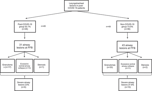
Table 2. Type and severity of airway lesions in matched post-COVID-19 and non-COVID patients.
Table 3. Classification of airway lesions based on localization in matched post-COVID-19 patients and non-COVID-19 patients.
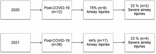
A sub analysis of the post-COVID-19 group was performed based on the period of SWC admission. During the first wave of COVID-19 in 2020, 12 patients were admitted: 75% (n = 9) had airway lesions and 22% (n = 2) had a severe lesion at bronchoscopy. During the second wave of COVID-19 in 2021, 38 patients were admitted: 44% (n = 17) had airway lesions and 23% (n = 4) had one lesion obstructing > 50% of the lumen ().
Figure 2. Analysis the post-COVID-19 group based on the period of SWC admission.
Discussion
To our knowledge, this is the first study to compare laryngotracheal lesions in non-COVID-19 adult tracheostomized patients admitted to an SWC compared to a post-COVID-19 cohort. The post-COVID-19 cohort was matched with a historical non-COVID-19 control group.
During the pandemic, the ICUs in Argentina showed an increase in the time required for IMV and, therefore, the duration of ETI and the need for tracheostomy [Citation1]. Other factors contributing to the risk of laryngotracheal lesions were the endotracheal tube size, sedation depth, preexisting comorbidities, and local infections [Citation4]. Following the first wave of the COVID-19 pandemic, the Laryngotracheal Stenosis Committee of the European Laryngological Society alerted the medical and scientific communities about the importance of early detection of lesions and potential sequelae, secondary to the artificial airway, and prompt referral of patients to a medical center specialized in airway lesions [Citation2]. Further, healthcare quality could have decreased during the pandemic due to the sustained high workload experienced by healthcare professionals. Despite these facts, however, statistically significant differences were found in the prevalence of laryngotracheal lesions in the historical control cohort of non-COVID-19 patients.
The finding of a lower percentage/of injuries in the COVID-19 group versus the historical control non-COVID-19 group may be caused by changes in care processes that occurred during the COVID-19 pandemic. It is possible that the patient had fewer care visits during their stay in the ICU, but these were more protocolized, minimizing strictly necessary procedures, including special care of the natural and artificial pathway. And this will result in a lower incidence of injuries. Another possible explanation is the difference in the time of the historical control cohort that is related to changes in usual procedures. However, the historical cohort was from four years prior the pandemic, which we do not believe has redundancy in the changes in care processes due to new advances in technology and knowledge. Another explanation for this lower incidence of injuries in the cohort of COVID-19 patients is the lowest percentage of COPD patients (26.5%), in contrast with the non-COVID-19 group, and its association with some types of airway injuries as described by Kandaswamy [Citation9].
Our findings showed that the prevalence of airway lesions in post-COVID-19 patients was 53.1%, similar to that reported by Bartier et al., where the prevalence of laryngeal lesions in decannulated post-COVID-19 patients was 55%. It is worth noting that in this study all bronchoscopic procedures were performed within the first week at admission to our SWC, and airway lesions were assessed along the entire tracheobronchial tree, not exclusively at the laryngeal level as in the work of Bartier [Citation10]. The most prevalent airway pathology was granuloma, similar to that reported in studies conducted on non-COVID-19 patients [Citation8] Our findings differ from that reported by Batiers et al., where the most prevalent pathology in decannulated post-COVID-19 patients was laryngeal edema, followed by unilateral vocal cord paralysis, hyposensitivity, and granuloma [Citation10].
Unless symptoms appear, airway lesions may remain undetected and hence underdiagnosed. In contrast to other studies [Citation11], all tracheostomized patients admitted to the SWC underwent a bronchoscopy, eliminating the risk of underdiagnosis and enabling us to foresee possible progressive airway lesions.
Our sub analysis, based on the period of SWC admission, showed that 75% of patients sustained an airway lesion in 2020 compared with only 44% in 2021. The results might be due to the lack of knowledge and uncertainty about the pathology during the first wave of COVID-19, the risk of aerosolization, and infection transmission among healthcare professionals, which caused a decrease in airway care intervention [Citation4,Citation12].
Hernández-García et al describe 90% of objectifiable organic lesions in the fiberscopic examination, but this was performed in symptomatic patients who had achieved extubation or decannulation [Citation13]
Other complications, including vocal fold paralysis or paresis and posterior glottic stenosis, which are two additional common complications of prolonged intubation, were not assessed.
Our study has some limitations. Data were analyzed in a single center in Buenos Aires, thus caution should be exercised in the generalization of the results. The lack of sample calculation is a limitation and may incur a type 1 error, accepting H0 being false. To partially correct this weakness, an ad-hoc power calculation was performed, which yielded a value of 84% to avoid falling into type 2 error and to detect a reproducible difference between both populations. Because patients from different institutions are admitted to our center after going through the acute stage of the disease, there are data from that period that were lost, for example: endotracheal tube size, cuff care, steroid use, type and technique of tracheostomy, among others.
Despite the limitations, a comparative study was conducted on a matched historical control group of non-COVID-19 patients, and a propensity score analysis was performed to adjust for baseline differences between groups and generate valid comparisons. No higher prevalence of laryngotracheal lesions was found among post-COVID-19 patients compared with non-COVID-19 patients admitted to the SWC.
Conclusion
Our findings did show a lower prevalence of laryngotracheal lesions in matched tracheostomized post-COVID-19 patients compared with historical control group of non-COVID-19 patients.
The most prevalent lesion in both groups was mild (luminal obstruction < 50%) subglottic granuloma.
Declaration
The present authors declare that they have no conflicts of interest.
This study does not have any funding.
Authors contribution
Carballo Juan Manuel confirms that the study objectives and procedures are honestly disclosed. Moreover, he has reviewed study execution data and confirms that procedures were followed to an extent that convinces all authors that the results are valid and generalizable to a population similar to that enrolled in this study.
CJM, VA, DBL, PMP, TP, PCE, TK, PF and VD contributed to study design and conception, development, and critical revision of the manuscript for important intellectual content.
PF contributed to study design and conception, critical revision of the manuscript and the performance of fiberoptic bronchoscopy.
DBL contributed to the study design and conception, statistical analysis, data interpretation, and critical review of the manuscript for important intellectual content.
Quick look
Current knowledge
We do not know if laryngotracheal lesions were more prevalent in post-COVID-19 tracheostomized adult patients compared with a group of non-COVID-19 adult patients.
What this paper contributes to our knowledge
We have compared, through propensity analysis, the prevalence of airway lesions in tracheostomized patients with COVID-19 versus a subgroup of patients without this pathology.
We have not found significant differences in the prevalence of airway lesions.
Disclosure statement
No potential conflict of interest was reported by the authors.
References
- Estenssoro E, Loudet CI, Ríos FG, et al. SATI-COVID-19 Study Group. Clinical characteristics and outcomes of invasively ventilated patients with COVID-19 in Argentina (SATICOVID): a prospective, multicentre cohort study. Lancet Respir Med. 2021;9(9):989–7
- Piazza C, Filauro M, Dikkers FG, et al. Long-term intubation and high rate of tracheostomy in COVID-19 patients might determine an unprecedented increase of airway stenoses: a call to action from the European laryngological society. Eur Arch Otorhinolaryngol. 2021 Jan;278(1):1–7. doi: 10.1007/s00405-020-06112-6
- Naunheim MR, Zhou AS, Puka E, et al. Laryngeal complications of COVID-19. Laryngoscope Investig Otolaryngol. 2020 Oct 30;5(6):1117–1124. doi: 10.1002/lio2.484
- Sandu K. Laryngotracheal complications in intubated COVID-19 patients. Clin Med Insights Case Rep. 2021 May 28;14:11795476211020590. doi: 10.1177/11795476211020590
- Félix L, Tavares TL, Almeida VPB, et al. Incidence of laryngotracheal lesions after orotracheal intubation in coronavirus disease patients. Laryngoscope. 2022 May;132(5):1075–1081. doi: 10.1002/lary.29862
- Epstein SK. Late complications of tracheostomy. Respir Care. 2005 Apr;50(4):542–549.
- Scrigna M, Plotnikow G, Feld V, et al. Decanulación después de la estadía en UCI: Análisis de 181 pacientes traqueotomizados. Rev amer med respiratoria Internet. 2013 Jun [cited 2022 Jul 4];13(2):58–63.
- Planells F, Villalba D, Viviana F, et al. Prevalence and Characteristics of Tracheal lesions Observed in tracheostomized patients. J Bronchology Interv Pulmonol. 2019 Apr;26(2):119–123. doi: 10.1097/LBR.0000000000000538
- Kandaswamy C, Balasubramanian V. Review of adult tracheomalacia and its relationship with chronic obstructive pulmonary disease. Curr Opin Pulm Med. 2009 Mar;15(2):113–119. PMID: 19532025. doi: 10.1097/MCP.0b013e328321832d.
- Bartier S, La Croix C, Evrard D, et al. Tracheostomies after SARS-CoV-2 intubation, performed by academic otorhinolaryngologists in the Paris area of France: preliminary results. Eur Ann Otorhinolaryngol Head Neck Dis. 2021 Dec;138(6):443–449. doi: 10.1016/j.anorl.2021.03.002
- Ayten O, Iscanli IGE, Canoglu K, et al. Tracheal stenosis after prolonged intubation due to COVID-19. J Cardiothorac Vasc Anesth. 2022 Aug;36(8 Pt B):2948–2953. doi: 10.1053/j.jvca.2022.02.009
- Tintinago LF, Victoria W, Escobar Stein J, et al. Laryngotracheal stenoses post-acute Respiratory distress syndrome due to COVID-19: clinical presentation, histopathological findings and management. A series of 12 cases. Indian J Otolaryngol Head Neck Surg. 2022 Apr;6(S2):1–6. doi: 10.1007/s12070-022-03076-3
- Hernández-García E, Hernández-Sandemetrio R, Quintana-Sanjuás A, et al. Laryngotracheal complications after intubation for COVID-19: a multicenter study. Life. 2023 May 18;13(5):1207. doi: 10.3390/life13051207