Keywords:
Introduction
According to the older literature, leiomyosarcoma (LMS) is the most common sarcoma of the uterus. But in 1993, the Gynecologic Oncology Group (GOG) found that the proportion of LMS of the uterus was only 16% of uterine sarcomas.Citation1,2 However, sarcomas comprise less than 1% of all cervical malignancies,Citation3 of which LMS is an extremely rare tumour, which follows an aggressive course. As per the world literature, only 22 casesCitation4 have been described. To our knowledge only two casesCitation4 have been reported in the Indian literature. We present a case study on a 40-year-old woman of Indian origin, who was diagnosed with cervical LMS.
Case study
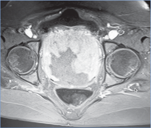
A-40-year-old married woman (gravida 2, para 2) presented with a foul-smelling, white vaginal discharge of two months’ duration. She also complained of abdominal pain and the retention of urine, experienced over the previous few days, and of menorrhagia, followed by spotting for three months. She had a previous history of lower segment Caesarean section. The abdominal examination revealed a uterus of 12-14 weeks gestational size. A haemorrhagic mass was seen on speculum examination. The cervix was not separately visualised. There was an absence of fresh bleeding. A fragment of mass was felt through the os, as per the vaginal examination. The patient underwent pelvic ultrasonography, which revealed a large, well defined heterogenous solid hypoechoic mass lesion in the region of the cervix, measuring 11.6 cm x 9.9 cm x 8.3 cm. Minimal internal vascularity was noted. The presence of pelvic lymphadenopathy or free fluid was not noted. A well defined pedunculated mass, measuring 11.8 cm x 8.9 cm x 8.5 cm, in the upper two thirds of the vagina, and arising from the anterior wall of the cervix (Figure ) was also seen on magnetic resonance imaging (MRI) of the pelvis.
Figure 1: A well defined, large pelvic mass filling the sacrum was shown on magnetic resonance imaging of the pelvis

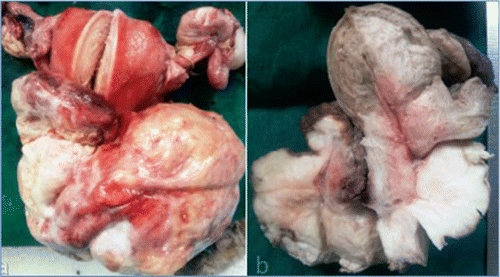
The endometrium, ovaries and fallopian tubes bilaterally were normal. Pelvic lymph nodes were not identified. The preoperative diagnosis was given as a large cervical polyp or submucosal fibroid prolapsing into the vagina. On haematological investigation, the patient was found to have a beta thalassaemia trait with haemoglobin (Hb) 10.7 g/dl, HbA of 93.5 g/ml and HbA2 of 6.3 g/ml. Her peritoneal fluid and Papanicolau smear were sent for cytological examination, which did not reveal any malignancy. The patient underwent dilatation and curettage with cervical biopsy. The histopathology report of the cervical biopsy was highly suggestive of LMS. Thereafter, the patient underwent a radical hysterectomy, followed by bilateral pelvic lymph node dissection under general anaesthaesia. The received pathology specimen consisted of the uterus with cervix with bilateral adnexa. A large mass was seen at the cervical region. The uterus with the cervix measured 7 cm x 6 cm x 2 cm. The endometrium was of normal thickness and the myometrium was unremarkable on cut section of the uterus. A large proliferative growth was seen arising from the cervix measuring 13 cm x 7 cm x 7 cm. The cut surface of the mass was solid, fleshy and tan in colour, with areas of haemorrhage and necrosis (Figure a and b).
Figure 2 a and b: The gross appearance of the resected tumour and the uterus, both intact (a), and as in the hemi-section (b).

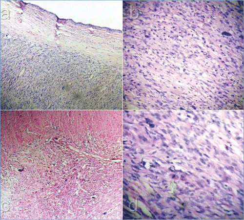
The ovaries, Fallopian tubes and pelvic lymph nodes appeared to be unremarkable. On microscopic examination, the tumour comprised spindle-shaped cells, arranged in fascicles and bundles. The tumour cells had pleomorphic blunt nuclei with moderate eosinophilic cytoplasm. Large areas of necrosis and mitotic activity of 10-12/10 hpf with atypical mitotic figures were noted (Figure a, b, c and d). Lymphovascular emboli were not seen in the cervical stroma. An immunohistochemical stain showed the tumour to be positive for smooth muscle actin and caldesmon. The uterus (endometrium and myometrium), both ovaries and the Fallopian tubes, pelvic lymph nodes and resected surgical margins were free of tumour. The patient underwent postoperative high-dose vaginal radiotherapy in order to improve local control and minimise recurrence risk. At the time of writing, after regular follow-up for a period of eight months, the patient was without evidence of persistent or recurrent disease.
Discussion
Cervical LMS is an extremely uncommon tumour, compared to uterine LMS. Cervical LMS occurs in the perimenopausal age group. The average age of diagnosis is 46 years. Abnormal vaginal bleeding is the most common presenting symptom.Citation5−7 Our patient was aged 40 years, in the premenopausal stage, and presented with abnormal vaginal bleeding. The size of the tumour on gross examination has been reported to be approximately 12 cm in various case reports,Citation8−11 whereas in our case, the tumour size was 11 cm.
The diagnosis is confirmed on histology, and the criteria proposed by Norris and Taylor,Citation8,12 which is similar to those for uterine LMS. Other criteria were proposed by Bell, Kempson and Hendrickson.Citation13 In our case, neoplastic smooth muscle cells were large, with cellular atypia having hyperchromatic nuclei. More than 10 mitosis/10 hpf were identified. Also, coagulative tumour cell necrosis was observed. These findings are consistent with both the criteria.
As very few cases have been reported in the literature, the optimal management of cervical LMS has not been fully established. Therefore, the management guidelines for the uterine LMS have to be consulted. These include total abdominal hysterectomy with bilateral salpino-oophorectomy. However, pelvic or reteroperitoneal lymph node dissection should be performed if found to be involved.Citation1
Adjuvant radiotherapy is given to decrease local recurrences, and adjuvant chemotherapy for the management of metastaic LMS.Citation1 Our patient was subjected to radiotherapy because of high-risk factors for local recurrence, including tumour size > 5 cm, high mitotic activity and a high grade of tumour.Citation1,14
Several favourable and unfavourable prognostic factors are observed in patients with LMS, which impact on patient survival rates. The premenopausal age group, low mitotic figures (< 10/10 hpf) and lower grades (grade 1 and 2) are favourable prognostic factors associated with a higher survival rate.Citation1,15 Being of an older age group (> 51 years) and having a larger tumour size (> 5 cm) are unfavourable factors, and are associated with a reduced likelihood of survival. Being of premenopausal age was the only favourable prognostic factor in our patient.
Because of the rarity of this tumour and the paucity of literature on the subject, clinicians and pathologists should look for guidance to the current accepted standards for staging, grading and the management of uterine LMS.
Declaration
Funding was not received for this study.
Conflict of interest
There was no conflict of interest to declare.
References
- Grover S, Mary A, Mahajan MK. Leiomyosarcoma of the cervix. J Obstet Gynecol India. 2009;59(4): 364–366.
- Major FJ, Blessings JA, Silverberg SG, et al. Prognostic factors in early stage uterine sarcoma. A Gynecologic Oncology Group study. Cancer. 1993;71(4 Suppl): 1702–1709.
- Fadare O. Uncommon sarcomas of the uterine cervix: a review of selected entities. Diagnostic Pathology. 2006;1: 30.
- Dhull AK, Kaushal V, Marwah N. The uncovered story of leiomyosarcoma of the cervix: a rare case report and review of literature. BMJ Case Rep. 2013;2013.
- Irvin W, Presley A, Andersen W, et al. Leiomyosarcoma of the cervix. Gynecologic Oncol. 2003;91(3): 636–642.
- Rothbard MJ, Markham EH. Leiomyosarcoma of the cervix: report of a case. Am J Obstet Gynecol. 1974;120(6): 853–854.
- Jawalekar KS, Zacharopoulou M, McCaffrey RM. Leiomyosarcoma of the cervix uteri. South Med J. 1981;74(4): 510–511.
- Kasamatsu T, Shiromizu K, Takahashi M, et al. Leiomyosarcoma of the uterine cervix. Gynecol Oncol. 1998;69(2): 169–171.
- Blaustein A, Immerman B. Leiomyosarcoma of the cervix. Obstet Gynecol. 1963;22: 224–227.
- Jawalekar KS, Zacharopoulou M, McCaffrey RM. Leiomyosarcoma of the cervix uteri. South Med J. 1981;74(4): 510–511.
- Abdul-Karim FW, Bazi TM, Sorensen K, Nasr MF. Sarcoma of the uterine cervix: clinicopathologic findings in three cases. Gynecol Oncol. 1987;26(1): 103–111.
- Taylor HB, Norris HJ. Mesenchymal tumor of the uterus. Arch Pathol. 1966;82(1): 40–44.
- Bell SW, Kempson RL, Hendrickson MR. Problematic uterine smooth muscle neoplasms. Am J Surg Pathol 1994;18(6): 535–558.
- Giuntoli RL 2nd, Metzinger DS, DiMarco CS, et al. Reterospective review of 208 patients with leiomyosarcoma of the uterus. Prognostic indicators, surgical management and adjuvant therapy. Gynecol Oncol. 2003;89(3): 460–469.
- O’Connor DM, Norris HJ. Mitotically active leiomyomas of the uterus. Hum Pathol. 1990;21(2): 223–227.

