
Abstract
Leiomyomas are classified as benign mesenchymal neoplasms and consist of smooth muscle cells with variable amounts of fibrous stroma. The tumours occur most frequently in the uterus, affecting 20–30% of women of reproductive age but vaginal leiomyomas are rare with only around 300 cases reported since the first case was described in 1733. These tumours are thought to arise from Müllerian smooth muscle cells in the sub-epithelium of the vagina. Vaginal leiomyomas are usually situated in the anterior vaginal wall. This article reports a case of primary leiomyoma arising from the left lateral vaginal wall, which presented with vaginal discharge and a lateral vaginal wall mass.
Introduction
Leiomyomas are benign mesenchymal neoplasms that can occur in the female genital tract, the uterus being the most frequent site. These tumours are thought to arise from Müllerian smooth muscle cells in the sub-epithelium of the vagina.Citation1 Leiomyomas affect 20–30% of women of reproductive age but leiomyomas occurring primarily in the vagina are rare, with only around 300 cases reported since the first case was described in 1733.Citation2,3 The anatomical location of a leiomyoma in the vagina is most commonly in the anterior vaginal wall.Citation4,5 We report a case of a primary leiomyoma arising from the left lateral vaginal wall, which presented with vaginal discharge and a lateral vaginal wall mass. The patient provided consent for the publication of the photographs used in this case report.
Case study
A 41-year-old woman, gravida 2, para 2, was referred to our gynaecological oncology unit with a large lateral vaginal wall mass. The referring hospital initially diagnosed this as a Bartholin’s abscess but during attempted surgery the mass was found to be solid. The surgery was abandoned without biopsy and the patient referred to our unit. The main presenting symptoms were discharge associated with minimal bleeding, and a lateral vaginal wall mass that had been present for the previous eight months and had been increasing in size. On examination at our unit the patient was generally well and systemic examination revealed no abnormalities. The uterus and adnexae appeared within normal limits on bimanual examination, as confirmed by transvaginal ultrasonography. The cervix was clinically normal and she had a normal Pap smear result. Vaginal examination revealed a mass in the left vaginal wall with erosion of the overlying vaginal epithelium and extending to the vulva. The mass was mobile, firm and well circumscribed with smooth borders and estimated to be at least 6 cm in largest diameter as the upper limit of the mass could not be delineated.
Magnetic resonance imaging (MRI) showed a solid tumour that extended from the left vulva into the left ischio-rectal and ischio-anal fossa, which displaced the anal canal but did not infiltrate any adjacent structures. The probable diagnosis after the imaging studies was a vaginal neoplasm, most likely a vaginal leiomyoma, but the possibility of a vaginal leiomyosarcoma was also considered. The outpatient punch biopsy showed extensive granulation tissue formation but no specific diagnosis. The decision was made to perform an excisional biopsy as the tumour appeared to be probably benign clinically and on imaging.
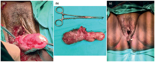
The tumour was surgically removed via a vulvo-vaginal incision. Complete excision was possible, with careful dissection in the posterior-medial border of the tumour from the anal sphincter to prevent damage. The defect was closed in layers to obliterate dead space and a good anatomical repair was achieved. Histological diagnosis revealed features in keeping with a benign leiomyoma (8 x 5 x 4 cm) with surface ulceration of the overlying epithelium (Figures and ).
Figure 1: MRI image of pelvis demonstrating a solid tumour in the left subvaginal soft tissue.
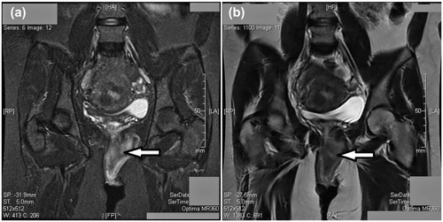
Figure 2: Intraoperative and postoperative pictures demonstrating removal of leiomyoma via vulvo-vaginal incision and closure with drainage of remaining dead space.
Discussion
The occurrence of vaginal leiomyomas is very rare. They are usually seen in the age group 35 to 50 yearsCitation6 and are reported to be more common among Caucasian women while uterine leiomyomas are more common among non-Caucasian groups.Citation2,5,7 Vaginal leiomyomas are usually situated in the midline anterior vaginal wall as a single, well-circumscribed mass;Citation8 our index case where the mass presented as a lateral vaginal wall mass is a rare occurrence.
Symptoms may include vaginal bleeding, lower abdominal pain, frequency of micturition, dyspareunia, dysuria or other features of urinary obstruction.Citation7–10 Symptoms seem to be dependent on size and location of the tumour and many patients complain only of a bulging mass.
Variation in consistency complicates making an appropriate clinical diagnosis. Vaginal leiomyomas are usually single, benign and slow growing. Sarcomatous change has, however, been reported.Citation8,11,12 If a lesion is reported to be fast growing, fixed to underlying structures with irregular borders on clinical examination, the diagnosis of a leiomyosarcoma becomes more likely. Appropriate preoperative imaging may be of value to help differentiate between a benign and a malignant process and aid in treatment planning. It is important to note, however, that no imaging modality is completely reliable in making this distinction and that clinical, radiologic features and intraoperative findings as well as postoperative histology must guide the ultimate treatment decision.
Preoperative imaging
Ultrasound (US) is recommended as the first-line imaging modality for vaginal masses in the American College of Radiology Appropriateness criteria.Citation13 Main limitations of US are operator dependence, patient obesity and the fact that, during typical routine transvaginal US, the perineum and the vagina are bypassed when the transducer is inserted into the anterior or posterior fornix prior to imaging.Citation14 Sonographic findings suggestive of a sarcoma include mixed echogenic and poor echogenic parts, central necrosis, colour flow Doppler findings of irregular vessel distribution and low impedance to flow.Citation15
Poor vaginal tissue characterisation excludes computerised tomography (CT) as the modality of choice in evaluating the female pelvis. Pelvic MRI is recommended if imaging is inconclusive or non-diagnostic during transvaginal US.Citation13 MRI is a more reliable diagnostic tool in evaluating normal vaginal anatomy and vaginal disease due to its soft-tissue characterisation and depiction of anatomical detail.Citation16 T2-weighted sequences are found to best depict the vaginal anatomy. A vaginal leiomyoma typically appears as a round homogenous lesion with similar signal to that of the myometrium, with multiple calcifications.Citation16 The absence of calcifications is a consistent finding in leiomyosarcomas.Citation17 The presence of intra-lesional haemorrhage makes the diagnosis of a sarcoma more likely.Citation18
Management
The treatment of choice for these tumours is surgical removal. The vaginal approach is usually feasible but at times an abdominal perineal approach may be required during excision of large tumours.Citation7,11 Preoperative planning is crucial and, as stated earlier, imaging may have a role in this. The approach must be decided upon, and the anticipated defect must be considered as well as the need for reconstruction of a large defect. This reconstruction may involve the utilisation of simple flaps or more complex myo-cutaneous flaps. Reconstructive procedures help repair anatomic defects, promote tissue healing and have a positive impact on psychological well-being, sexual functioning and quality of life of the patient.
If the vaginal route is chosen, the anatomical location will determine the surgical incision in the vagina. In the majority of cases the leiomyoma is located in the anterior vaginal wall.Citation8 In this instance the anterior vaginal wall is incised and the tumour dissected off the vaginal wall and underlying structures. Care must be taken not to injure the urethra and bladder – surgical planes should be developed and the tumour is then carefully resected from the underlying structures. If the leiomyoma is located in the posterior aspect of the vagina, the posterior vaginal wall must be incised and the tumour carefully resected from the rectum and anal sphincter to avoid injury. In this case study the leiomyoma was situated in the lateral vaginal wall, extending into the pararectal space. Entrance into this space was achieved by incising the posterior vaginal wall and undermining the vaginal mucosa. The rectum was separated from the posterior vaginal wall and the pararectal space was developed. The tumour was located, and dissected off the anal sphincter and rectum up to its origin extending into the pararectal space.
After removal of the leiomyoma (irrespective of location), haemostatic sutures are placed and the empty space obliterated. The vaginal mucosa must be re-approximated.
The diagnosis is confirmed by histopathology, which must rule out malignancy. In cases where malignancy is found, further surgical treatment and adjuvant management is mandatory. The histological features of leiomyosarcoma include the presence of two of the following three histological features: cytological atypia, coagulative tumour cell necrosis and 10 or more mitoses per 10 high-power microscopic fields. More recent criteria regard the presence of coagulative tumour cell necrosis and cytological atypia as the most important features in distinguishing a benign from a malignant smooth muscle tumour.Citation19
Conclusion
Vaginal leiomyoma is a rare entity and preoperatively a definitive diagnosis may be difficult. Imaging using transvaginal ultrasound and MRI can assist by demonstrating the consistency of the tumour and the absence of normal tissue invasion. MRI may also assist to plan the surgical approach and to evaluate the extent of the lesion. The practical approach to a suspected vaginal leiomyoma entails careful excision with or without prior biopsy to confirm histologic diagnosis. Histologic evaluation confirms the diagnosis and guides the need for further treatment.
References
- Delmore J, Benign Neoplasms of the vagina. The global library of women’s medicine [online]. 2008 [cited 12 Oct 2016]. doi: 10.3843/GLOWM.10005. Available from: https://www.glowm.com/section_view/heading/Benign%20Neoplasms%20of%20the%20Vagina/item/5#8231
- Östör AG. Chapter 6. Tumours of the vagina: Mesenchymal tumours. In: Tavassoli FA, Devilee P, editors. World Health Organization classification of tumours. Pathology and genetics of tumours of the breast and female genital organs. Lyon: IARC Press; 2003 [cited 22 Sept 2016]. Available from: http://www.iarc.fr/en/publications/pdfs-online/pat-gen/bb4/bb4-chap6.pdf
- Young SB, Rose PG, Reuter KL. Vaginal fibromyomata: two cases with preoperative assessment, resection and reconstruction. Obstet Gynecol. 1991;78:972–4. PMID: 1923243.
- Kaufman RH, Gardner HL. Tumors of the vulva and vagina. Benign mesodermal tumors. Clin Obstet Gynecol. 1965;8(4):953–81 . PMID: 5323603.10.1097/00003081-196512000-00014
- Nel CP, Tiltman AJ. Leiomyoma of the vagina. S Afr Med J. 1978;54:816–7.
- Bennett HG, Ehrlich MM. Myoma of the vagina. Am J Obstet Gynecol. 1941;42:314–20.10.1016/S0002-9378(16)40639-3
- Pulfus E, Newcomer J. Vaginal wall mass. Obstet Gynecol Surv. 1999;54:149–50. PMID: 10071837.10.1097/00006254-199903000-00001
- Chakrabarti I, De A, Pati S. Vaginal leiomyoma. J Midlife Health. 2011;2(1):42–3. doi:10.4103/09767800.83274.
- Sim CH, Lee JH, Kwak JS, et al. Necrotizing ruptured vaginal leiomyoma mimicking a malignant neoplasm. Obstet Gynecol Sci. 2014;57(6):560–3. doi: 10.5468/ogs.2014.57.6.560.
- Imai A, Furui T, Hatano Y, et al. Leiomyoma and rhabdomyoma of the vagina. Vaginal myoma. J Obstet Gynaecol. 2008;28(6):563–6. doi: 10.1080/01443610802310333.
- Gowri R, Soundararaghavan S, Oumachigui A, et al. Leiomyoma of the vagina: an unusual presentation. J Obstet Gynaecol Res. 2003;29(6):395–8.10.1111/j.1341-8076.2003.00135.x
- Çobanoğlu Ö, Zorlu C, Ergun Y, et al. Leiomyosarcoma of the vagina. Eur J Obstet Gynecol Reprod Biol. 1996;70:205–7. doi: 10.1016/S0301-2115(95)02554-5.
- American College of Radiology. ACR appropriateness criteria. [cited 25 Aug 2016]. Available at: http://www.acr.org/secondarymainmenucategories/quality_safety/app_criteria.aspx
- Walker DK, Salibian RA, Salibian AD, et al. Overlooked diseases of the vagina: a directed anatomic-pathologic approach for imaging assessment. RadioGraphics. 2011;31:1583–98. doi: 10.1148/rg.316115531.
- Amant F, Coosemans A, Debiec-Rychter M, et al. Clinical management of uterine sarcomas. Lancet Oncol. 2009;10:1188–98.10.1016/S1470-2045(09)70226-8
- Elsayes KM, Narra VR, Dillman JR, et al. Vaginal masses: magnetic resonance imaging features with pathologic correlation. Acta Radiol. 2007;48(8):921–33. doi:10.1080/02841850701552926.
- Van den Bosch T, Coosemans A, Morina M, et al. Screening for uterine tumours. Best Pract Res Clin Obstet Gynaecol. 2012;26:257–66.10.1016/j.bpobgyn.2011.08.002
- Samual A, Fennessy FM, Tempany CM, et al. Avoiding treatment of leiomyosarcomas: the role of magnetic resonance imaging in focused ultrasound surgery. Fertil Steril. 2008;90:850.
- Watanabe K. Suzuki. Uterine leiomyoma versus leiomyosarcoma: a new attempt at differential diagnosis based on their cellular characteristics. Histopathology. 2006;48:563–8. doi:10.1111/j.1365-2559.2006.02368.