
Abstract
Mitotically active cellular fibroma (MACF) is a category of fibromatous tumours of the ovary described in the WHO classification of tumours of female reproductive organs. The case of a 54-year-old female who presented with lower limb swelling due to deep vein thrombosis (DVT) is reported. On examination, she had a 5 x 8 cm fixed pelvic mass. She underwent optimal cytoreductive surgery. Grossly the tumour, which was found to occupy the left pelvic space, was hard and irregular in shape and was adherent to the surrounding structures. Histopathology revealed collagen-producing spindle cells showing mitotic activity of 6–8/10 HPF with minimal nuclear atypia and Ki 67 labelling index of 10%. A final diagnosis of MACF was made. In view of sparse evidence regarding its management and due to residual tumour (< 1 cm), she received adjuvant radiotherapy. She has remained disease free over a period of two years.
Introduction
Mitotically active cellular fibroma (MACF) is a recently described category of fibromatous tumours of the ovary, which is defined as an ovarian cellular fibrous tumour with mitotic activity of ≥ 4/10 HPF (high power field) without moderate or severe atypiaCitation1 and is less aggressive than fibrosarcoma.
As this is a recent entry into the World Health Organization classificationCitation2 and since very few cases have been reported in the literature, we present this case and review the literature.
Case report
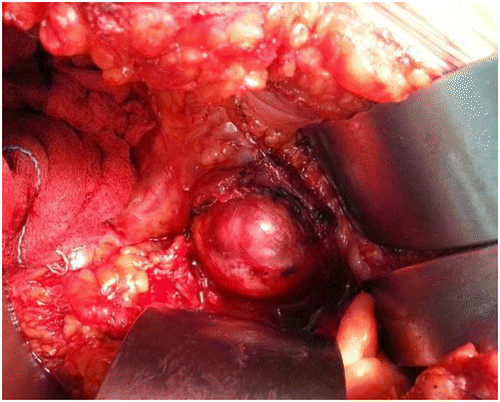
This report discusses a 54-year-old female with history of total hysterectomy undertaken for fibroid uterus 12 years ago, who presented to us with lower limb swelling and pain. On evaluation she had a 5 x 8 cm hard pelvic mass that was adherent to the left pelvic sidewall. She also had DVT (deep vein thrombosis) extending from the left popliteal vein to the common iliac vein. Positron emission tomography/computerised tomography (PET/CT) revealed a solid lobulated mass in the region of the left adnexa with coarse nodular calcifications with standardised uptake value (SUV) of 8.1, left mild hydroureteronephrosis and filling defects in the left distal main pulmonary artery. An inferior venacaval filter was placed and she was started on anticoagulants. She underwent cytoreductive surgery, left ureterotomy and double J stenting. Intraoperatively we found a 5 x 8 cm hard, irregular left adnexal mass which was adherent to the left ureter, bladder, rectum and iliac vessels (Figure ). She underwent optimal cytoreductive surgery with residual tumour of < 1 cm.
Figure 1: Intraoperative image showing a left pelvic mass adherent to the iliac vessels.
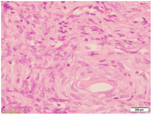
Histopathology revealed collagen-producing spindle cells in most places forming areas of intersecting bundles and with a storiform pattern. Large acellular hyalinised areas and intercellular oedema was also noted. Six to eight per 10 HPF mitoses with numerous large scattered ectatic blood vessels were noted, but nuclear atypia was minimal. Features of necrosis and lymphovascular space invasion were absent (Figure ). Proliferative index by immunohistochemistry showed a Ki67 labelling index of 10% and a final diagnosis of MACF of ovary was made. Following a multidisciplinary team discussion and due to lack of evidence on management of patients with residual tumour, a consensus regarding the need for adjuvant radiation therapy to prevent local recurrence was made. She received 30 fractions of external beam radiotherapy, and has now been disease free for two years.
Figure 2: Microscopic appearance of mitotically active cellular fibroma.
Discussion
Tumours derived from gonadal stroma account for about 5% of all the ovarian tumors.Citation3 Historically since 1981, as described by Pratt and ScullyCitation,4 mitotic activity of the cells was given equal importance as nuclear atypia in predicting the aggressiveness of an ovarian fibromatous tumour and classifying it as fibrosarcoma. The tumours exhibiting a mitotic activity of ≤ 3/10 HPF were labelled fibromas, while those with ≥ 4/10 HPF mitotic figures were considered malignant in nature. As time elapsed and more cases were reported, tumours that had high mitotic counts of ≥ 4/10HPF but minimal nuclear atypia were found to be less aggressive and followed a more indolent clinical course. Hence in 2006, Irving et al.Citation1 proposed the reclassification of fibromatous tumours, introducing MACF for the first time, which fitted into the afore-mentioned criteria. This has been included in the WHO histological classification since 2014.
To date, only a handful of MACF cases have been reported in the literature. After a search of English-language literature, we found seven more cases. All eight patient characteristics including ours are listed in Table . Mean age at presentation was 42.3 years (ranging from 13 to 76 years). More commonly, patients presented with symptoms of pain or mass per abdomen. Our case was unique in its presentation with lower limb swelling due to DVT being the sentinel event. Mean size of the tumour considering all the cases was 11 cm (ranging from 6 to 19 cm). Surgical resection of the tumour was the mainstay of treatment for all cases and fertility-sparing procedures were performed for young patients. All eight cases showed densely cellular fibromatous tumours, with few exhibiting herringbone and storiform pattern as in our case. Mitotic figures ranged from 4 to 17 with a mean of 7.42/10 HPF. The mean Ki67 of these tumours was 9.14%, ranging from 8% to a description of strongly positive. Prognostic features, which have been linked to the tumour behaviour, included intraoperative rupture, dense adherence, histological features of tumour necrosis and haemorrhage. Of the seven cases described in the literature, six cases had complete cytoreduction and had not received any adjuvant treatment with no recurrence. One caseCitation5 received adjuvant hormonal therapy possibly due to incomplete resection of the tumour. In spite of adjuvant treatment, she had recurrence at six months for which secondary cytoreductive surgery was performed and had remained disease free for a further six months until reporting of the case. Our patient received adjuvant radiotherapy due to the presence of poor prognostic factors (adherent nature of the disease, tumour rupture and residual tumour of < 1 cm) and has now been disease free for over 2 years.
Table 1: Patient characteristics
Conclusion
The distinction of MACF from fibrosarcoma is of paramount importance. Despite high mitotic count when nuclear atypia is low, it confers a relatively favourable outcome. This particular tumour can present in various ways with an exceptional presentation of DVT and pulmonary embolism as in our case. Due to paucity of evidence regarding adjuvant treatment in cases that have undergone primary complete cytoreduction, careful follow-up with high degree of vigilance is a sound option. Until clear guidelines have been established, for those patients with poor prognostic factors, such as postoperative residual tumour, tumour rupture or adherence, adjuvant treatment has to be individualised.
References
- Irving JA, Alkushi A, Young RH, Clement PB. Cellular fibromas of the ovary: a study of 75 cases including 40 mitotically active tumors emphasizing their distinction from fibrosarcoma. Am J Surg Pathol. 2006;30: 929–938.10.1097/00000478-200608000-00001
- McCluggage WG, Kiyokawa T, Staats PN, Young RH. Sex cord-stromal tumours- pure stromal tumours. In: Kurman RJ, Carcangiu ML, Herrington CS, Young RH. WHO classification of tumours of female reproductive organs. 4th ed. International agency for research on cancer Lyon; 2014. p. 44.
- Monteiro SB, Costa A, Paiva V. Mitotically active cellular ovarian fibroma with Meigs’ syndrome and elevated CA-125: towards fertility preservation. J Pediatr Adolesc Gynecol. 2012;25: e107–109.10.1016/j.jpag.2012.05.012
- Prat J, Scully RE. Cellular fibromas and fibrosarcomas of the ovary: a comparative clinicopathologic analysis of seventeen cases. Cancer. 1981;47: 2663–2670.10.1002/(ISSN)1097-0142
- Bucella D, Limbosch JF, Buxant F, Simon P, Fayt I, Anaf V, et al. Recurrence of mitotically active cellular fibroma of the ovary. Obstet Gynecol Int. 2009;2009: 803062.
- Kaku S, Takeshima N, Akiyama F, Furuta R, Hirai Y, Takizawa K. A unique fibrous tumor of the ovary: fibrosarcoma or mitotically active cellular fibroma? Anticancer Res. 2007;27: 4365–4369.
- Zong L, Lin M, Fan X. Mitotically active cellular fibroma of ovary should be differentiated from fibrosarcoma: a case report and review of literature. Int J Clin Exp Pathol. 2014;7: 7578–7582.
- Wu H, Xie J, Huang W, Wu J. Mitotically active cellular fibroma of the ovary: a case report and a review of the literature. Eur J Gynaecol Oncol. 2014;35: 81–83.
- Yamada T, Hattori K, Satomi H, Hirose Y, Nakai G, Daimon A, et al. Mitotically active cellular fibroma of the ovary: a case report and literature review. J Ovarian Res 2015;8: 65.10.1186/s13048-015-0191-x
- Yildirim N, Clock B, Akalin F, Gynecology C, Obuz H. Ugur. Mitotically active cellular fibroma of the ovary: a case report. J Turk Soc Obstet Gynecol 2015;1: 53–55.10.4274/tjod