
Keywords:
Case study
A 48-year-old right-hand-dominant cleaner presented to her general practitioner with several months history of right wrist pain. There was no history of previous wrist trauma, and she did not have a history of inflammatory arthropathy. She neither drank nor smoked. An X-ray of the wrist was reported to be normal. A diagnosis of de Quervain’s tenosynovitis was made and she was treated with anti-inflammatory drugs and physiotherapy, and received an injection of corticosteroid. Six months’ later, the patient complained of worsening symptoms and was referred to an orthopaedic surgeon. The patient reported wrist pain and a longstanding problem of a wrist that “clicked”, with deterioration of function over the preceding 12 months. In particular, she had difficulty with power movements of the wrist.
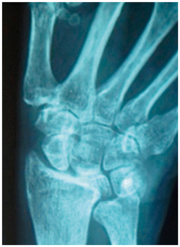
The examination revealed swelling around the volar and dorsal aspect of the radiocarpal joint and tenderness around the scaphotrapezotrapezoidal joint. The range of motion of the wrist was limited, and there was pain on stressing the scapholunate articulation. A plain new X-ray was taken (Figure ).
Figure 1: A plain anteroposterior X-ray of the patient’s right wrist.
Questions
What abnormalities can be seen on the X-ray (Figure )?
What is the most likely diagnosis?
What treatment options are available?
Answers to the questions
Q1
The radiographs in Figure demonstrate sclerosis of the lunate, joint space narrowing of the radiolunate joint and collapse of the lunate, as well as negative ulnar variance.
Q2
The most likely diagnosis is Kienböck’s disease, an avascular necrosis of the lunate bone first described in 1910.Citation1 The condition is thought to be due to vascular impairment as a result of chronic injury, and results in progressive collapse and degenerative changes within the radiocarpal joint.Citation2 Frequently, it is unilateral, affecting the right wrist. Predisposed individuals are often aged 20–40 years and involved in manual labour, with repeat episodes of minor trauma. Negative ulnar variance (relative shortening of the ulnar in relation to the radius) is usually associated with the condition.Citation3 However, in some cases, patients may exhibit a neutral ulnar, or even a positive ulnar variance.Citation4Clinical features are progressive pain and swelling of the wrist.
The condition is staged radiographically using Lichtman’s classification:
Stage I: Normal radiographs
Stage II: Sclerosis of the lunate
Stage III: Lunate collapse
Stage IV: Degenerative changes around the lunate.Citation5
Q3
In the early stages of the disease, when the wrist pain is localised to the radial aspect of the wrist, the symptoms may resemble those of de Quervain’s tenosynovitis.
Pain and swelling may be managed with anti-inflammatory drugs, activity modification and splinting. Unfortunately, since Kienböck’s disease is progressive, the condition may eventually require surgical treatment.
There are several surgical options for the treatment of Kienböck’s disease.Citation6 The choice of procedure depends on several factors, including the patient’s activity level, personal goals, surgeon experience with various procedures, and most importantly, how far the disease has progressed.
In stages I and II, treatment options include revascularisation procedures using a vascularised graft from the radius that is inserted into the lunate. A joint levelling procedure may be carried out to treat negative ulnar variance. In this situation, the ulna may be lengthened using a bone graft, or the radius shortened by removing a section of bone. The levelling procedure reduces the forces that compress the lunate and can halt progression of degeneration of the joint.
Proximal row carpectomy can be performed for more advanced stage III and IV disease. This relieves pain and maintains partial wrist motion. Alternatively, the carpal bones surrounding the lunate may be fused to relieve the pain and to preserve some motion. All of the wrist bones may have to be fused to the radius in end-stage disease accompanied by lunate collapse and wrist arthritis. Although this relieves pain, wrist motion is lost but forearm motion preserved.
Patient outcome
The patient underwent a wrist fusion and is currently satisifed with her condition.
Conclusion
Kienböck’s disease is a relatively rare disorder,Citation7 and is of ongoing interest as the precise cause of the condition is still unknown and there is no strong evidence on the most appropriate treatment for the disorder prior to the development of secondary wrist osteoarthritis. Advanced cases usually require surgical fixation, and as outlined in this case, this procedure can result in a satisfactory improvement with respect to the pain, but at the cost of a decrease in wrist movement and function. Our case highlights the importance of the need to monitor patient disorders, and if conditions fail to settle within an appropriate timeframe, for an alternative diagnosis to be considered, appropriately investigated and referred.
Additional information
Funding
References
- Kienböck R. Uber traumatische Malazie des Mondbeins und ihre Folgezustande: (Entartungsformen und Kompressionfrakturen) [Concerning traumatic malacia of the lunate and its consequences: degeneration and compression fractures.]. Fortschr Geb Roentgen. 1910;16:78–103.
- Keith PPA, Nuttall D, Trail I. Long-term outcome of nonsurgically managed Kienböck’s disease. J Hand Surg Am. 2004;29(1):63–67.10.1016/j.jhsa.2003.10.016
- Bonzar M, Firrell JC, Hainer M, et al. Kienböck disease and negative ulnar variance. J Bone Joint Surg Am. 1998;80(8):1154–7.
- Chung KC, Spilson SV, Kim HM. Is negative ulnar variance a risk factor for Kienböck’s disease? A meta-analysis. Ann Plast Surg. 2001;47(5):494–9.10.1097/00000637-200111000-00004
- Lichtman DM, Mack GR, MacDonald RI, et al. Kienböck’s disease: the role of silicone replacement arthroplasty. J Bone Joint Surg Am. 1977;59(7):899–908.
- Lichtman DM, Lesley NE, Simmons SP. The classification and treatment of Kienböck’s disease: the state of the art and a look at the future. J Hand Surg Eur. 2010;35(7):549–54.10.1177/1753193410374690
- Office of Rare Diseases Research, US Department of Health and Human Services. 2013 May 7 [cited 2014 Nov 12]. Available from: http://rarediseases.info.nih.gov/gard/9690/kienbocks-disease/resources/1