
Abstract
Paediatric patients make up a significant portion of the patient load at primary care level. Children can present at the primary care physician with a wide range of eye problems, some of which are serious enough to impair the quality of the child’s life. The aim of this review article was to highlight serious paediatric visual disorders of which the primary care practitioner should be aware. The article includes a brief discussion of the features of amblyopia, strabismus, retinopathy of prematurity, allergic conjunctivitis, ophthalmia neonatorum and retinoblastoma. Causes of sight-threatening conditions are highlighted, and methods to detect them at primary care level described. Paediatric eye disorders are relatively common and need to be identified and managed as early as possible in order to prevent a potential lifetime of visual morbidity, blindness or worse.
Introduction
Paediatric patients make up a significant portion of the patient load at primary care level. Children can present to the primary care physician with a wide range of eye problems, some of which are serious enough to impair the quality of the child’s life. Reduced vision can affect academic performance, choice of occupation and socio-economic status. The aim of this review article was to highlight serious visual disorders of which the primary care practitioner should be aware. The need for an update in this field became apparent following a recent study on the primary health eye care knowledge of general practitioners.Citation1 This article is the sixth in a series which attempts to address this issue.
Approach to the patient
Examining a child with an ocular disorder requires patience and skill. The diagnosis can often be suspected from the history provided by the parents. Symptoms reported by parents include persistently holding objects and books up to the face, sitting close to the television, behavioural problems and failure of the child in reaching developmental milestones. A family history of squinting or wearing spectacles from a young age is also relevant. It is important to enquire about other potentially inherited ocular conditions, i.e. congenital cataracts, retinitis pigmentosa, juvenile glaucoma and retinoblastoma, or genetic diseases with ocular manifestations, such as collagen vascular disorders or phakomatoses.Citation2 If any of these conditions are suspected, the child requires visual screening, and referral to an ophthalmologist if the screening reveals any abnormalities.
Clinical conditions
Amblyopia
This is a disorder which is characterised by reduced vision in an eye that appears to be normal, or loss of vision out of proportion to the structural abnormalities found in the eye. It is estimated to affect 1-5% of the population.Citation2 Bilateral amblyopia is sometimes seen in those with severe refractive errors, such as a high degree of hyperopia, astigmatism or myopia.Citation3 The lateral geniculate body (LGB) receives input from both eyes, so if the input is disrupted during the critical period of visual development (the first 6-8 years of life), layers of the LGB undergo atrophy, as do the corresponding areas in the cerebral cortex. The eye may appear to be anatomically normal, but it will not have normal vision unless the underlying cause and the amblyopia are treated early.
Amblyopia is usually classified according to the presenting visual condition causing the impaired visual development:Citation4
Strabismic amblyopia, if a squint is present in one eye.
Anisometropic amblyopia, if there is a difference in refractive error between the two eyes.
Mixed, if anisometropic and strabismic amblyopia coexist.
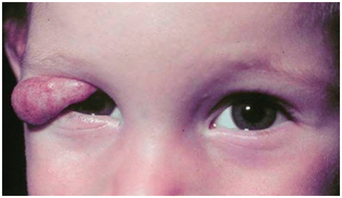
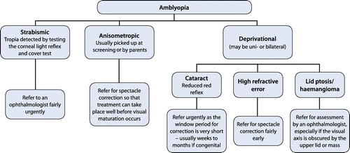
Stimulus deprivation, if there is some obstruction of vision by a cataract, high refractive error, upper lid ptosis (Figure ) or lid haemangioma (Figure ).
Figure 1: Bilateral upper lid ptosis
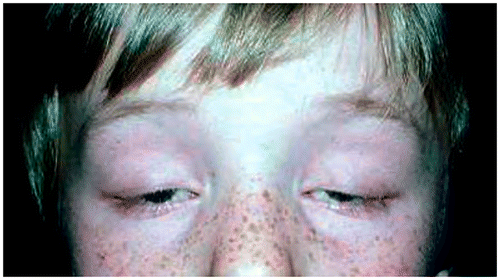
Figure 2: Upper lid haemangioma
When evaluating a child for possible amblyopia, the primary care doctor should be able to:
Estimate visual acuity in the child: Estimating visual acuity in a child depends on his or her age. The response to patching one eye in very young children can be assessed. A child whose good eye is occluded and who is forced to use the weaker eye may strongly object to this by crying or trying to remove the patch. If the child is old enough, offer him or her very small sweets like “100s and 1 000s” or silver balls, and see if he or she can pick them up. It is possible to measure visual acuity from the age of 2-3 years by using special picture charts or matching cards.Citation5
Detect strabismus by testing the corneal light reflex and conducting a cover test: The child looks at a penlight, or the baby’s attention is attracted with a light. If there is a squint, the corneal light reflex will appear asymmetrical on the two sides, otherwise it should fall just nasal from the centre of each pupil (see Figures and , and later, under the section on Strabismus, for an illustration of symmetrical versus asymmetrical corneal light reflexes). A cover test involves covering the fixing eye and watching the other eye to check if it moves to take up the fixation. If there is no movement, it means that the other eye was also fixing on the target.
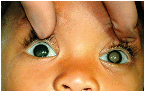
Examine the red reflex and carry out fundoscopy: An absent red reflex may indicate a cataract or intraocular tumour (Figure ).
Figure 3: Left esotropia and hypertropia
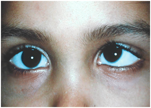
Figure 4: Epicanthic folds, causing pseudoesotropia
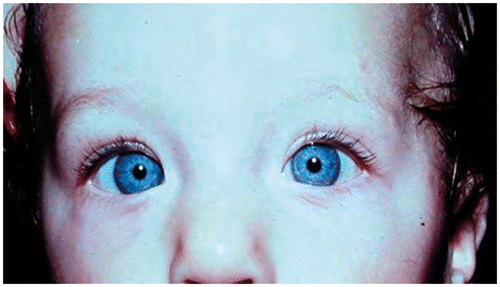
Figure 5: Reduced red reflex
Management
Management depends on the cause of the reduced vision (Figure ). It is treatable if detected early, but needs treatment before the age of visual maturation. It is important to refer the patient to an ophthalmologist if there is concern about any of the following:Citation5
A cataract.
Other causes of leukocoria.
Refractive error.
A squint.
If a mother says that her child “does not see”.
Figure 6: Management of amblyopia
Strabismus (squint)
Strabismus is defined as a misalignment of the eyes (visual axis). Most paediatric squints are comitant, i.e. the angle of deviation remains the same in all directions of gaze.Citation4 By contrast, if the angle of deviation changes with different directions of gaze, then the squint is incomitant. Cranial nerve palsies are incomitant. Incomitance in a patient with a left sixth nerve palsy is illustrated in Figure and . On looking to the right, misalignment is not visible, but on looking to the left, a large-angle squint is apparent. A “tropia” is a manifest squint that is present when both eyes are open. A “phoria” is a latent squint that only appears when one eye is covered. Most people have a small, inconsequential phoria.Citation5 As soon as both eyes are uncovered, the brain “realises” that one eye has drifted off and corrects the phoria. Deviation inwards, i.e. towards the nose is esotropia, while deviation outward is exotropia, up is hyperphoria and down is hypotropia or hypophoria.
Figure 7a: Incomitance in a patient with a left sixth nerve palsy. On looking to the right, misalignment is not visible
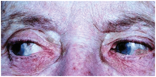
Figure 7b: Incomitance in a patient with a left sixth nerve palsy. On looking to the left, a large-angle squint is apparent
Comitant squints are often idiopathic, but may have other causes. Squints may be hereditary. They may also be associated with developmental factors, such as mental retardation and foetal alcohol syndrome.Citation2 Hypermetropia (far-sightedness) is a common cause of esotropia. In order to produce a clear image for near, a hypermetropic child exerts very powerful accommodation, eventually resulting in an esotropia due to over-convergence. Correcting the hypermetropia relieves this reflex and “straightens” the eyes. Visual loss may also cause a squint. Before the age of two years, any cause of visual loss may result in esotropia. After the age of five years, visual loss usually results in exotropia.Citation5
Pathophysiology
The mechanism of development of comitant squints is unclear when there is no obvious underlying cause.
Symptoms and signs
Diplopia (double vision) does not occur in early childhood due to suppression of the second image by the brain, and the squint is recognised by the abnormal position of the eyes. Most childhood squints are convergent [esotropia (Figure )]. It is important to look for epicanthic folds which may cause a pseudoesotropia (Figure ). The normal position of the corneal light reflexes will differentiate a tropia from a pseudosquint.
A cover test confirms the presence of a tropia and it may also help to detect a phoria:
In the presence of a tropia, there will be a corrective movement of the deviating eye when the normal eye is covered. This test only works if the eye has reasonable vision and a good range of movement.
A phoria will be detected if an alternate cover test is conducted. The deviating eye (under the cover) will move to take up fixation as soon as it is uncovered.
Management
Any squint which is present after the age of three months is abnormal, and should be referred for assessment with respect to an underlying cause, treatment of any associated amblyopia and possible surgery.Citation5
Retinopathy of prematurity
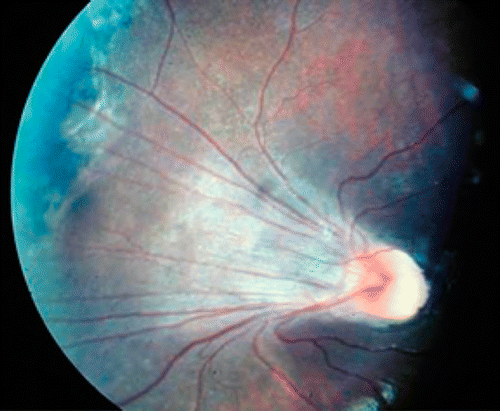
The retina is only fully vascularised at term. At four months of gestation, vascularisation is only up to the disc. At eight months of gestation, vascularisation is up to the nasal half of the retina. At nine months, the temporal retina is completely vascularised. Neonatal insults, such as prematurity and exposure to supplemental oxygen, can cause abnormal retinal vascularisation. The avascular peripheral retina is very sensitive to blood oxygen levels. Terminal branches of retinal vessels may constrict and cause ischaemia, which, in turn, can lead to abnormal vessel growth, retinopathy and retinal scarring (Figure ). Oxygen saturation in preterm neonates receiving supplemental oxygen should run between 88% and 92% to reduce the chances of retinopathy of prematurity (ROP) developing.
Figure 8: Dragged macula as a result of scarring following retinopathy of prematurity
Guidelines for screeningCitation6
Screening for ROP applies as follows:
Of neonates born before 32 weeks gestation.
Of preterm infants weighing < 1 500 g.
Of preterm infants weighing 1 500–2 000 g with increased risk of ROP, i.e. a family history of ROP, cardiac arrest and multiple (> 2) blood transfusions.
It should be performed at 4-6 weeks chronological age, or 31-33 weeks post-conception age (whichever comes first).
After the initial screening, follow-up is determined by the ophthalmologist.
Management
Severe ROP is treated with laser or cryotherapy to the ischaemic retina to prevent retinal detachment and blindness.Citation6,7 Squints and myopia also occur more frequently in premature babies. It is important to remember this, and to refer as necessary.
Retinoblastoma
This is the most common primary intraocular malignancy in children. It is a tumour of the embryonal retinal cells. It is caused by a mutation in the tumour suppressor gene, RB1.Citation7
Symptoms and signs
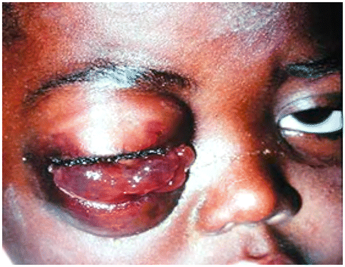
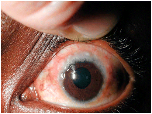
Patients usually present before the age of three years with leucocoria (a white pupil) and a squint (Figure ). Occasionally, a red eye that does not get better may be the initial symptom (Figure ). Signs of orbital inflammation may be present in advanced cases (Figure ).
Figure 9: Leucocoria and esotropia in a patient with retinoblastoma
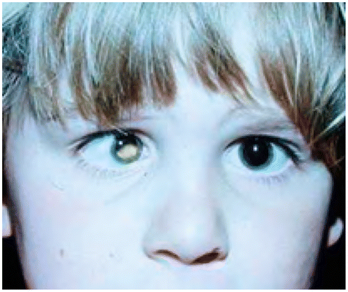
Figure 10: Leucocoria and a persistent red eye
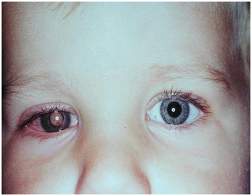
Figure 11: Orbital involvement with late presentation
Management
Refer to an ophthalmologist. The type of treatment (chemotherapy, radiotherapy and surgery) depends on whether or not the tumour is localised to the eye, or whether or not there has been metastatic spread.
Ophthalmia neonatorum
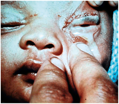
This is defined as any conjunctivitis which occurs within the first postnatal month. It is commonly caused by gonococcus, chlamydia or other pathogens, such as Staphylococcus aureus and Streptococcus pneumoniae.Citation5 It is usually acquired during the passage through the birth canal of an infected mother. It is serious because of lack of immunity in the infant and the immaturity of the ocular surface.
Symptoms and signs
Signs and symptoms include:
Bilateral conjunctival injection and eyelid oedema.
Discharge, which is initially mucoid, and later becomes mucopurulent (Figure ).
Corneal complications, such as ulcer or perforation, are more likely if infected with gonococcus.
Figure 12: Ophthalmia neonatorum
Management
This condition is notifiable. All patients should be referred to hospital for further management. A conjunctival pus swab must be taken and sent for microscopy and culture. Treatment depends on the cause. Chlamydial infection is treated with oral erythromycin for two weeks.Citation5 Gonococcal infection requires ceftriaxone intravenously or intramuscularly.Citation7
It is important to note that if chlamydia or gonorrhoea is confirmed, the parent and partner must be investigated and treated.
Allergic eye disease
Acute allergic rhinoconjunctivitis
This is the most common form of ocular and nasal allergy, and affects approximately 20% of the population.Citation7 It usually presents with transient acute attacks of redness, watering and itching associated with sneezing and nasal discharge. An ocular examination reveals bilateral red eyes, with a clear watery or stringy mucoid discharge. There may also be lid oedema. Patients respond to topical antihistamines and mast cell stabilisers.Citation5
Vernal keratoconjunctivitis
This is a bilateral chronic disease that affects children with a history of atopy. Symptoms peak prior to the onset of puberty, and gradually resolve over a period of 5–10 years. Children may present with palbebral (involving the upper tarsal conjunctiva), or limbal (corneal) disease (Figure ), or a combination of both. Patients usually require a combination of topical steroids and mast cell stabilisers.Citation7 They should be referred to an ophthalmologist for management, and they also have an increased risk of developing keratoconus.
Figure 13: Limbal vernal keratoconjunctivitis
Atopic keratoconjunctivitis
This condition is characterised by bilateral inflammation of the conjunctiva and eyelids, which is often associated with severe atopic dermatitis. Unlike vernal keratoconjunctivitis, it tends to be chronic and unremitting.Citation7 Management is difficult because patients require long-term steroid therapy, which can result in other ocular complications, such as glaucoma and cataract. Therefore, it is associated with significant visual morbidity.
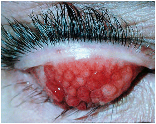
Giant papillary conjunctivitis
This is an inflammatory disorder of the superior tarsal conjunctiva. It occurs secondary to an ocular foreign body, such as a contact lens, an ocular prosthesis or exposed sutures.Citation7 Symptoms include a foreign body sensation, redness and itching. Large papillae are seen on eversion of the upper lid (Figure ). Treatment consists of removing the allergen and using topical mast cell stabilisers. Topical steroids are rarely indicated.
Figure 14: Everted upper lid with giant papillary conjunctivitis
Conclusion
Paediatric eye disorders are relatively common and the primary care physician should be familiar with serious childhood eye problems so that they can be identified and managed as early as possible in order to prevent a potential lifetime of visual morbidity, and in some cases, even mortality.
Conflict of interest
The authors declare that there were no financial or personal relationships which might have inappropriately influenced them when writing this paper.
Acknowledgements
The Division of Ophthalmology, University of Cape Town, is thanked for providing permission to use the clinical photographs. Permission for the use of the photographs was obtained from all patients (parents).
References
- Van Zyl LM, Fernandes N, Rogers G, et al. Primary health eye knowledge among general practitioner’s in the Cape Town metropole. SA Fam Pract. 2011;53(1):52–5.
- Walker S, Harris Z. Detecting the serious visual disorders of childhood. Paediatrics Child Health. 2012;22(1):25–30.10.1016/j.paed.2011.04.009
- Davidson S, Quinn GE. The impact of pediatric vision disorders in adulthood. Pediatrics. 2011;127(2):334–9.10.1542/peds.2010-1911
- Webber AL, Wood J. Amblyopia: prevalence, natural history, functional effects and treatment. Clin Exp Optom. 2005;88:365–75.10.1111/cxo.2005.88.issue-6
- Du Toit N, Cook C. Paediatric ophthalmology. In: Lecture notes for medical students. Cape Town: Juta 2009, 66–70.
- Visser L, Singh R, Young M, et al. Guideline for the prevention, screening and treatment of retinopathy of prematurity (ROP). S Afr Med J. 2013;103:116–25.
- Kanski JJ. Clinical ophthalmology. A systematic approach. 6th ed. Butterworth Heinemann Elsevier; 2007.