
Abstract
Background: Abdominal compartment syndrome is a severe condition of tension generated within the closed abdominal cavity from perforation of peptic ulcer. Due to the chemical reaction and pressure consequences it has poor prognosis with very high mortality > 80%.
Study method: We present a case study of one patient who developed abdominal compartment syndrome and survived as a result of prompt intervention. The patient had acute abdomen due to perforated gastric ulcer that resulted in leakage of gastric contents into the peritoneal cavity and build up of tension due to chemical irritation and gases. Urgent laparotomy with correction of the electrolyte imbalance and supporting intensive care was provided. The patient recovered from cardiac arrest that was reversed with some residual brain damage.
Conclusion: Effective, prompt recognition and appropriate emergency interventions are key to the survival of victims of abdominal compartment syndrome that otherwise has a very high mortality. The primary care physician and emergency care practitioners play a critical role in the early recognition and initial management of patients with this condition.
Introduction
Peptic ulcer disease is a common disorder of the gastrointestinal mucosa, affecting 3.7 million people globally every year.Citation1 It is a problem of the gastrointestinal tract, characterised by mucosal damage secondary to pepsin and gastric acid secretion. It is commonly associated with Helicobacter pylori infection and the use of nonsteroidal anti-inflammatory drugs. It usually occurs in the stomach and proximal duodenum. It occurs less commonly in the lower oesophagus, the distal duodenum or the jejunum.Citation2 It is usually seen in patients aged 25–64 years.Citation3
The natural progression of a peptic ulcer ranges from development of the disease to its resolution or the occurrence of complications which contribute to significant morbidity and mortality. The most common complications of peptic ulcer are perforation, bleeding and obstruction of the hollow visceral organs. Abdominal compartment syndrome is a condition which describes the tension generated within the closed abdominal cavity from perforation of the peptic ulcer. Abdominal compartment syndrome is uncommon owing to recent advances in the diagnosis, which result in a timely recognition and intervention.Citation4 However, it has very poor prognosis because of leakage of the gastric contents into the abdominal cavity, resulting in chemical and pressure consequences.Citation5 Abdominal compartment syndrome is associated with very high mortality. The survival rate is reported to be as low as 20%.Citation6
Case study
Mr D, a 32-year-old man, was brought into the accident and emergency unit of the regional hospital in Bojanala District, North West province, by an ambulance, with the history of severe abdominal pain. He reported that he had experienced severe, sharp abdominal pain in the epigastrium, starting at 5h00, which rapidly spread to the entire abdomen. He took some sodium bicarbonate (baking soda) suspension, which he had been using for a long time whenever he experienced heartburn, but experienced no relief this time. He indicated that he had been suffering from heartburn for four years, and that the epigastric pain was usually worse at night. He had no other significant medical or surgical history. He had been smoking 20 cigarettes a day since he was a teenager.
The following vital signs were noted at the triage station: a heart rate of 100 beats per minute, blood pressure of 155/116 mmHg, oxygen saturation of 98% on room air, a respiratory rate of 22/minute, a temperature of 35 °C, haemoglobin of 16.6 g/dl and random blood sugar of 12.8 mmol/l. He passed considerable flatus and the abdomen was noted to be uniformly distended, very tender and hard. Oxygen of 10 litres/minute was given immediately, using a mask. An infusion of 2 l of Ringer’s lactate was performed intravenously. While these procedures were being conducted, Mr D complained of a tingling sensation in both legs, difficulty breathing and suddenly became restless. He was rushed into the resuscitation room, where blood gas analysis was performed. The oxygen saturation had dropped from 98% to 78%, and the blood gas result showed a pH of 7.07, partial pressure of carbon dioxide (PCO2) of 67 mmHg, with a base excess of −12.3 mmol/l. Ventilation with a bag, valve and mask and 100% oxygen was immediately carried out, followed by endotracheal intubation.
After 10 minutes of resuscitation, it was noted that there was no improvement in the oxygen saturation and that the blood pressure had dropped further to 113/54 mmHg. The second blood gas result after the intubation was worse than before (the PCO2 was 91 mmHg, with a base excess of −19.9 mmol/l). The abdomen became more tense and distended, and more resonant on percussion. The X-ray examination revealed a reduction in lung capacity and massive pneumoperitoneum, which pushed the lateral border of the liver towards the midline (Figure ). Abdominal compartment syndrome was suspected, and immediate decompression performed with two large-bore needles.
Figure 1: An X-ray of the chest and abdomen (taken in the sitting position)
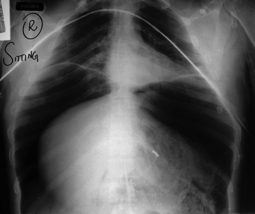
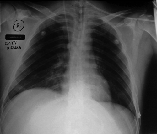
Figure 2: The chest X-ray after the operation
Mr D was rushed into theatre for an emergency laparotomy. During the operation, a perforation measuring 4 cm was found on the lesser curvature of the stomach, with copious frothy fluid and food in the peritoneal cavity. The perforation was repaired, and a thorough peritoneal washout performed with warm sterile fluid. The blood gas results immediately after the surgery revealed a pH of 7.05, PCO2 of 50 mmHg, bicarbonate of 13.8 mmol/l, standard bicarbonate of 12.0 mmol/l, and base excess of −16.6 mmol/l (Table ).
Table 1: The blood gas results
The patient was admitted to the intensive care unit (ICU) after the surgery. That same night at 23h00, he experienced cardiac arrest. Cardiac massage was performed immediately, and adrenalin of 1 mg given every five minutes. Cardiac activity was resumed after four doses of adrenalin and continuous cardiac massage. Adrenalin infusion and the intensive care continued.
Mr D developed swelling of the left lower limb the day after the surgery. The investigations revealed deep venous thrombosis, which was treated with clexane. The thrombosis resolved within a week. Thereafter, Mr D was transferred from the ICU unit to the surgical ward and placed under the care of a multidisciplinary team, consisting of doctors, nurses, physiotherapists, speech therapists and dietitians. Although the surgery was successful, he suffered from hypoxic brain injury. He became physically and mentally disabled. Ongoing care was provided by the occupational therapist, speech therapist, physiotherapist and psychologists.
Severe respiratory and metabolic acidosis was noted before decompression of the abdomen. The PCO2 improved markedly after the operation. The patient was discharged from the hospital 18 days after admission. Radiological abnormalities were not found on a follow-up computed tomography scan of the brain two months later. Mr D received the treatment on an outpatient basis by the rehabilitation team. Eight months after the incident, he had recovered remarkably, except for the loss of his short-term memory, which was noted to be improving.
Discussion
This case study describes the rare occurrence of abdominal compartment syndrome arising from a tension and chemical reaction that resulted from the gastric contents leaking through the perforated gastric ulcer. Abdominal compartment syndrome is usually associated with intra-abdominal pathology. Correction of hypovolaemic shock with large amounts of fluid results in leakage of the fluid into the peritoneal cavity, with increased abdominal pressure (IAP). The series of events which compromise the cardiovascular, urogenital, respiratory and central nervous systems occurs due to the increased IAP.
Normal intra-abdominal pressure occurs at 0 cm of water.Citation7 Paula et al. described the grading of IAP into four categories:
| • | Grade 1: 10–15 cmH2O. | ||||
| • | Grade 2: 15–25 cmH2O. | ||||
| • | Grade 3: 25–35 cmH2O. | ||||
| • | Grade 4: > 35 cmH2O. | ||||
Organ damage has been associated with an IAP of 20–40 cm of water.Citation8
The diaphragm is elevated and total lung capacity reduced due to increased IAP. The functional residual capacity and the residual volume also decrease. There is diminished alveolar oxygen tension and increased intrathoracic pressure, which causes greater pulmonary vascular resistance. This series of events clinically presents as reduced oxygen saturation, hypercapnia and increased ventilator pressure.Citation7
The high intra-abdominal pressure compresses the inferior vena cava, which causes less venous return to the heart. This results in reduced stroke volume, which clinically manifests as an increased heart rate and low blood pressure. A reduction in renal blood flow and the glomerular filtration rate results in a reduction in urine output and the derangement of renal function. There is rise in intracranial pressure due to increased central venous pressure and pleural pressure as a result of the IAP.Citation7
Reperfusion syndrome may occur after abdominal compartment syndrome because of the release of chemicals, such as cytokines, bradykinin, oxygen-free radicals and products of anaerobic metabolism. These chemicals are known to have a toxic effect on the tissue.Citation8,9 After the release of IAP, the chemicals are washed out into the system, causing damaging effects to the organs.Citation10 Mr D experienced cardiac arrest, which may have been the result of reperfusion syndrome which occurred approximately 11 hours after the surgery.
Several home remedies and over-the-counter medication are used to relieve peptic ulcer pain. Sodium bicarbonate (baking soda), which has been used for centuries, is a common home remedy. Sodium bicarbonate is a powerful antacid which neutralises the stomach acid.Citation11 Sodium choloride, water and carbon dioxide are the end products of this neutralisation.
In this case, the gas produced by the reaction with the sodium bicarbonate in the stomach might have escaped into the intra-abdominal cavity through the perforation, causing a rapid increase in intra-abdominal pressure. Positive pressure ventilation of the patient with the Ambu® bag and face mask might also have aggravated the IAP as the gas that was directed to the gastrointestinal tract escaped into the abdominal cavity through the perforation. Our patient developed respiratory acidosis, and metabolic acidosis with cardiovascular collapse (Table ). Immediate recognition of abdominal compartment syndrome and decompression of the abdomen improved the respiratory and cardiovascular system. The blood pressure became normal, and the saturation improved to 99%. The blood results revealed the insult to the renal system due to IAP, which improved remarkably after the laparotomy (Table ).
Table 2: The laboratory results
Early recognition of and timely intervention in abdominal compartment syndrome is key to a successful outcome. Emergency medical practitioners should be aware of the signs and symptoms of abdominal compartment syndrome, and have adequate skills in order to provide immediate life-saving decompression and definitive management. A high index of suspicion is essential for the survival of such patients.
Conclusion
This case study provided an opportunity to highlight the possibly devastating consequences of abdominal compartment syndrome, arising as a complication of peptic ulcer disease, a condition commonly seen in primary healthcare and accident and emergency units. Effective, prompt recognition and appropriate emergency intervention are key to the survival of patients with this complication, without which high mortality results.
Acknowledgement
The authors extend their sincere gratitude to the hospital management and the patient for giving permission to publish this important case study. Special thanks are also given to the accident and emergency team and surgical team of the hospital for their prompt intervention in this emergency.
References
- Prevalence of peptic ulcer. 2003 June [cited 2014 Jun 8]. Available from: http://www.cureresearch.com/p/peptic_ulcer/stats-country_printer.htm.
- Parsak CK, Sakman G. Acute gastric dilatation and abdominal compartment syndrome as complication of pyloric stenosis. J Gastroenterol. 2007;5(2).
- De Backer D. Abdominal compartment syndrome. Crit Care. 1999;3:R103–4.
- Ramakrishnan K, Salinas RC. Peptic ulcer disease, Oklahoma. Am Fam Phys. 2007 Oct 1;76(7):1005–12.
- Lynn JJ, Weng YM, Weng CS. Perforated peptic ulcer associated with abdominal compartment syndrome. Am J Emerg Med. 2008;26:1071–5.
- Sonnenberg A, Everhart JE. The prevalence of self-reported peptic ulcer in the United States. Am J Public Health. 1996;86:200–5.10.2105/AJPH.86.2.200
- Vegar-Brozovic V, Stoic- Brezak J. Pathophysiology of abdominal compartment syndrome. Transplant Proceed. 2006;38(3):833–5. doi: 10.1016/j.transproceed.2006.01.077(9).
- Paula R. Abdominal compartment syndrome. 2014 Sep [cited 2014 May 29]. Available from: emedicine.medscape.com/article/829008.
- Vidal MG, Weisser JR, Gonzalez F, et al. Incidence and clinical effects of intra-abdominal hypertension in critically ill patients. Crit Care Med. 2008;36:1823–31.10.1097/CCM.0b013e31817c7a4d
- Zimmerman BJ, Granger DN. Mechanisms of reperfusion injury. Am J Med Sci. 1994;307:284–92.10.1097/00000441-199404000-00009
- Remedies for peptic ulcer. 2015July [cited 2014 Jun 05]. Available from: http://www.ncbi.nlm.nih.gov/pubmedhealth/PMHT0012146/.