
 ?Mathematical formulae have been encoded as MathML and are displayed in this HTML version using MathJax in order to improve their display. Uncheck the box to turn MathJax off. This feature requires Javascript. Click on a formula to zoom.
ABSTRACT
Introduction
Ocular abnormalities among down syndrome (DS) patients are a sensitive issue since the DS patients is a minor group in the general population.
Objectives
This study determined the prevalence of visual acuity, refractive errors, and ocular diseases among the DS patients; analyzed the association between refractive errors and (age, gender, visual acuity, and type of squint); and compared the results with similar studies in other countries.
Methods
Clinical examination of the eye included visual assessment with cyloplegic refraction and ocular motility. The visual acuity was evaluated according to the patient’s intelligence and responsiveness. The sample size was 50 children. All members of the Palestinian Association of DS patients were subject to ocular investigation after giving consent form from the parents.
Results
Results showed that 27 boys and 23 girls in the age range of 10–16 years were suffering from DS. The majority of visual acuity was 6/60 (22%) and minority was 6/6 (8%). Astigmatism (40%) was the highest ocular abnormality followed by hyperopia (36%), whereas the low prevalence was with myopia (14%) among both genders.
Conclusion
The study revealed the association between age and hyperopia and astigmatism. These results suggest the influence of environmental quality. This study recommends investigating the influence of environmental quality in the ocular abnormalities in DS and general population.
1. Introduction
Down syndrome children (DSC) is a widespread feature worldwide. It is the most frequent chromosomal aberration associated with mild to moderate mental disability, a characteristic facial appearance, and poor muscle tone in infancy [Citation1]. DSC may have mild to severe ocular abnormalities worldwide. Considerable attention has been given for ocular abnormalities among DSC worldwide. For instance, high prevalence of refractive error (Ref Error) has been reported among DSC from China (56%) [Citation2], Hong Kong (86.3%) [Citation3], the UK [Citation4], Turkey (85%) [Citation5], Egypt (41) [Citation6], and Israel [Citation7].
Additionally, high prevalence of epicanthal fold has been reported among DSC from Italy (84%) [Citation8] and Brazil (61%) [Citation9].
Optic nerve atrophy has been reported among DSC from The Netherlands [Citation10] and Belgium [Citation11]. Blindness has been reported among DSC from the US [Citation12]. Additionally, high prevalence of exotropia, bilateral cataracts, and retinitis pigmentosa has been reported among DSC from the USA [Citation13]. A high prevalence of looking patterns has been reported among DSC from the UK [Citation14], whereas a high prevalence of strabismus has been reported among DSC form Canada [Citation15]. Furthermore, a high prevalence of blepharitis (81.9%) has been reported among DSC form Iran [Citation16].
A high prevalence of congenital ocular abnormalities has been reported among DSC form Lomé [Citation17], whereas DSC from Egypt [Citation18] have a high prevalence of ocular abnormalities in patients with vitiligo (vitiligo is an acquired pigmentary disorder affecting 0.5–2.0% of the population worldwide and may develop at any age).
On the other hand, low prevalence of nystagmus (29.2%), esotropia (26.1%), epiphora (21.5%), Brushfield spots (16.9%), and lens opacities (12.3%) has been reported in Slovenia [Citation1]. DSC form Iran [Citation19] have a high prevalence of prolate cornea.
As shown above, different types of ocular abnormalities have been reported worldwide. It can be noticed that refractive error is the most common ocular abnormality found worldwide followed by epicanthal fold, whereas other ocular abnormalities are less reported worldwide. Furthermore, the abovementioned reports have a wide information of ocular abnormalities in DSC from many countries worldwide, but a few reports from Arab countries are found besides the fact that no information from Palestine was reported. This study was designed to report the ocular abnormalities in DSC from Gaza Strip, Palestine, and compare the results with those mentioned in the literature and discuss the reasons of differences.
2. Materials and methods
2.1. Description of the study
The study included 50 members of DSC in the age range of 10–16 years old with no restrictions on gender. The children were registered in the national Down syndrome association in Gaza (NDSA). They were born in 2005–2021. These children were subject to annual routine medical examination by local pediatricians in the NDSA who requested a full ocular examination test for all children. From an ethical point view, the medical investigation was done based on consent form completion by at least one parent of the child. The study excluded DSC suffering from chronic eye disease, history of known ear disorder, and history of autoimmune disease. The children were examined between 3 and 5 times. The clinical characteristics of their ocular pathology, including nystagmus, were investigated by performing a complete ophthalmological examination, expanded according to the child’s needs. The small size of the study population is due to the small number of DSC in Gaza Strip, Palestine, who are accommodated by the NDSA. Additionally, many parents of the DSC refused to sign the consent form to participate in the study. Moreover, the study excluded DSC who had previous eye disease.
Following the procedure described previously [Citation1], visual acuity was assessed monocularly in cooperative children; otherwise it was assessed binocularly using Teller Acuity Cards, Cambridge Crowding Cards, Lea symbols, and various preverbal optotypes. Furthermore, refractive error was performed at all examinations by retinoscopy and automatic refraction done with Nikon Retinomax Plus K2 [Citation1].
Strabismus was evaluated with the Hirschberg test, the cover test, prisms, or other orthoptic methods, if possible. Eye motility was evaluated by observing eye movements when fixating a moving target.
Slit-lamp biomicroscopy was performed on eye lid, conjunctiva, cornea, and iris. Lens abnormalities were examined such as blepharitis, conjunctivitis, keratitis, and cataract.
The DSC were classified into groups according to gender and age. The percentage of each group was calculated. Ref Error was classified into three groups according to the following range: group 1 has a range of 1–2.75 D, mild case, group 2 has Ref E range of 3–5.75D, mandate case, and group 3 has a Ref E ≥ 6D, severe case.
2.2. Statistical analysis
Data analysis was conducted using IBM SPSS (version 22.SPSS Inc., Chicago, IL, USA). Univariate analysis was used to evaluate the correlations between the Ref Error and visual acuity, age, gender and type of squint, and correlation between type of squint and corneal curvature.
Central corneal curvature was measured by automated keratometry with paired readings taken in two orthogonal meridia. An average of three pairs of readings including axes was calculated. Corneal power in diopters (D) = 337.5/keratometry in mm (where 337.5 is the hypothetical refractive index of the cornea).
For comparison of eye abnormalities among countries, the following statistical analysis was used.
Calculation of reference average and relative average of Ref Error among countries.
Reference average (Ref Aver) of Ref Error was calculated according to recent published results [Citation20] with slight modifications:
where ,
, and
are the value of Ref Error reported in country 1, country 2, and country n (the last country), which can be an average or percentage reported in the country. N is the total number of Ref Error values reported in all countries.
Similarly, reference average of other eye abnormality such as epicanthal fold can be calculated if necessary (if the parameter was reported in four countries).
Then, Relative average (Rel Aver) of Ref Error and/or eye abnormality, in each country can be calculated according to EquationEquation (2)(2)
(2) .
Based on the results of EquationEquation (2)(2)
(2) , Ref Error from different countries was subdivided into three categories as follows.
Category I includes countries having Rel Aver value below 0.1 to <0.7, category II includes countries having Rel Aver values between 0.7–0.99, and category III includes countries having Rel Aver values ≥ 1.
To estimate the significant differences among the Rel Aver values of countries, the reference standard deviation (Ref Stdev) and Reference standard error (Ref Std Error) were calculated from the general formula of the calculation using the general statistics.
Then, a country Std Error was calculated from EquationEquation 3(3)
(3) as follows:
where n is the number of cases in a country having the eye abnormality. The values of standard errors of each country were used to indicate the error bars in the corresponding figure. The overlapping error bars indicate no significant differences in the relative means at p-value ≤ 0.05, whereas no overlapping indicates significant differences in the Rel Aver in the studied countries.
Additionally, forest plot was drawn for Ref Error to compare the distribution among country countries.
Eye abnormalities reported in less than four countries were not compared because the data are not enough for pooling.
3. Results
3.1. Social parameter of the study
The study included 27 boys and 23 girls in the age range of 10–16 years having DS characteristics. The DSC are members in the Palestinian DS association in Gaza. They receive daily care by the association and their families.
3.2. Prevalence of ocular abnormalities (POA)
The POA among DSC in Gaza Strip, Palestine, is presented in . It can be noticed that ocular abnormalities include several eye diseases such as epicanthus, astigmatism, hyperopia, cataract, strabismus, ptosis, myopia, conjunctivitis, nystagmus, blepharitis, and madarosis with different frequencies.
Table 1. Ocular abnormalities among DSC.
It has been shown that epicanthus is the most common ocular abnormality (24 patients, 48%) among all cases followed by astigmatism (20 patients, 40%), whereas hyperopia was reported in 18 cases. Additionally, cataract and strabismus were reported in 16 cases. On the other hand, minor eye abnormalities such as ptosis, myopia, conjunctivitis, nystagmus, blepharitis, and madarosis were reported in less than 10 cases each.
Definitions of the ocular abnormalities listed in are shown below:
Astigmatism is as an eye condition where the cornea is not perfectly round, so it cannot focus light evenly onto the retina located in the back of the eye.
Myopia, is an eye disease where a distant object appears blurry, whereas close object appears clear.
Hyperopia is an eye condition where a distant object is seen clearly, whereas a near object appears blurry.
Epicanthus is a skin fold covering the medial angle of the eye.
Cataract is a cloudy area in the lens of your eye.
Strabismus is a misalignment of an eye, causing one eye to deviate inward (esotropia) toward the nose, or outward (exotropia), while the other eye remains focused.
Ptosis is the medical name for the drooping of the upper eyelid, which can happen in one or both eyes.
Conjunctivitis (pink eye) is an eye inflammation or infection of the transparent membrane (conjunctiva) that lines an eyelid and covers the white part of the eyeball. It can cause the white of the eye to take on a pink or red color.
Nystagmus is an involuntary rhythmic side-to-side, up and down or circular motion of the eyes that occurs with a variety of conditions.
Blepharitis is a common eye condition that makes eyelids red, swollen, irritated, and itchy.
Madarosis is a terminology that refers to loss of eyebrows or eyelashes.
3.3. Visual acuity
Visual acuity is a measure of the ability of the eye to distinguish shapes and the details of objects at a given distance. Its prevalence among DSC is presented in . It can be noticed that the majority of the DSC have visual acuity from 6/60 to 6/36, whereas 4 of them have a visual acuity of group 6/6.
Table 2. Visual acuity groups among DSC.
3.4. Refractive error
Refractive errors are a type of vision problem that makes it hard to see clearly. The prevalence of refractive errors among DSC is presented in . It can be noticed that refractive error among DS patients includes three types of ocular abnormalities: astigmatism, hyperopia, and myopia. It is obvious that astigmatism was the highest incidence of abnormality (40%) followed by hyperopia (36%), whereas myopia was the lowest percentage found (14%) among DS patients.
Table 3. Types of refractive error among DSC.
Detailed descriptions of astigmatism, hyperopia, and myopia are shown in . It can be noticed that astigmatism cases are 28% at a Ref E range of (±1 to ±2.75 D) mild cases, whereas 12% of the astigmatism cases are in the ref E (≥ ±3 D) moderate cases.
Table 4. Frequency of astigmatism, hyperopia, and myopia among DSC.
On the other hand, 14% of hyperopia cases have a Ref E range of (+1 to +2.75 D) mild cases, whereas 5% of the cases have a Ref E range of (+3 to +5.75 D) moderate cases, and only 6% of hyperopia cases have a Ref E value above +6 D, severe cases.
Furthermore, myopic cases are quite different. For instance, only one case (2%) has a Ref E range of (−1 to < −2.75 D) mild cases, whereas three cases (6%) have Ref E range of (−3 to < −5.75 D) moderate cases and only three cases (6%) have a Ref E range (Ref E ≤ −6.00 D).
Detailed descriptions of Ref E among the cases presented in are presented in . It can be noticed that mild, moderate, and severe Ref E are found in hyperopia and myopia cases, whereas severe Ref E was not noticed in astigmatism cases. This is probably due to the fact that hyperopia and myopia cases are optic nerve problem, whereas astigmatism cases are muscles problem.
The manifest threshold of astigmatism, hyperopia, and myopia are ±1 D, +1.0 D, and −1 D, respectively.
3.5. Strabismus
Detailed description of strabismus among DSC is shown in .
Table 5. Distribution of strabismus in DSC.
Sixteen children (32%) had strabismus among all cases reported (). Among them, seven cases have esotropia (14%) where this type was the most common type among all cases. Additionally, six cases have alternative esotropia and three cases have exotropia ().
3.6. Ocular manifestations
Ocular manifestations are presented in . Twenty-four DSC (48%) had prominent epicanthic fold, whereas 16 DSC (32%) had cataract in both eyes. Nine children (18%) had ptosis, whereas six children (12%) had conjunctivitis in the right eye. Additionally, four children (8%) had nystagmus. Another small group (four children) had blepharitis and one DSC (2%) had madarosis in both eyes.
Table 6. Ocular manifestations among DSC.
3.7. Statistical relation between variables
The authors performed the statistical analysis to detect if ocular abnormalities listed in are dependent or independent parameters. In other words, to detect if the DSC who have one parameter should have another one. If yes, p-value should be <0.05, if not p-value should be >0.05, indicating no association. In our cases, statistical analysis showed a p-value < 0.05 between refractive errors and visual acuity, indicating an association between them. This suggests that if the child has Ref E he should have a problem with visual acuity. This is quite true in this study.
On the other hand, statistical analysis detected a high p-value (p-value ≥ 0.05) between refractive errors and age or any parameter of those listed in . This indicates no association between Ref E and other parameters except vial acuity. This suggests that the majority of the ocular parameters are independent.
4. Discussion
4.1. Prevalence of ocular abnormalities
clearly shows the prevalence of ocular abnormalities among DSC in Gaza Strip, Palestine. It has been shown that epicanthus is the most common ocular abnormality followed by astigmatism. Additionally, conjunctivitis, nystagmus, blepharitis, and madarosis less frequently occurred among DSC. These abnormalities may occur in DSC due to the fact that they had physiological disability in some muscles or nerves that may result in smoothly curved cornea or the eye lens not evenly working. In this case, the incident light rays are not refracted properly. This may result in having a refractive error. This is in agreement with previous reports [Citation16,Citation19] that found similar results. The high incidence of hyperopia in the eyes is in consensus with previous studies that reported similar findings with DSC [Citation21].
On the other hand, the incidence of cataract and strabismus in the current study is 32%, which coincides with a previous study [Citation22].
Furthermore, clearly indicates that the majority of DS cases have a visual acuity of 6/60–6/24, and the minority of them have a visual acuity of 6/9 and 6/6. The explanation of these results is probably due to the fact that visual acuity is directly connected to the optical nerves of the eye.
Additionally, low vision among DSC may occur due to injuries affecting the eye and/or disorder such as diabetes and glaucoma that affects the entire body. Furthermore, DSC may have a low physical activity due to their health status and/or social parameters. This low activity may have a negative impact on their visual acuity and the general health status of the eye. This explanation is in accordance with Ong [Citation23], who revealed that several eye conditions, including visual acuity, low vision, glaucoma, age-related macular degeneration, and diabetic retinopathy, are associated with low activity levels. Noteworthy, Agrawal [Citation24] revealed that glaucoma is projected to affect the visual acuity of more than 11 million individuals worldwide by the year 2020. Moreover, low vision may occur due to macular degeneration. Our explanation is in accordance with a previous report [Citation25] that revealed the occurrence of low vision among populations of different age groups. Low vision is not an age-related disorder, it can affect any one due to a variety of conditions and injuries. This is in accordance with a previous report [Citation26] that revealed low vision in different age groups.
Additionally, clearly shows the types of Ref E among the DSC, where mild, moderate, and severe cases were reported only on hyperopic and myopic cases only. This is probably due to the fact that the mentioned cases are connected directly to the optical nerve and the brain, whereas astigmatism is connected directly to the eye muscles. This is in agreement with Taylor [34], who found that hyperopic and myopic case are linked to the optical nerve of the eyes.
clearly shows the frequency of astigmatism, hyperopia, and myopia among DSC.
The prevalence of astigmatism is 14% in the range ±1 to ±2.75 D, whereas only 12% of the cases have a Ref E range above ±3 D. The prevalence of hyperopia is 14% in the range of Ref E + 1 D to +2.75 D, whereas hyperopia cases above +6 D are 12%. These results may appear among DSC because refractive error is the most common cause of subnormal visual acuity. Furthermore, refractive errors may be transient phenomena, as in diabetics whose blood glucose has recently been elevated. Additionally, myopia may be caused by the reversible osmotic swelling of the crystalline lens that accompanies hyperglycemia. Moreover, previous infection or trauma may decrease the visual acuity. Noteworthy, retinal disorders may cause irreversible visual loss especially at age-related macular degeneration. Diabetic retinopathy may cause visual acuity loss, whereas nondiabetic retinovascular disorders may decrease visual acuity. This explanation is in accordance with Walker [Citation27], who revealed similar observation for other cases.
In this study, the percentage of myopia between 1.00 Ds to 3.00 Ds was found to be (2%) for both eyes, then from 3 Ds to 5.00 Ds was found to be 6%. Furthermore, 6% myopia was more than 5.00 Ds.
The incidence of myopia in this study (14%, ) is not significantly different from a previous study [Citation6]. This indicates the validity of our results.
Moreover, clearly demonstrates the strabismus classes among DS cases. This classification of strabismus is in accordance with a recent published work [Citation28]. Furthermore, it appears that esotropia (14%) was more commonly observed than other types of strabismus, whereas exotropia was less commonly observed (6%) among all cases. Our results are in accordance with Harrison [Citation29], who found similar observation in a different location. In this study, the rate of unilateral exotropia was 4% and the rate of alternative exotropia was 2%.
clearly shows ocular manifestation among DS cases in Gaza Strip, Palestine. It can be noticed that prominent epicanthic fold was the most common ocular manifestation in this study, and that was similar to last studies [Citation30,Citation31]. The percentage of nystagmus had 8% in the present study, in accordance with a previous study [Citation32]. The incidence of the blepharitis in this study was 8%, which is similar to the result of Paudel et al. [Citation33], and Creavin and Brown [Citation4] reported the results of 18 studies, the prevalence was 5% or less in eight of these studies, while two studies found the prevalence to be 20–37%. In this study, the frequency of cataract was 32%.
The high percentage of cataract among DSC is probably due to the fact that they did not make any surgery because health insurance does not cover surgical operation of eyes such as cataract.
The quite common results of ocular manifestation among DSC worldwide are probably due to the fact that those population (DSC) have quite a common feature and physiological aspects and/or quite similar responses to the external stress. These issues may stand behind common results worldwide.
4.2. Comparison between our results and those in the literature
A comparison between our results and those from the literature is presented in . It can be noticed that this study detected 12 ocular abnormalities, one of them eye and environment. All studies presented in tested both gender (boys and girls) with different age groups and up to 18 years. Regardless of the target groups and population size, the results have quite a common feature.
Table 7. Comparison between our results and some of those from the literature. The correct symbole and dash indicate object done and not done respectively.
On the other hand, this study found 14 ocular abnormalities among Down syndromes, whereas other studies [Citation1], [Citation2,Citation8] found fewer ocular abnormalities. This suggests the influence of environmental conditions, living conditions, food types, and may be socioeconomic factor. Although this study did not investigate the influence of environmental conditions, it emphasized it through the discussion section and provided scientific evidences from the literature. This may open a window for scientist elsewhere to integrate the influence of the environmental conditions, contamination, and ocular abnormalities.
Furthermore, regardless of the smallest population size of this study, it detected 12 ocular abnormalities, whereas the study of Wong and Ho [Citation2] and Fimiani et al. [Citation8] detected fewer cases of ocular abnormalities. This may highlight the influence of age group since those studies have a larger number of very young children. Additionally, this suggests that ocular abnormalities may be developed at a later stage at 5 years old. It can be concluded that this study integrates the picture of ocular abnormalities among DS population and opens a new area of investigation (age group and influence of environment on the development of ocular abnormalities).
Furthermore, statistical analysis of data from some countries having the same parameter (Refractive error) is presented in .
Figure 1. Forest plot of data from five countries having different values of Refractive error.
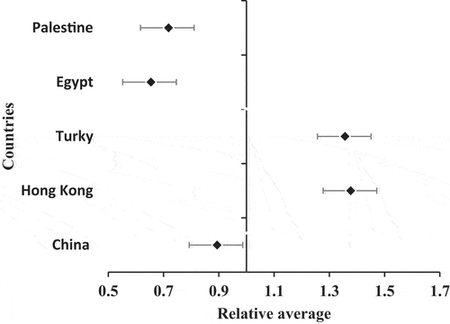
It can be seen that Refractive error in Palestine, Egypt, and China has values of relative average value of Ref error less than 1, whereas Turkey and Hong Kong have values greater than 1. Additionally, the error bars in Palestine Egypt and China are not contacting the Y-ax (the value of reference average of Ref Error) and can be overlapped together indicating no significant difference among each other. Similarly, Turkey and Hong Kong. On the other hand, the relative average subdivides the country into two groups: left side (Palestine, Egypt, and China) and right side (Turkey and Hong Kong). Since the error bars in the left side and the right side are not contacting the bar of relative average and do not overlap, this indicates significant differences. The similarity of the results in the left and right sides indicates the validity of results, whereas the differences between both sides may be attributed to the different sample size and physical environment.
5. Conclusion
The rationale of this study is that it investigates the ocular abnormalities among sensitive group of population. DSC demonstrated a high incidence of epicanthic fold, cataract, and strabismus, especially esodeviation. The study reports ocular abnormalities among DSC for the first time in Gaza Strip, Palestine. Epicanthus, astigmatism, and hyperopia were the major ocular abnormalities among DSC, whereas nystagmus, blepharitis, and madarosis were the minor abnormalities. Comparing the results among countries shows that Ref Error is quite similar in Palestine, Egypt, and China and different from Turkey and Hong Kong. Furthermore, this study investigated many eye abnormalities and compared them to other countries (). This indicates that this study fills the gap on missing knowledge. It can be concluded that the study provided data that can advance our understanding in the field of ocular abnormalities among DSC. These data can be a suitable reference for young interested scientists that can better improve the quality of eye disease among DSC. On the other hand, the weak point of the study is that it dealt with a small population size of DSC. Additionally, DSC need special cooperation and coordination team beyond the medical team. This study recommends the following points: (1) early eye investigation for children of DS characters (1–12 months age) followed by frequent investigations each year; (2) investigation of the influence of environmental quality in the ocular abnormalities in the DS and general population; (3) investigation of the risk of eye rubbing; and (4) investigation of the reasons of cataract formation.
Disclosure statement
No potential conflict of interest was reported by the author(s).
Additional information
Notes on contributors
Khalid Awad
Khalid Awad is a Doctor and assistant Professor at the optometry department.
Yasser El-Nahhal
Yasser El-Nahhal is a Professor Dr of Toxicology at Faculty of Science.
References
- Stirn Kranjc B. Ocular abnormalities and systemic disease in Down syndrome. Strabismus. 2012;20(2):74–77.
- Wong V, Ho D. Ocular abnormalities in Down syndrome: an analysis of 140 Chinese children. Pediatr Neurol. 1997;16(4):311–314.
- Fong AHC, Shum J, Ng ALK, et al. Prevalence of ocular abnormalities in adults with Down syndrome in Hong Kong. Br J Ophthalmol. 2013;97(4):423–428.
- Creavin AL, Brown RD. Ophthalmic abnormalities in children with Down syndrome. J Pediatr Ophthalmol Strabismus. 2009;46(2):76–82.
- Kaplan AT, Oral YA, Kaymak ZN, et al. Analyzing ocular and systemic findings of patients with Down syndrome. South Clin Ist Euras. 2019;30(3):232–237.
- Afifi HH, Abdel Azeem AA, El-Bassyouni HT, et al. Distinct ocular expression in infants and children with Down syndrome in Cairo, Egypt: myopia and heart disease. JAMA Ophthalmol. 2013;131(8):1057–1066.
- Merrick J, Koslowe K. Refractive errors and visual anomalies in Down syndrome. Down’s Syndr Res Pract. 2001;6(3):131–133.
- Fimiani F, Iovine A, Carelli R, et al. Incidence of ocular pathologies in Italian children with Down syndrome. Eur J Ophthalmol. 2007;17(5):817–822.
- da Cunha RP, Moreira JB. Ocular findings in Down’s syndrome. Am J Ophthalmol. 1996;122(2):236–244.
- Morava E, Wosik HN, Sykut-Cegielska J, et al. Ophthalmological abnormalities in children with congenital disorders of glycosylation type I. Br J Ophthalmol. 2009;93(3):350–354.
- Postolache L. Abnormalities of the optic nerve in Down syndrome and associations with visual acuity. Front Neurol. 2019;10:633.
- Kranz C, Basinger AA, Güçsavaş-Çalıkoğlu M, et al. Expanding spectrum of congenital disorder of glycosylation Ig (CDG-Ig): sibs with a unique skeletal dysplasia, hypogammaglobulinemia, cardiomyopathy, genital malformations, and early lethality. Am J Med Genet. 2007;143A(12):1371–1378.
- Esfandiari H, Mets MB, Kim KH, et al. Ocular abnormalities in a patient with congenital disorder of glycosylation type Ig. Ophthalmic Genet. 2019;40(6):549–552.
- Van Herwegen J, Ranzato E, Karmiloff-Smith A, et al. Eye movement patterns and approximate number sense task performance in Williams syndrome and Down syndrome: a developmental perspective. J Autism Dev Disord. 2019;49(10):4030–4038.
- Umfress AC, Hair CD, Donahue SP. Prevalence of ocular pathology on initial screening and incidence of new findings on follow-up examinations in children with trisomy 21. Am J Ophthalmol. 2019;207:373–377.
- Makateb A, Hashemi H, Farahi A, et al. Ocular alignment, media, and eyelid disorders in Down syndrome. Strabismus. 2020;28(1):42–48.
- Diatewa BM, Maneh N, Domingo AS, et al. Les anomalies congénitales oculaires au Centre Hospitalier Universitaire-Campus de Lomé, Togo [Congenital ocular anomalies at the University Hospital Campus in Lomé, Togo]. Pan Afr Med J. 2021;38:79.
- Genedy R, Assal S, Gomaa A, et al. Ocular and auditory abnormalities in patients with vitiligo: a case-control study. Clin Exp Dermatol. 2021;46(6):1058–1066.
- Asgari S, Mehravaran S, Fotouhi A, et al. Total corneal refractive power and shape in Down syndrome. Eur J Ophthalmol. 2021;31(1):69–77.
- El-Nahhal I, El-Nahhal Y. Data on estimation of health hazards associated with pesticide residues in drinking water. Data Brief. 2022;41:107830.
- Ljubic A, Trajkovski V, Stankovic B. Strabismus, refractive errors and nystagmus in children and young adults with Down syndrome. Ophthalmic Genet. 2011;32(4):204–211.
- Ljubic A, Trajkovski V, Tesic M, et al. Ophthalmic manifestations in children and young adults with Down syndrome and congenital heart defects. Ophthalmic Epidemiol. 2015;22(2):123–129.
- Ong SR, Crowston JG, Loprinzi PD, et al. Physical activity, visual impairment, and eye disease. Eye (Lond). 2018;32(8):1296–1303.
- Agrawal A, Singh A, Mittal SK. Glaucoma in Asia- an epidemiological perspective. Nepal J Ophthalmol. 2017;9(18):208–211.
- GBD 2019 Blindness and Vision Impairment Collaborators, & Vision Loss Expert Group of the Global Burden of Disease Study. Causes of blindness and vision impairment in 2020 and trends over 30 years, and prevalence of avoidable blindness in relation to VISION 2020: the right to sight: an analysis for the global burden of disease study. Lancet Glob Health. 2021;9(2):e144–160.
- Cleveland Clinic.Low vision: causes, treatment and prevention. 2022. [cited 2022 Dec3]. https://my.clevelandclinic.org/health/diseases/8585-low-vision
- Walker HK, Hall WD, Hurst JW, Eds. Clinical methods: the history, physical, and laboratory examinations. 3rd ed. Boston: Butterworths; 1990.
- Gisselbaek S, Hoeckele N, Klainguti G, et al. Clinical classification of acquired concomitant Esotropia. Klinische Klassifikation von erworbenen konkomitanten Esotropien. Klin Monbl Augenheilkd. 2021;238(4):482–487.
- Harrison A, Allen L, O’Connor A. Strabismus surgery for Esotropia, Down syndrome and developmental delay; is an altered surgical dose required? A literature review. Br Ir Orthoptic J. 2020;16(1):4–12.
- Fried K. A score based on eight signs in the diagnosis of Down syndrome in the newborn. J Mental Deficiency Res. 1980;24(3):181–185.
- Pfeiffer MJ. Chirurgische Behandlung des medialen Epikanthus durch Hautersatz [Surgical treatment of medial epicanthus by skin replacement]. Klin Monbl Augenheilkd. 2016;233(1):50–53.
- Weiss AH, Kelly JP, Phillips JO. Infantile Nystagmus and Abnormalities of conjugate eye movements in Down syndrome. Invest Ophthalmol Visual Sci. 2016;57(3):1301–1309.
- Paudel N, Leat SJ, Adhikari P, et al. Visual defects in Nepalese children with Down syndrome. Clin Exp Optometry. 2010;93(2):83–90.