ABSTRACT
Immunoinformatics tools were used to predict human leukocyte antigen (HLA) class II-restricted T cell epitopes within the envelope glycoproteins and nucleocapsid proteins of Ebola virus (EBOV) and Sudan virus (SUDV) and the structural proteins of Venezuelan equine encephalitis virus (VEEV). Selected epitopes were tested for binding to soluble HLA molecules representing 5 class II alleles (DRB1*0101, DRB1*0301, DRB1*0401, DRB1*0701, and DRB1*1501). All but one of the 25 tested peptides bound to at least one of the DRB1 alleles, and 4 of the peptides bound at least moderately or weakly to all 5 DRB1 alleles. Additional algorithms were used to design a single “string-of-beads” expression construct with 44 selected epitopes arranged to avoid creation of spurious junctional epitopes. Seventeen of these 44 predicted epitopes were conserved between the major histocompatibility complex (MHC) of humans and mice, allowing initial testing in mice. BALB/c mice vaccinated with the multi-epitope construct developed statistically significant cellular immune responses to EBOV, SUDV, and VEEV peptides as measured by interferon (IFN)-γ ELISpot assays. Significant levels of antibodies to VEEV, but not EBOV, were also detected in vaccinated BALB/c mice. To assess immunogenicity in the context of a human MHC, HLA-DR3 transgenic mice were vaccinated with the multi-epitope construct and boosted with a mixture of the 25 peptides used in the binding assays. The vaccinated HLA-DR3 mice developed significant cellular immune responses to 4 of the 25 (16%) tested individual class II peptides as measured by IFN-γ ELISpot assays. In addition, these mice developed antibodies against EBOV and VEEV as measured by ELISA. While a low but significant level of protection was observed in vaccinated transgenic mice after aerosol exposure to VEEV, no protection was observed after intraperitoneal challenge with mouse-adapted EBOV. These studies provide proof of concept for the use of an informatics approach to design a multi-agent, multi-epitope immunogen and provide a basis for further testing aimed at focusing immune responses toward desired protective T cell epitopes.
Introduction
Vaccines that can safely and effectively protect against aerosol exposure to the alphavirus Venezuelan equine encephalitis virus (VEEV) and filoviruses such as Ebola virus (EBOV) and Sudan virus (SUDV) are of great importance for biodefense. We previously developed and tested DNA vaccines expressing the envelope glycoproteins of these viruses in mice and nonhuman primates (NHPs) and demonstrated protection against both peripheral and aerosol challenges with VEEVCitation1-4 and peripheral challenges with EBOV.Citation5-7 Ideally, it would be possible to confer protection against all 3 viruses with a single vaccine formulation, but this raises the concern of non-protective, immunodominant epitopes limiting the generation of beneficial immune responses to each pathogen. For VEEV, a strong neutralizing antibody response is generally correlated with protective immunity; however, a role for CD4+ T cells in controlling VEEV infection of the central nervous system has also been demonstrated.Citation8-11 Although both humoral and cellular immune responses have been shown to be required for protection from EBOV peripheral challenge, the immune responses required for aerosol protection remain unclear.Citation12-15
Acute viral infection results in the generation of antigen-specific CD4+ T cells with various effector functions. In particular, it has long been recognized that a robust CD4+ T cell response is critical for optimal B cell antibody production. One subset of CD4+ T cells, the T follicular helper cells (TFH), is a specialized CD4+ T cell subset that migrates to the B cell follicle and provides the proper signals for antibody production. In the follicle, TFH cells activate B cells to form germinal centers (GC), which are required for isotype class switching, affinity maturation, and B cell memory. Additionally, it has been demonstrated that specific subsets of the TFH cells migrate to sites of peripheral infection where they provide potent memory responses.Citation16,17,18 These 2° effector CD4+ T cells, which were found to have different gene profiles than their naïve counterparts, are able to respond much more quickly than central 1° CD4+ effector T cells to a viral infection; thus, they are a highly desirable outcome of a preventive vaccine.Citation18
Factors leading to the generation of 2° tissue-resident memory CD4+ T cells are not well defined; however, a recent study of influenza A virus in mice demonstrated that most activated CD4+ T cells undergo contraction within 4 weeks of infection, but that it is possible to prevent CD4+ apoptosis and direct them into the memory pool by providing a second exposure to activated antigen presenting cells (APCs) and the induction of high levels of IL-2 within 5–7 d of the initial priming dose of a subunit vaccine.Citation19 It was further suggested that a second vaccination during this time more closely mirrors antigen exposure to the immune system during an actual infection, and could drive an improved CD4+ memory response. Logically, it follows that it should be possible to stimulate a specific memory pool of CD4+ effector cells by exposure to selected class II-restricted epitopes.
DNA vaccines offer a convenient means to present the immune system with tailored antigens or epitopes for immune focusing. DNA constructs can be designed to not only present desirable epitopes for memory responses, but to also avoid undesirable immunodominant epitopes or to broaden the response by including epitopes from multiple viral variants or diverse viruses. With the development of new immunoinformatics tools, it has become possible to accurately predict human class I- and class II–restricted T cell epitopes, avoid epitopes that resemble “self-antigens,” and design a construct with epitopes arranged for optimal presentation by APC processing machinery. This approach has been tested for creating immunogens for several pathogens to include HIV, influenza virus, variola virus, M. tuberculosis, F. tularensis, and H. pylori.Citation20-26 In the study reported here, we used in silico prediction algorithms to design a DNA construct consisting of human leukocyte antigen (HLA) class II-restricted T cell epitopes derived from the envelope glycoproteins (GP) and nucleocapsid proteins (NP) of EBOV and SUDV and the structural proteins (C-E3-E2–6K-E1) of VEEV. We then vaccinated mice with the multi-epitope DNA vaccine and assessed their immune responses. This proof-of-concept work provides a foundation for further studies using such constructs for immune focusing, enhancing memory responses, or for broadening vaccine responses against multiple pathogens.
Results
Immunoinformatic identification of putative class II T cell epitopes of EBOV, SUDV, and VEEV
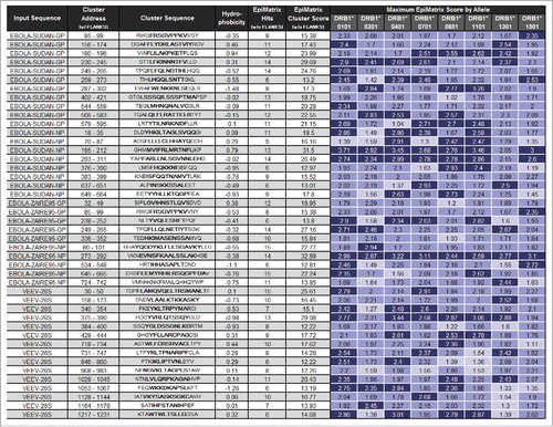
The amino acid sequences derived from the GP and NP genes of EBOV and SUDV and the structural proteins of VEEV were downloaded in GenPept format, where the accession number and corresponding amino acid sequence of each of the 5 antigens were exported and then uploaded to an in-house database. Per antigen, the amino acid sequence was parsed into 9-mer peptides overlapping by 8 amino acids and analyzed using the EpiMatrix 1.2 algorithm to identify putative T cell epitopes.Citation27 9-mer sequences were used for the analysis, because they represent the minimal peptide length required for HLA binding. The matrix coefficients were then summed to produce a raw score for each 9-mer and then normalized. Peptides scoring ≥ 1.64 on the EpiMatrix “Z” scale, typically the top 5% of any given sample, are likely to be HLA ligands and were considered “hits” (). Class II epitopes were identified for 8 supertype alleles (DRB1*0101, DRB1*0301, DRB1*0401, DRB1*0701, DRB1*0801, DRB1*1101, DRB1*1301 and DRB1*1501) that cover >90% of the human population.Citation28 The ClustiMer algorithm generated an initial list of 84 class II T cell epitope clusters, which are regions of high epitope density characteristic of ‘promiscuous’ epitopes.Citation29 These epitopes were manually reviewed to confirm properly centered putative binding motifs, and several of the algorithm-generated peptides were either split or trimmed to facilitate synthesis and in vitro testing. A few peptides were rejected at this point based on extreme hydrophobicity and/or low score profiles. The selected epitopes were rescored and BLASTed against the non-redundant database of human sequences on file at Genbank. Additional sequences were rejected because they were derived from a signal sequence, homologous to human protein sequences, or duplicated within the input sequences. Forty-four sequences were selected for further analysis based on the criteria described above, and 4 of the selected epitopes identified were conserved between SUDV GP and EBOV GP ().
Table 1. EpiMatrix analysis of amino acid sequences.
Figure 1. Peptides selected for inclusion in the class II DNA vaccine construct. Input sequence describes the antigen from which each peptide was derived and Cluster Address describes the location of each peptide within its source antigen. Under Cluster Sequence, the core peptide (underscored middle amino acids in bold) defines the actual cluster that was identified during the analysis. The stabilizing flanks (N-terminal and C-terminal, not bold) are included for use with the core sequence. Flanks are necessary stabilizing factors in any peptide synthesized for analysis in immunoassays. Hydrophobicity relates the grand average of hydropathy (GRAVY) score for each complete peptide. Peptides with hydrophobicity scores above +2 tend to be difficult to synthesize. HLA alleles considered in cluster analysis are the supertype representatives DRB1*0101, DRB1*0301, DRB1*0401, DRB1*0701, DRB1*0801, DRB1*1101, DRB1*1301, and DRB1*1501. The number of hits is the number of significant 9mer-to-allele assessments (EpiMatrix Z scores above 1.64, or the top 5% of random peptides, shaded medium blue) found within the sequence. The top 1% of random peptides is shaded dark blue, while top 10% assessments are shaded light blue. The EpiMatrix Cluster Score is derived from the number of hits normalized for the length of the cluster. Thus, Cluster Score is the excess or shortfall in predicted aggregate immunogenicity relative to a random peptide standard. Cluster Scores above +10 indicate significant potential for promiscuous response. Selected peptides each contain between 6 and 15 EpiMatrix hits and register Cluster Scores between 12.22 and 32.89.
Verification of HLA binding of the predicted epitopes
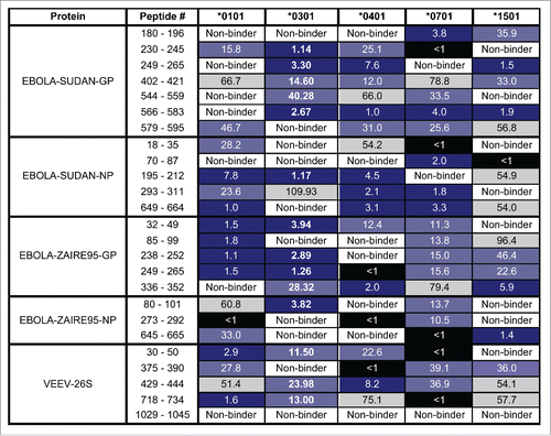
To confirm that the in silico predictions resulted in epitopes able to bind to HLA molecules, in vitro binding assays were performed on the top 5 scoring peptides from each of the input sequences and HLA alleles DRB1*0101, DRB1*0301, DRB1*0401, DRB1*0701, and DRB1*1501 as described in the Methods. The peptides were previously selected for predicted binding to more than one HLA DR allele. Twenty-one of the 25 peptides bound strongly or very strongly to at least one allele in the binding assay (). All the peptides tested except VEEV_1029–1045 bound at least moderately or weakly to at least one of the 5 alleles. This peptide contains a single relatively low-scoring binding motif for alleles *0301, *0401, and *0701 and 2 low-scoring motifs for allele *1501. Fifteen of the 25 peptides bound strongly or very strongly to at least 2 alleles. Apart from VEEV_1029–1045, all peptides bound at least moderately or weakly to at least 2 alleles. In addition, 5 of the 25 peptides bound strongly or very strongly to at least 3 alleles and 22 of the 25 peptides bound at least moderately or weakly to at least 3 alleles. EBOLA-SUDAN-GP_566–583 was the only peptide to bind strongly or very strongly to 4 alleles, while 15 of the 25 peptides bound at least moderately or weakly to at least 4 alleles. Although none of the peptides bound strongly or very strongly to all 5 alleles, 4 of the 25 peptides bound at least moderately or weakly to all 5 alleles. Overall, 96% percent of these predicted peptides were validated as ligands for at least one tested allele.
Figure 2. In vitro HLA binding of peptides representing identified putative epitopes. The binding affinities of the peptides were calculated in competitive binding assays. The input protein, location of the epitope cluster in its source antigen, HLA DR allele tested, and calculated IC50 value in µM units are listed, respectively. Peptide binding affinity is shown according to the following: IC50 < 1 µM (Black), IC50 1 µM-10 µM (Dark Blue), IC50 10 µM-50 µM (Light Blue), IC50 50 µM-100 µM (Gray). Non-binders (White) are those peptides with IC50 values too high to accurately measure under binding conditions.
Immune responses of BALB/c mice vaccinated with the multi-epitope DNA construct
To create the multi-epitope construct, the 44 selected epitopes were initially arranged in order by EpiMatrix score and then were reordered using the VaccineCAD algorithm to eliminate the formation of junctional epitopes. The final multi-epitope open reading frame was codon-optimized for expression in Homo sapiens and synthesized along with a tissue plasminogen activator (tPA) leader sequence and a C-terminal 3X FLAG tag for protein detection. The synthesized gene was cloned into the pWRG7077 mammalian expression plasmid,Citation30 and gene expression resulting in a polypeptide of the expected size of 89 kDa was confirmed by Western blot using an anti-FLAG M2 monoclonal antibody (data not shown).
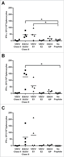
Because the predicted epitopes were selected based on binding to HLA class II molecules, we expected that immunogenicity studies would require the use of HLA-transgenic mice. However, analysis of the selected peptides revealed 17 epitopes that were predicted to also bind to the class II major histocompatibility complex (MHC) alleles of wild-type BALB/c mice (). Therefore, we were able to initially assess the immunogenicity of the multi-epitope DNA construct in an immunocompetent and cost-effective animal model that is routinely used for studying vaccines against EBOV or VEEV. The 17 predicted 9-mer epitopes included 2 from SUDV GP, 1 from SUDV NP, 2 from EBOV GP, 4 from EBOV NP, and 8 from the VEEV structural proteins. For this, groups of mice (N = 4) were vaccinated by intramuscular (IM) electroporation (EP) 3 times at 3-week intervals with either 20, 35, or 50 µg of the multi-epitope DNA construct, and splenocytes were isolated one week after the third and final vaccination (day 49). Splenocytes from mice receiving 20 µg () or 35 µg () of the multi-epitope DNA construct produced statistically significant cellular responses (p = 0.0322 and p = 0.0003, respectively) when stimulated with pooled peptides representing the identified class II EBOV and SUDV epitopes as measured by interferon (IFN)-γ ELISpot assay. Splenocytes from mice in the 20 µg dose group also demonstrated a significant cellular response (p = 0.031) to pooled peptides representing the VEEV E1 protein as compared with the no peptide controls (). Although cellular responses to the class II EBOV and SUDV and VEEV E1 peptides were detected in some of the mice receiving 50 μg of the multi-epitope construct, statistical significance was not achieved for any of the peptide pools at this dose level ().
Table 2. Murine T cell epitopes contained within the class II multi-epitope construct.
Figure 3. IFN-γ ELISpot responses elicited in BALB/c mice vaccinated with the class II multi-epitope DNA vaccine. Splenocytes isolated on day 49 from individual mice vaccinated on days 0, 21, and 42 by IM EP with 20 (A), 35 (B), or 50 µg (C) of the class II multi-epitope DNA vaccine were stimulated for 48 h with a pool of peptides representing the identified class II VEEV epitopes, a pool of peptides representing the identified class II EBOV and SUDV epitopes, pools of overlapping peptides spanning the E1 or E2 envelope glycoprotein of VEEV, a pool of overlapping peptides spanning the GP of EBOV, or no peptide. Data are presented as the spot forming cells (SFC) per million splenocytes for individual mice with black horizontal bars representing the means for each group. Statistically significant responses (*, p ≤ 0.05) are indicated.
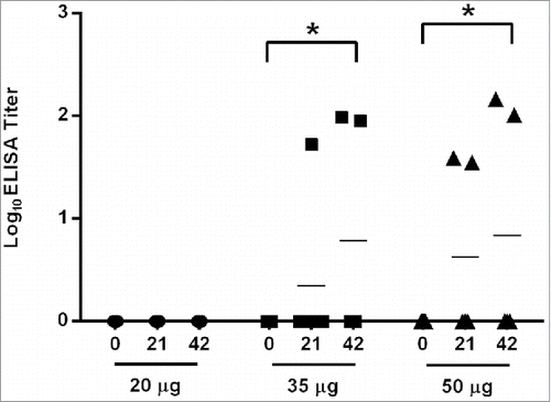
To determine if the multi-epitope DNA construct also has the potential to elicit humoral immune responses in mice, serum samples obtained 3 weeks after the first and second vaccination (days 21 and 42) were assayed by ELISA for anti-VEEV and anti-EBOV total IgG antibodies. Although antibody responses to EBOV were not detected in any of the mice (data not shown), half of the mice receiving either 35 or 50 μg of the multi-epitope DNA vaccine had detectable antibody responses against VEEV after a single vaccination, and the mean log10 titers were statistically significant compared with pre-vaccination sera (p = 0.0492 and p = 0.0356, respectively) after 2 vaccinations ().
Figure 4. VEEV-specific antibody responses elicited by class II multi-epitope DNA construct in BALB/c mice. Serum samples obtained on days 0, 21, and 42 from mice (N = 4) vaccinated by IM EP with 20, 35, or 50 µg of the class II multi-epitope DNA vaccine were assayed for total IgG anti-VEEV antibodies by ELISA. The log10 mean titers for each dose group at each time point are represented by the black bars. Total IgG anti-VEEV responses were detected on day 42 from mice receiving 35 (*, p = 0.049) or 50 µg (*, p = 0.036) of the multi-epitope DNA vaccine as compared with day 0.
Epitope-specific cellular and humoral immune responses in vaccinated HLA-DR3 mice
To evaluate the immune responses generated by the multi-epitope DNA construct in the context of an HLA class II molecule, an additional study was performed using HLA-DR3 transgenic mice. These mice lack murine MHC class II and express both the HLA DRB1*0301α and the HLA DRB1*0301β genes.Citation31 A group of HLA-DR3 mice (N = 6) received 2 priming doses of 20 µg of the multi-epitope DNA construct administered by IM EP 2 weeks apart (days 0 and 14). To further augment the immune responses to the class II-restricted T cell epitopes elicited by the multi-epitope DNA vaccine, we also gave 2 boosting doses each consisting of a total of 50 μg of the 25 peptides representing the class II-restricted T cell epitopes used in the HLA binding studies formulated with incomplete Freund's adjuvant (IFA) and 10 µg each of immunostimulatory CpG oligodeoxynucleotide 1826, muramyl dipeptide (MDP) and CL097 adjuvants administered by subcutaneous injection on days 28 and 42. Of these 25 peptides, 3 VEEV peptides and 11 EBOV and SUDV peptides bound strongly or moderately to the DRB1*0301 allele in the in vitro binding assays (). A negative control group received 2 vaccinations with 20 µg of the pWRG7077 empty vector followed by 2 injections with the adjuvants alone following the same schedule as for the experimental vaccine. An additional group received 2 vaccinations of 20 µg of a mixture of the whole-antigen DNA plasmids expressing the codon-optimized EBOV or SUDV GP or VEEV E3-E2–6K-E1 genes administered by IM EP on days 0 and 14, but did not receive peptide/adjuvant boosts.
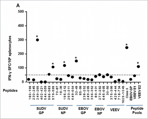
Two weeks following the final vaccination (day 56), splenocytes were isolated from the mice in all groups and cellular immune responses to individual peptides and peptide pools were measured by IFN-γ ELISpot assays. Mice receiving the multi-epitope DNA prime and peptide boost had statistically-significant responses to 4 of the 25 (16%) individual class II peptides tested, which included Sudan GP 249–260 (p < 0.0001), Sudan GP 579–595 (p = 0.0274), Sudan NP 195–212 (p = 0.0263), and EBOV GP 32–49 (p = 0.0011) (). These mice also developed significant responses against the peptide pool consisting of all 25 class II peptides (p < 0.0001) and against the pooled 15-mer peptides spanning VEEV E2 (p = 0.0246). For mice receiving the whole-antigen DNA vaccines, significant responses were detected against the pools of overlapping 15-mer peptides spanning the E2 (p < 0.0001) or E1 (p = 0.0442) envelope glycoprotein of VEEV (data not shown).
Figure 5. IFN-γ ELISpot responses elicited against the class II epitopes in vaccinated HLA-DR3 mice. Splenocytes isolated from groups of HLA-DR3 mice (N = 6) vaccinated with the multi-epitope construct followed by a peptide boost were stimulated for 48 h with individual peptides representing each epitope in the class II multi-epitope DNA vaccine, a pool of all the class II peptides, pools of overlapping peptides spanning the E1 or E2 envelope glycoprotein of VEEV, a pool of overlapping peptides spanning the GP of EBOV, or no peptide. Data are presented as the mean SFC per million splenocytes and are the average of 3 groups of 2 mice. Individual epitope and pooled epitope responses in vaccinated mice showing statistical significance when compared with the negative control group are indicated (*, p ≤ 0.05). A dotted line denotes the cutoff of 50 SFC over background per million splenocytes.
Pooled sera from each of the vaccine groups collected on day 56 were analyzed for IgG antibody responses against the individual class II peptides by ELISA. Significant peptide-specific antibody responses as compared with the negative controls were elicited against a single peptide, SUDV NP 70–87 (p = 0.0005), as well as the pool of 25 class II peptides (p = 0.0019) for mice receiving the multi-epitope vaccine (data not shown). Although they were not statistically significant, antibody responses against some of the other class II peptides were also observed for these mice (data not shown).
Virus-specific antibody responses in vaccinated HLA-DR3 mice
In 2 separate experiments, groups of HLA-DR3 mice (N = 10) were vaccinated with empty vector and adjuvants, the whole-antigen DNA vaccines, or the multi-epitope DNA prime and peptide boost as described above. Sera were collected from all of the mice on day 56 and EBOV GP- and VEEV-specific total IgG antibody responses were assessed by ELISA. Mice receiving the whole-antigen DNA vaccines developed significant levels of antibodies against EBOV () and VEEV () as compared with the negative control mice (p < 0.0001). Six of the 10 mice receiving the multi-epitope prime and peptide boost regimen also developed detectable levels of EBOV-specific antibodies, and the mean log10 titer was also significantly above that of the negative control samples (p = 0.001) (). However, there was a significant difference in mean log10 titers of mice receiving the whole-antigen DNA vaccines as compared with those that were given the multi-epitope DNA prime and peptide boost (p < 0.0001). Mice receiving the multi-epitope vaccine DNA developed similar levels of VEEV-specific IgG antibodies as those receiving the whole-antigen VEEV DNA vaccine and the mean log10 titer was also significantly above that of the negative control samples (p < 0.0001).
Figure 6. Virus-specific antibody responses elicited in vaccinated HLA-DR3 mice. Serum samples obtained on day 56 from groups of HLA-DR3 mice (N = 10) vaccinated as described in the Methods section were analyzed for anti-EBOV or -VEEV total IgG antibodies by ELISA or for VEEV-neutralizing antibodies by PRNT. Log10 ELISA titers for each mouse are indicated by symbols and group mean log10 titers are represented by black horizontal bars. (A) Significant total IgG anti-EBOV responses were detected in mice receiving the whole-antigen DNA vaccines (*, p < 0.0001) and the multi-epitope DNA vaccine (p = 0.0055) as compared with the negative control vaccine. (B) Significant total IgG anti-VEEV responses were detected in mice receiving the whole-antigen DNA vaccines (*, p < 0.0001) or the multi-epitope DNA vaccine (*, p < 0.0001) as compared with mice receiving the negative control vaccine. (C). Log10 PRNT80 titers for individual mice are indicated by symbols and group mean log10 PRNT80 titers are represented by black horizontal bars. Significant neutralizing antibody responses were generated in mice receiving the whole-antigen DNA vaccines (*, p = 0.0002) or multi-epitope DNA vaccine (*, p = 0.0033) as compared with mice receiving the negative control vaccine. The neutralizing antibody titers of mice that survived VEEV challenge are shown as open symbols.
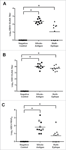
Because neutralizing antibodies are generally thought to be the main correlate of protection against VEEV infection, we also measured the neutralizing antibody responses of the vaccinated mice by plaque reduction neutralization test (PRNT). All mice receiving the whole-antigen DNA vaccines and 6 of the 10 mice receiving the multi-epitope DNA vaccine developed significant neutralizing antibody responses to VEEV (p < 0.0001, p = 0.0378, respectively) ().
EBOV and VEEV protective efficacy in vaccinated HLA-DR3 mice
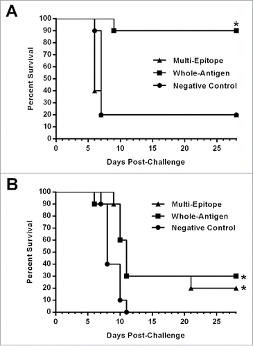
Although a multi-epitope DNA construct such as the one we developed would not likely be used as a stand-alone vaccine, but rather as a means to focus or broaden an immune response, the cellular and humoral immune responses that we observed in the HLA-DR3 mice vaccinated with the multi-epitope vaccine suggested that it may elicit some level of protective immunity against viral challenge. Consequently, the vaccinated HLA-DR3 mice described above were challenged with ∼1,000 LD50 of mouse adapted (ma)-EBOV by intraperitoneal (IP) injection or with ∼10,000 LD50 of aerosolized VEEV as described previously.Citation3,4,6,7 Consistent with our earlier studies with the EBOV DNA vaccine in BALB/c mice, the whole-antigen DNA vaccines provided significant protection against ma-EBOV challenge (90%; p < 0.01) ().Citation6 In contrast, the multi-epitope vaccinated mice and the negative control mice all exhibited clinical signs of disease after ma-EBOV challenge including ruffled fur, lethargy, and dehydration, and both groups had a 20% survival rate (). All mice in the vector/adjuvant only control group challenged with aerosolized VEEV displayed visible signs of disease including ruffled fur, inactivity and hunched posture, and all succumbed to disease or were euthanized due to morbidity (). Unlike our earlier studies in BALB/c mice and nonhuman primates in which the VEEV whole-antigen DNA vaccine elicited complete protective immunity against aerosolized VEEV,Citation4 only 30% of the HLA-DR3 mice vaccinated with the whole-antigen DNA vaccines survived the aerosol VEEV challenge. Similarly, 20% of the multi-epitope vaccinated mice survived aerosol VEEV challenge. Although these survival rates are low, both the whole-antigen DNA vaccines and the multi-epitope DNA vaccine provided statistically significant protection as compared with vector/adjuvant only controls (p = 0.0095 and p = 0.0031, respectively).
Figure 7. Survival of vaccinated HLA-DR3 mice after EBOV or VEEV challenge. Groups of HLA-DR3 mice (N = 10) vaccinated as described in the Methods section were challenged 4 weeks after the final vaccination with 103 PFU of ma-EBOV by IP injection or with 104 PFU of VEEV by aerosol. Kaplan-Meier survival curves indicating the percentage of surviving mice at each day of the 28-day observation period are shown. (A) Significantly increased survival was observed for mice receiving the whole-antigen DNA vaccines (*, p < 0.01). No significant protection against EBOV challenge was observed for mice receiving the multi-epitope vaccine. (B) Significantly increased survival against VEEV challenge was observed for mice receiving the whole-antigen DNA vaccine (*, p = 0.0095) or the multi-epitope vaccine (*, p = 0.0031) as compared with those receiving the negative control vaccine.
Discussion
Antiviral effector CD4+ T cells are produced when APCs take up viruses or virus-infected cells and then process the viral proteins to yield peptides that complex with MHC class II molecules for presentation to naïve CD4+ T cells, which triggers differentiation into subsets of antiviral effector cells.Citation17 DNA vaccines can generate MHC class II-restricted peptides either by direct transfection of APCs or by transcription and translation in bystander cells before antigen uptake by APCs.Citation32-35 It is also possible to engineer DNA constructs to include lysosomal targeting signals (e.g., lysosomal associated membrane protein, LAMP), although in our experience with numerous DNA vaccines, this has not been necessary for eliciting protective immune responses in animals or humans when the vaccines are delivered to skin by electroporation or gene gun or to muscle by electroporation.Citation1,36-40 Despite the strong immunogenicity that we have obtained with DNA vaccines encoding complete viral proteins, we hypothesized that it might be possible to improve and/or broaden the overall response by presenting the immune system with a subset of epitopes that specifically stimulate a particular arm of the immune system. This approach would have the added benefit of allowing for a single DNA vaccine plasmid expressing epitopes of multiple antigenically distinct pathogens, thereby limiting production costs and shot burden. Such a vaccine could be used either to provide a priming dose for a whole-antigen vaccine or as a boost following a whole-antigen vaccine to stimulate specific host memory subset populations.
As an initial test of this hypothesis, we used EpiMatrix in silico prediction tools to identify MHC class II-restricted epitopes in genes that we previously tested as whole-antigen DNA vaccines, namely the GP and NP genes of EBOV and SUDV and the structural proteins of VEEV. To assess the validity of the EpiMatrix algorithm binding predictions, we evaluated the top 5 scoring epitopes from each viral protein in HLA-binding studies with 5 of the 8 supertype class II alleles. We found that 96% of the predicted peptides bound at least moderately to one allele, thus demonstrating the accuracy of the EpiMatrix algorithm. Consequently, we constructed the multi-epitope DNA vaccine to span all 44 epitopes predicted to bind well, and assembled the vaccine coding region with the VaccineCAD algorithm to generate an optimal polypeptide, which did not have spurious junctional epitopes or similarity to self-antigens.
To confirm the immunogenicity of our multi-epitope DNA vaccine, we initially vaccinated a small cohort of immunocompetent, wild-type BALB/c mice. In silico analysis predicted that the MHC class II alleles expressed in BALB/c mice would overlap with 17 of our 44 EpiMatrix-identified peptides. Vaccination of BALB/c mice via IM EP with the class II multi-epitope DNA vaccine alone elicited low but measureable antigen-specific IFN-γ+ T cell responses at all tested doses. It is not surprising that these epitope-specific responses were modest given the very limited number of epitopes contained in the construct that were predicted to bind mouse MHC. Comparing the responses of the BALB/c mice receiving 20 μg to those receiving 35 μg of the multi-epitope construct appeared to show a dose response. However, mice in the group receiving 50 μg did not show a similarly increased response. As our previous studies have demonstrated that the VEEV DNA vaccine is highly immunogenic in mice, it is possible that the increased dose of the multi-epitope vaccine skewed the response toward immunodominant VEEV epitopes and limited the immune response to the filovirus epitopes. This is supported by the fact that there was no change in VEEV-specific T cell numbers between the 20 μg and 35 μg groups, but there was a trend toward increased responses in the 50 μg group. Furthermore, the multi-epitope vaccinated mice developed humoral responses only to the VEEV antigens, suggesting that either the development of EBOV- and SUDV-specific B cell responses was also impaired by immunodominant VEEV epitopes or that the selected EBOV and SUDV T cell epitopes don't overlap with B cell epitopes to the same degree as those for VEEV. Further experiments are necessary to determine the possible effects of VEEV epitopes on immune responses to the filovirus epitopes encoded within the multi-epitope DNA vaccine.
Of particular interest, while some mice in all 3 vaccine dose groups generated detectable immune responses to the MHC class II EBOV peptide pool, no cellular immune responses were detected for the peptide pool representing the MHC class II-restricted VEEV epitopes. However, cellular responses to peptides contained within the VEEV E1 overlapping peptide pool were detected in all of the multi-epitope vaccine groups. One possible explanation for these data are that the breadth of the cellular response generated in wild-type BALB/c mice through vaccination with the class II multi-epitope DNA vaccine is underrepresented by our in silico prediction algorithm, suggesting that multi-epitope DNA vaccines have the potential to induce broader cellular immune responses than previously expected. Although the mechanism behind this result is still unclear, it is conceivable that the MHC class II-restricted epitopes encoded within our vaccine are also capable of priming MHC class I-dependent responses. Thus, stimulation with a more diverse peptide pool may elicit stronger T cell responses than those seen in pools containing only the encoded MHC class II peptides. Such an effect would greatly increase the utility of multi-epitope vaccines by allowing for the priming of non-dominant or cryptic epitope-specific immune populations without sacrificing the ability of the host to respond to the whole antigen. Transient in vivo depletion of CD8+ T cells with a monoclonal antibody may allow for greater understanding of the ability of our MHC class II-restricted vaccine to stimulate MHC class I-dependent responses. Alternatively, the VEEV E1 overlapping peptide pool may have bound to BALB/c MHC II molecules with greater affinity than did the class II VEEV peptides, resulting in higher levels of CD4+ T cell stimulation.
In addition to quantifying the immunogenicity of our multi-epitope DNA vaccine, we compared the protective efficacy of the multi-epitope based approach to that of our whole-antigen DNA vaccines against both VEEV and EBOV viral challenge in transgenic HLA-DR3 mice. Unexpectedly, whole-antigen vaccination of HLA-DR3 mice only resulted in 30% protection against VEEV aerosol challenge, which is a dramatic departure from the 100% protection we have routinely observed in BALB/c mice and NHP.Citation3,4 A similar level of protection (20%) was observed in mice vaccinated with the class II multi-epitope DNA construct. This lower level of protection may be due to the use of a C57Bl/6 background of the transgenic mice, as several studies have demonstrated that mouse strain background can influence VEEV infection and progression.Citation8,41 In one study, however, VEEV infection was found to progress similarly in both BALB/c and C57Bl/6 mice.Citation41 Perhaps a more plausible explanation is that the VEEV epitopes encoded within both DNA vaccines are suboptimal for inducing immunity with regards to presentation by a single HLA-DRB1 allele. Our multi-epitope DNA vaccine was designed to provide coverage against the 8 supertype HLA class II alleles. The EpiMatrix algorithm predicted that only 2 VEEV peptides (1053–1067 and 1164–1178) were expected to be high DRB1*0301 binders (), suggesting that vaccination with the multi-epitope DNA construct could yield a limited anti-VEEV immune population. This hypothesis is supported by the lack of IFN-γ response elicited against the VEEV class II-restricted epitopes in HLA-DR3 mice (). It is likely that vaccination of transgenic mice expressing multiple HLA-DRB1 MHC class II alleles would allow for increased epitope presentation, thereby broadening both the cellular and humoral immune responses and providing greater protection. Despite these inherent limitations of the transgenic HLA-DR3 mouse system, which likely contributed to reduced protective efficacy, our finding that partial protection against the extremely high VEEV aerosol challenge dose (10,000 LD50) with only a small number of epitopes expected to be presented in the context of DRB1*0301 is an extremely encouraging result in support of this multi-epitope based approach.
Similar to the VEEV challenge, the outcome of the ma-EBOV challenge study indicated that the immune responses generated in the HLA-DR3 mice vaccinated with the MHC class II multi-epitope DNA vaccine were not sufficient for protection. These results were not unexpected based on the results of the immunogenicity studies in the HLA-DR3 mice. Cellular responses were only generated to a single EBOV epitope (EBOV GP 32–49), and the EBOV-specific antibody responses induced by the class II multi-epitope DNA vaccine were reduced compared with those generated by the whole-antigen DNA vaccine. However, these responses are still noteworthy given the relative lack of high-binding DRB1*0301 epitopes encoded within the vaccine (). Additionally, we were able to generate cellular responses to 3 SUDV epitopes and antibody responses to a single SUDV peptide, suggesting that protection from SUDV challenge may also be obtainable using such an approach.
Conclusion
Our studies confirmed the immunogenicity of some of the identified class II-restricted epitopes when delivered by IM EP in a DNA vaccine-only formulation or in a heterologous DNA prime-peptide boost regimen. Both vaccine strategies generated detectable cellular and humoral immune responses, demonstrating the potential for T cell epitope-based vaccines to induce a multi-arm response. Further studies aimed at testing class II-restricted, multi-epitope constructs for immune focusing and eliciting tissue-resident 20 effector CD4+ T cells are needed to determine if the immunoinformatics approach used here has the potential to improve vaccine design for humans. Finally, the design of epitope-based vaccines could also include class I-restricted epitopes, and it is also possible that identifying class I-restricted epitopes from each virus and using a 2-pronged approach by generating both CD8+ and CD4+ T cell responses would improve the breadth of the protective immune responses and lead to increased efficacy against both VEEV and EBOV.
Methods
Peptide synthesis
Selected HLA class II-restricted peptides were synthesized at >80% purity using standard solid-phase 9-fluoronylmethoxycarbonyl (Fmoc) chemistry (21st Century Biochemicals). Peptide sequence and mass were confirmed by collision-induced dissociation and tandem mass spectrometry (CID-MS/MS).
HLA class II binding assay
In vitro HLA class II binding assays were performed as described previously.Citation29 Briefly, experimental peptides were solvated in 100% DMSO at 5 concentrations and mixed with binding reagents in aqueous solutions to yield final peptide concentrations of 100, 50, 25, 5, and 1 μM. In 96-well plates, soluble HLA molecules representing 5 class II alleles (DRB1*0101, DRB1*0301, DRB1*0401, DRB1*0701, and DRB1*1501) were mixed with non-biotinylated test peptides at each concentration and biotinylated control peptides and incubated for 24 h at 37°C. The HLA-peptide complexes were then captured on ELISA plates coated with pan-anti-human DR antibodies (L243), developed by addition of streptavidin-europium, and bound HLA-labeled control peptide complexes were assessed on a time-resolved fluorescence plate reader at 615 nm. Concentrations of peptide leading to a 50% inhibition of biotinylated peptide binding (IC50) were then determined. Binders were defined as very strong (IC50 < 1 μM), strong (IC50 < 10 μM), moderate (IC50 < 50 μM), or weak (IC50 < 100 μM). Non-binders were defined as peptides with IC50 ≥ 100 μM.
DNA and peptide vaccines
The VEEV, EBOV, and SUDV DNA vaccines were generated as described previously.Citation4,6 Briefly, the codon-optimized Venezuelan equine encephalitis virus IAB structural genes minus capsid and the codon-optimized GP gene of EBOV and SUDV were synthesized by GeneArt and cloned into the NotI and BglII restriction sites of the pWRG7077 eukaryotic expression vector. The multi-epitope DNA vaccine was engineered by linking all 44 HLA class II epitopes that were previously identified by the EpiMatrix program into one “string-of-beads” open reading frame.Citation29 To avoid creation of new epitopes at epitope junctions, the sequence was analyzed using the VaccineCAD algorithm and epitopes were re-ordered as needed. Insertion of spacer sequences was not required at any epitope junctions. The tPA leader sequence was placed upstream of the multi-epitope sequence to target the expressed protein to the secretory pathway, and a 3X FLAG tag (DYKDHDGDYKDHDIDYKDDDDK) was placed downstream of the epitope sequences for detection of this protein product. The multi-epitope open reading frame was codon optimized for Homo sapiens, synthesized, and cloned into the pWRG7077 eukaryotic expression vector (GeneArt).Citation30 Research-grade preparations of all plasmids were then manufactured (Aldevron).
Peptides corresponding to the epitopes in the HLA class II multi-epitope DNA vaccine were emulsified at 2 µg/µl per peptide in incomplete Freund's adjuvant (IFA) and 10 µg each of immunostimulatory CpG oligodeoxynucleotide 1826, muramyl dipeptide (MDP), and CL097.
Mice
Female BALB/c mice were obtained from National Cancer Institute-Frederick. Female HLA-DR3 transgenic mice were obtained from Dr. Chella David (Mayo Medical School) under commercial license. These mice express the both the HLA DRB1*0301 α and HLA DRB1*0301 β genes and are negative at the mouse H2Ab0 class-II locus.Citation31 All mice were 6 to 8 weeks old at initiation of studies. All animal research was conducted in compliance with the Animal Welfare Act and other federal statutes and regulations relating to animals and experiments involving animals and adheres to principles stated in the “Guide for the Care and Use of Laboratory Animals,” Institute for Laboratory Animal Research, Division of Earth and Life Studies, National Research Council, National Academies Press, Washington, DC, 2011. The USAMRIID facility where some of this animal research was conducted is fully accredited by the Association for the Assessment and Accreditation of Laboratory Animal Care International. All animal research conducted at EpiVax was approved by the Institutional Animal Care and Use Committee and was conducted in compliance with the Animal Welfare Act.
Vaccinations
BALB/c mice were vaccinated 3 times at 3-week intervals with plasmid DNA diluted to specified concentrations in calcium- and magnesium-free PBS (Invitrogen) by IM EP using the Ichor Medical Systems TriGrid™ Delivery System (TDS) as described previously.Citation4,6 Briefly, mice were placed in an IMPAC6 chamber and anesthetized with straight isoflurane gas. Anesthetized mice were then injected in one tibialis anterior muscle with 20 µl of a DNA solution using a 3/10cm3 U-100 insulin syringe inserted into the center of the TriGrid™ electrode array with 2.5 mm electrode spacing. Injection of DNA was followed immediately by electrical stimulation at an amplitude of 250 V/cm, and the total duration was 40 ms over a 400 ms interval. HLA-DR3 transgenic mice were vaccinated 2 times at a 2-week interval with plasmid DNA by IM EP as described above. On days 28 and 42, the negative control and vaccine groups received 100 µl of an IFA/adjuvant emulsion or an IFA/adjuvant/peptide emulsion, respectively, by subcutaneous injection at the base of the tail.
Immunological assays
Epitope-specific cellular immune responses were detected and analyzed by IFN-γ enzyme-linked immunospot (ELISpot) assays using kits purchased from Mabtech or R&D Systems. All assays were performed according to the manufacturer's directions. Briefly, splenocytes were isolated from individual animals and resuspended in complete RPMI 1640 medium (Gibco). Splenocytes isolated from HLA-DR3 mice were added in triplicate at a concentration of 2.5 × 10⁵ cells per well. Individual or pooled target peptides were added at a concentration of 10 µg/ml. Pooled 15-mer peptides with an 11-base overlap spanning the E2 or E1 envelope glycoprotein of VEEV IAB (Pepscan) and pooled 15-mer peptides with a 10-base overlap spanning the envelope glycoprotein of EBOV (Mimotopes) were also added at 10 µg/ml. Stimulation with 2 µg/ml concanavalin A (Sigma-Aldrich) in 3 wells was used as a positive control, and 6 wells were plated with cells and media only as a background control. Splenocytes isolated from BALB/c mice were plated in duplicate at a concentration of 1.0 × 10⁵ cells/well. Peptide pools were added at a concentration of 10 µg/ml. Cells were incubated for 48 hours at 37°C in 5% CO2. Plates were sent to ZellNet Consulting, Inc. where raw spot counts were recorded using a Zeiss high-resolution automated ELISpot reader. The average number of spots per peptide or peptide pool was calculated and adjusted to spots per million cells.
Total IgG anti-EBOV and anti-VEEV end point antibody titers were determined for serum samples by standard ELISA using sucrose gradient-purified, irradiated whole EBOV or VEEV IAB antigen as described.Citation6,42 Briefly, 2-fold serial dilutions of test sera, starting at 1:100, were incubated with 250 ng of VEEV IAB antigen or 700 ng of EBOV antigen per well in 96-well plates. A heavy chain-specific goat anti-mouse horseradish peroxidase (HRP)-conjugated secondary antibody (Sigma-Aldrich) and ABTS (2,2′-Azinobis [3-ethylbenzothiazoline-6-sulfonic acid]-diammonium salt) peroxidase substrate (KPL) were used for detection of VEEV-specific responses. The same secondary antibody and TMB (3,3′,5,5′-Tetramethylbenzidine) 2 peroxidase substrate (KPL) were used for detection of EBOV-specific responses. Total IgG epitope-specific antibody titers were measured as described above with a few modifications. Individual and pooled peptides were diluted in 1x carbonate buffer (Sigma-Aldrich) and added at 250 ng per well in 96-well plates. The same secondary antibody and the ABTS peroxidase substrate were used for detection of peptide-specific responses. The optical density was measured at 405 nm for all plates using ABTS and at 450 nm for all plates using TMB 2 with a SpectraMax M2e microplate reader (Molecular Devices) and end point titers were determined using Softmax Pro v5.4.1 (Molecular Devices).
VEEV IAB-neutralizing antibody titers were determined by PRNT for each serum sample as described previously.Citation2 Briefly, 2-fold serial dilutions of sera were mixed with equal volumes of Hanks Balanced Salt Solution (HBSS) w/ HEPES supplemented with phenol red, 2% Fetal Bovine Serum (FBS), and 1% penicillin-streptomycin containing 200 PFU of virus and incubated at 4°C overnight. The mixtures were used to infect monolayers of Vero 76 cells for 1 h at 37°C in 5% CO2. The monolayers were then overlaid with 2 ml of 0.6% agarose in complete Basal medium Eagle with Earle's Salts (EBME) (Invitrogen). Plates were stained after 24 h at 37°C in 5% CO2 with 2 ml of overlay consisting of 0.6% agarose in complete EBME containing 5% neutral red. Plaques were enumerated 24 h after staining and the antibody titer required for an 80% reduction in the number of plaques as compared with controls (PRNT80) was calculated.
Virus challenges
Mice were challenged with ma-EBOV by IP injection as described previously.Citation43 VEEV IAB (strain Trinidad donkey) was prepared as described previouslyCitation4 and mice were challenged via the aerosol route as described previously.Citation8 Briefly, mice were placed in a whole-body aerosol chamber within a class III biologic safety cabinet and exposed for 10 min to a VEEV aerosol created by a Collision nebulizer. Samples of the generated aerosol were collected from the all-glass impinger (AGI) attached to the aerosol chamber. AGI samples were analyzed by standard plaque assay to determine inhaled dose as described previously.Citation44 Mice were monitored for 28 d post challenge for clinical symptoms and death. Any moribund animals were euthanized. All challenge studies involving the use of VEEV or ma-EBOV were performed at USAMRIID in Animal Biosafety Level 3 or 4 laboratories, respectfully.
Statistical analysis
GraphPad Prism software v6 for Windows (Graph, Inc.) was used to graph and conduct statistical analysis of all data. Briefly, Kaplan-Meier survival curve analysis using a long-rank test was performed for the HLA-DR3 mouse challenge data, one-way analysis of variance with Tukey's post hoc tests was used to compare ELISA titers between challenge groups at each time point, 2-way analysis of variance with Tukey's post hoc tests was used to compare peptide ELISA titers and ELISpot counts between HLA-DR3 challenge groups for each peptide, 2-way analysis of variance with Dunnet's post hoc tests was used to compare ELISA titers between dose study BALB/c groups at each time point, and one-way analysis of variance with Dunnet's post hoc tests was used to compare ELISPOT counts for each BALB/c dose study group. Log10 transformations were applied to peptide ELISA titers, whole virus ELISA titers and PRNT80 titers. Probability (p) values < 0.05 were considered statistically significant.
Abbreviations
| EP | = | electroporation |
| ELISpot | = | enzyme-linked immunospot |
| ELISA | = | enzyme-linked immunosorbent assay |
| EBOV | = | Ebola virus |
| HLA | = | human leukocyte antigen |
| IM | = | intramuscular |
| MHC | = | major histocompatibility complex |
| PRNT | = | plaque reduction neutralization test |
| SUDV | = | Sudan virus |
| VEEV | = | Venezuelan equine encephalitis virus |
Disclosure of potential conflicts of interest
Anne S. De Groot and William D. Martin are founders and majority owners of EpiVax, Inc., a bio-technology company that provides access to immunoinformatics tools and designs vaccines for commercial clients. Leonard Moise holds options at EpiVax, Inc., and both he and Frances Terry are employees of EpiVax, Inc. Due to this relationship with EpiVax, these authors acknowledge that there is a potential conflict of interest inherent in the publication of this manuscript and assert that they made an effort to reduce or eliminate that conflict, where possible.
Acknowledgments
We would like to thank Michelle Richards, Daniel Mitchell, Rebecca Grant-Klein, and Nicole Van Deusen for their assistance in performing the animal studies and immunogenicity analyses. This work was performed while Callie Bounds was a National Research Council postdoctoral Associate.
Funding
The studies described herein were supported by Grant R.R.0001_07_RD_B to USAMRIID from the Joint Science and Technology Office for Chemical and Biological Defense of the Defense Threat and Reduction Agency. The opinions, interpretations, conclusions, and recommendations contained herein are those of the authors and are not necessarily endorsed by the US. Army.
References
- Riemenschneider J, Garrison A, Geisbert J, Jahrling P, Hevey M, Negley D, Schmaljohn A, Lee J, Hart MK, Vanderzanden L, et al. Comparison of individual and combination DNA vaccines for B. anthracis, Ebola virus, Marburg virus and Venezuelan equine encephalitis virus. Vaccine 2003; 21:4071-80
- Dupuy LC, Locher CP, Paidhungat M, Richards MJ, Lind CM, Bakken R, Parker MD, Whalen RG, Schmaljohn CS. Directed molecular evolution improves the immunogenicity and protective efficacy of a Venezuelan equine encephalitis virus DNA vaccine. Vaccine 2009; 27:4152-60; PMID:19406186; https://doi.org/10.1016/j.vaccine.2009.04.049
- Dupuy LC, Richards MJ, Reed DS, Schmaljohn CS. Immunogenicity and protective efficacy of a DNA vaccine against Venezuelan equine encephalitis virus aerosol challenge in nonhuman primates. Vaccine 2010; 28:7345-50; PMID:20851089; https://doi.org/10.1016/j.vaccine.2010.09.005
- Dupuy LC, Richards MJ, Ellefsen B, Chau L, Luxembourg A, Hannaman D, Livingston BD, Schmaljohn CS. A DNA vaccine for venezuelan equine encephalitis virus delivered by intramuscular electroporation elicits high levels of neutralizing antibodies in multiple animal models and provides protective immunity to mice and nonhuman primates. Clin Vaccine Immunol 2011; 18:707-16; PMID:21450977; https://doi.org/10.1128/CVI.00030-11
- Vanderzanden L, Bray M, Fuller D, Roberts T, Custer D, Spik K, Jahrling P, Huggins J, Schmaljohn A, Schmaljohn C. DNA vaccines expressing either the GP or NP genes of Ebola virus protect mice from lethal challenge. Virology 1998; 246:134-44; PMID:9657001; https://doi.org/10.1006/viro.1998.9176
- Grant-Klein RJ, Van Deusen NM, Badger CV, Hannaman D, Dupuy LC, Schmaljohn CS. A multiagent filovirus DNA vaccine delivered by intramuscular electroporation completely protects mice from ebola and Marburg virus challenge. Hum Vaccin Immunother 2012; 8:1703-6; PMID:22922764; https://doi.org/10.4161/hv.21873
- Grant-Klein RJ, Altamura LA, Badger CV, Bounds CE, Van Deusen NM, Kwilas SA, Vu HA, Warfield KL, Hooper JW, Hannaman D, et al. Codon-Optimized Filovirus DNA Vaccines Delivered by Intramuscular Electroporation Protect Cynomolgus Macaques from Lethal Ebola and Marburg Virus Challenges. Hum Vaccines Immunotherapeutics 2015; 11:1991-2004, in press; https://doi.org/10.1080/21645515.2015.1039757
- Hart MK, Pratt W, Panelo F, Tammariello R, Dertzbaugh M. Venezuelan equine encephalitis virus vaccines induce mucosal IgA responses and protection from airborne infection in BALB/c, but not C3H/HeN mice. Vaccine 1997; 15:363-9; PMID:9141206; https://doi.org/10.1016/S0264-410X(96)00204-6
- Phillpotts RJ, Jones LD, Howard SC. Monoclonal antibody protects mice against infection and disease when given either before or up to 24 h after airborne challenge with virulent Venezuelan equine encephalitis virus. Vaccine 2002; 20:1497-504; PMID:11858855; https://doi.org/10.1016/S0264-410X(01)00505-9
- Phillpotts RJ, O'Brien L, Appleton RE, Carr S, Bennett A. Intranasal immunisation with defective adenovirus serotype 5 expressing the Venezuelan equine encephalitis virus E2 glycoprotein protects against airborne challenge with virulent virus. Vaccine 2005; 23:1615-23; PMID:15694514; https://doi.org/10.1016/j.vaccine.2004.06.056
- Yun NE, Peng BH, Bertke AS, Borisevich V, Smith JK, Smith JN, Poussard AL, Salazar M, Judy BM, Zacks MA, et al. CD4+ T cells provide protection against acute lethal encephalitis caused by Venezuelan equine encephalitis virus. Vaccine 2009; 27:4064-73; PMID:19446933; https://doi.org/10.1016/j.vaccine.2009.04.015
- Dye JM, Herbert AS, Kuehne AI, Barth JF, Muhammad MA, Zak SE, Ortiz RA, Prugar LI, Pratt WD. Postexposure antibody prophylaxis protects nonhuman primates from filovirus disease. Proc Natl Acad Sci U S A 2012; 109:5034-9; PMID:22411795; https://doi.org/10.1073/pnas.1200409109
- Qiu X, Audet J, Wong G, Pillet S, Bello A, Cabral T, Strong JE, Plummer F, Corbett CR, Alimonti JB, et al. Successful treatment of ebola virus-infected cynomolgus macaques with monoclonal antibodies. Sci Transl Med 2012; 4:138ra81; PMID:22700957; https://doi.org/10.1126/scitranslmed.3003876
- Warfield KL, Olinger G, Deal EM, Swenson DL, Bailey M, Negley DL, Hart MK, Bavari S. Induction of humoral and CD8+ T cell responses are required for protection against lethal Ebola virus infection. J Immunol 2005; 175:1184-91; https://doi.org/10.4049/jimmunol.175.2.1184
- Warfield KL, Olinger GG. Protective role of cytotoxic T lymphocytes in filovirus hemorrhagic fever. J Biomed Biotechnol 2011; 2011:984241; https://doi.org/10.1155/2011/984241
- Chapman TJ, Topham DJ. Identification of a unique population of tissue-memory CD4+ T cells in the airways after influenza infection that is dependent on the integrin VLA-1. J Immunol 2010; 184:3841-9; https://doi.org/10.4049/jimmunol.0902281
- Swain SL, McKinstry KK, Strutt TM. Expanding roles for CD4(+) T cells in immunity to viruses. Nat Rev Immunol 2012; 12:136-48; PMID:22266691
- Strutt TM, McKinstry KK, Kuang Y, Bradley LM, Swain SL. Memory CD4+ T-cell-mediated protection depends on secondary effectors that are distinct from and superior to primary effectors. Proc Natl Acad Sci U S A 2012; 109:E2551-60; PMID:22927425; https://doi.org/10.1073/pnas.1205894109
- McKinstry KK, Strutt TM, Bautista B, Zhang W, Kuang Y, Cooper AM, Swain SL. Effector CD4 T-cell transition to memory requires late cognate interactions that induce autocrine IL-2. Nat Commun 2014; 5:5377; PMID:25369785; https://doi.org/10.1038/ncomms6377
- De Groot AS, Marcon L, Bishop EA, Rivera D, Kutzler M, Weiner DB, Martin W. HIV vaccine development by computer assisted design: the GAIA vaccine. Vaccine 2005; 23:2136-48; PMID:15755584; https://doi.org/10.1016/j.vaccine.2005.01.097
- De Groot AS, McMurry J, Marcon L, Franco J, Rivera D, Kutzler M, Weiner D, Martin B. Developing an epitope-driven tuberculosis (TB) vaccine. Vaccine 2005; 23:2121-31; PMID:15755582; https://doi.org/10.1016/j.vaccine.2005.01.059
- Gregory SH, Mott S, Phung J, Lee J, Moise L, McMurry JA, Martin W, De Groot AS. Epitope-based vaccination against pneumonic tularemia. Vaccine 2009; 27:5299-306; PMID:19616492; https://doi.org/10.1016/j.vaccine.2009.06.101
- McMurry JA, Kimball S, Lee JH, Rivera D, Martin W, Weiner DB, Kutzler M, Sherman DR, Kornfeld H, De Groot AS. Epitope-driven TB vaccine development: a streamlined approach using immuno-informatics, ELISpot assays, and HLA transgenic mice. Curr Mol Med 2007; 7:351-68; PMID:17584075; https://doi.org/10.2174/156652407780831584
- Moise L, Buller RM, Schriewer J, Lee J, Frey SE, Weiner DB, Martin W, De Groot AS. VennVax, a DNA-prime, peptide-boost multi-T-cell epitope poxvirus vaccine, induces protective immunity against vaccinia infection by T cell response alone. Vaccine 2011; 29:501-11; PMID:21055490; https://doi.org/10.1016/j.vaccine.2010.10.064
- Moise L, Tassone R, Latimer H, Terry F, Levitz L, Haran JP, Ross TM, Boyle CM, Martin WD, De Groot AS. Immunization with cross-conserved H1N1 influenza CD4+ T-cell epitopes lowers viral burden in HLA DR3 transgenic mice. Hum Vaccin Immunother 2013; 9:2060-8; PMID:24045788; https://doi.org/10.4161/hv.26511
- Moss SF, Moise L, Lee DS, Kim W, Zhang S, Lee J, Rogers AB, Martin W, De Groot AS. HelicoVax: epitope-based therapeutic Helicobacter pylori vaccination in a mouse model. Vaccine 2011; 29:2085-91; PMID:21236233; https://doi.org/10.1016/j.vaccine.2010.12.130
- De Groot AS, Bosma A, Chinai N, Frost J, Jesdale BM, Gonzalez MA, Martin W, Saint-Aubin C. From genome to vaccine: in silico predictions, ex vivo verification. Vaccine 2001; 19:4385-95; PMID:11483263; https://doi.org/10.1016/S0264-410X(01)00145-1
- Southwood S, Sidney J, Kondo A, del Guercio MF, Appella E, Hoffman S, Kubo RT, Chesnut RW, Grey HM, Sette A. Several common HLA-DR types share largely overlapping peptide binding repertoires. J Immunol 1998; 160:3363-73
- Moise L, McMurry JA, Buus S, Frey S, Martin WD, De Groot AS. In silico-accelerated identification of conserved and immunogenic variola/vaccinia T-cell epitopes. Vaccine 2009; 27:6471-9; PMID:19559119; https://doi.org/10.1016/j.vaccine.2009.06.018
- Schmaljohn C, Vanderzanden L, Bray M, Custer D, Meyer B, Li D, Rossi C, Fuller D, Fuller J, Haynes J, et al. Naked DNA vaccines expressing the prM and E genes of Russian spring summer encephalitis virus and Central European encephalitis virus protect mice from homologous and heterologous challenge. J Virol 1997; 71:9563-9; PMID:9371620
- Kong YC, Lomo LC, Motte RW, Giraldo AA, Baisch J, Strauss G, Hämmerling GJ, David CS. HLA-DRB1 polymorphism determines susceptibility to autoimmune thyroiditis in transgenic mice: definitive association with HLA-DRB1*0301 (DR3) gene. J Exp Med 1996; 184:1167-72; PMID:9064334; https://doi.org/10.1084/jem.184.3.1167
- Amante DH, Smith TR, Kiosses BB, Sardesai NY, Humeau LM, Broderick KE. Direct transfection of dendritic cells in the epidermis after plasmid delivery enhanced by surface electroporation. Hum Gene Therapy Methods 2014; 25:315-6; PMID:25470335; https://doi.org/10.1089/hgtb.2014.061
- Smith TR, Schultheis K, Kiosses WB, Amante DH, Mendoza JM, Stone JC, McCoy JR, Sardesai NY, Broderick KE. DNA vaccination strategy targets epidermal dendritic cells, initiating their migration and induction of a host immune response. Mol Therapy Methods Clin Dev 2014; 1:14054; https://doi.org/10.1038/mtm.2014.54
- Brave A, Nystrom S, Roos AK, Applequist SE. Plasmid DNA vaccination using skin electroporation promotes poly-functional CD4 T-cell responses. Immunol Cell Biol 2011; 89:492-6; PMID:20838412; https://doi.org/10.1038/icb.2010.109
- Howarth M, Elliott T. The processing of antigens delivered as DNA vaccines. Immunol Rev 2004; 199:27-39; PMID:15233724; https://doi.org/10.1111/j.0105-2896.2004.00141.x
- Hannaman D, Dupuy LC, Ellefsen B, Schmaljohn CS. A Phase 1 clinical trial of a DNA vaccine for Venezuelan equine encephalitis delivered by intramuscular or intradermal electroporation. Vaccine 2016; 34:3607-12; PMID:27206386; https://doi.org/10.1016/j.vaccine.2016.04.077
- Schmaljohn C, Custer D, VanderZanden L, Spik K, Rossi C, Bray M. Evaluation of tick-borne encephalitis DNA vaccines in monkeys. Virology 1999; 263:166-74; PMID:10544091; https://doi.org/10.1006/viro.1999.9918
- Spik K, Shurtleff A, McElroy AK, Guttieri MC, Hooper JW, SchmalJohn C. Immunogenicity of combination DNA vaccines for Rift Valley fever virus, tick-borne encephalitis virus, Hantaan virus, and Crimean Congo hemorrhagic fever virus. Vaccine 2006; 24:4657-66; PMID:16174542; https://doi.org/10.1016/j.vaccine.2005.08.034
- Hooper JW, Moon JE, Paolino KM, Newcomer R, McLain DE, Josleyn M, Hannaman D, Schmaljohn C. A Phase 1 clinical trial of Hantaan virus and Puumala virus M-segment DNA vaccines for haemorrhagic fever with renal syndrome delivered by intramuscular electroporation. Clin Microbiol Infect 2014; 20(Suppl 5):110-7; PMID:24447183; https://doi.org/10.1111/1469-0691.12553
- Boudreau EF, Josleyn M, Ullman D, Fisher D, Dalrymple L, Sellers-Myers K, Loudon P, Rusnak J, Rivard R, Schmaljohn C, et al. A Phase 1 clinical trial of Hantaan virus and Puumala virus M-segment DNA vaccines for hemorrhagic fever with renal syndrome. Vaccine 2012; 30:1951-8; PMID:22248821; https://doi.org/10.1016/j.vaccine.2012.01.024
- Steele KE, Twenhafel NA. REVIEW PAPER: pathology of animal models of alphavirus encephalitis. Vet Pathol 2010; 47:790-805; PMID:20551475; https://doi.org/10.1177/0300985810372508
- Hodgson LA, Ludwig GV, Smith JF. Expression, processing, and immunogenicity of the structural proteins of Venezuelan equine encephalitis virus from recombinant baculovirus vectors. Vaccine 1999; 17:1151-60; PMID:10195627; https://doi.org/10.1016/S0264-410X(98)00335-1
- Bray M, Davis K, Geisbert T, Schmaljohn C, Huggins J. A mouse model for evaluation of prophylaxis and therapy of Ebola hemorrhagic fever. J Infect Dis 1998; 178:651-61; PMID:9728532; https://doi.org/10.1086/515386
- Pratt WD, Gibbs P, Pitt ML, Schmaljohn AL. Use of telemetry to assess vaccine-induced protection against parenteral and aerosol infections of Venezuelan equine encephalitis virus in non-human primates. Vaccine 1998; 16:1056-64; PMID:9682359; https://doi.org/10.1016/S0264-410X(97)00192-8
