
ABSTRACT
Many types of cancers are prevalent in India and worldwide. Monoclonal antibodies (MAbs) are one of the major types of cancer therapeutics, which have included MAbs of hybridoma, chimeric, humanized, or human origin. MAbs are mostly generated currently by direct cloning from B cells. Bispecific antibodies (BAbs), as the name suggests, have two different antigen-binding domains in a single molecule and thus have dual functionality/specificity combined in a single antibody. In addition to the detection of two different antigenic molecules, the dual functionality of BAbs can be utilized to mount T-cell-mediated killing of tumor cells wherein one Fv binds to the tumor-specific antigen and the another recruits T cells to the site of action. Breast cancer and prostate cancer are among the most prevalent cancers in women and men, respectively. Biomarkers such as HER2 and ER/PR are expressed in breast cancer, while overexpression of hepsin and prostate-specific membrane antigen is observed in prostate cancer. Developing BAbs against these biomarkers may be a potent therapeutic option to target breast and prostate cancer, respectively. Therefore, an efficient method using recombinant DNA technology and mammalian cell culture platform is required to generate BAbs against specific diseases as biomarkers as well as for the generation of antibody-based therapeutics.
KEYWORDS:
Introduction
Monoclonal antibodies (MAbs) have revolutionized cancer treatment. Over the last few decades, they have evolved as significant contributors in cancer therapeutics that have been effective in addressing various oncogenic malignancies. More than 70 licensed MAbs have reached the market and are being widely used in treatment and diagnosis. According to Cancer Research UK, the global market for MAb drugs stood at 102 USD billion in 2018, and it is expected to grow at a CAGR of 8.5% to 2025.Citation1
With their high specificity and sensitivity, MAbs have emerged as a successful treatment option for cancers. MAbs work by killing the tumor cells either by direct action through activation of different signaling pathways such as Vascular Endothelial Growth Factor (VEGF) signaling, Endothelial Growth Factor-Related (EGFR) signaling, etc., or by the indirect pathway that involves stimulating the immune system components to act on the tumor cells. In another mode of action, MAbs work by vascular and stromal ablation of tumors.Citation2 Various mechanisms of action of MAbs are listed in .
Table 1. Mechanisms of action of MAbs (Adapted from Scott et al.)Citation2
MAbs have proven to be highly potent, specific, and relatively safe therapeutics for specific killing of tumor cells as opposed to conventional radiotherapy and chemotherapy, which are ‘systemic’ in treatment and produce various side effects in the patient. MAbs have certain shortcomings in terms of relatively high cost and short-lived response. Generating MAbs has been a slow process, although direct cloning from B cells has accelerated the discovery process. Much research is being applied toward devising efficient options for the diagnosis and treatment of cancer that can be used in combination with MAbs for better prognosis.
Cancer as a multi-factorial disease is not always treated as effectively as possible by single-target immunotherapy under various circumstances.Citation3 Furthermore, the ability of cancer cells to constantly mutate can lead to resistance or lack of responsiveness to targeted therapeutics.Citation4 Bispecific antibodies (BAbs) represent the next line of potential therapeutics that can be used in combination with the existing treatment options for better cancer prognosis and cure.
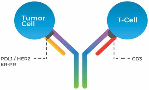
The concept of BAbs that do not occur in nature has its roots from the study in which Staerz et al. Citation5 demonstrated cancer cell lysis by engaging T cells.Citation5 It was only after 25 y that research on BAbs was boosted when the first BAb, Blinatumomab/MT103 (bispecific for CD3 and CD19), was being tested in clinical trials.Citation6,Citation7 This BAb, which is a bispecific T-cell engager (BiTE) in its structure,Citation8 has been approved for acute lymphoblastic leukemia (ALL). BAbs have a dual functionality combined in a single antibodyCitation9 as depicted in .
Figure 1. Basic architecture of a bispecific antibody. BAbs have two arms complementary to different antigens, which imparts dual specificity to the molecule. MAbs have both Fab fragments with the same specificity; i.e., they bind to a single antigen. The dual specificity enables BAbs to affect cell-specific drug delivery, cytotoxic T-cell interactions with tumor cells, and other immune-mediated actions on foreign antigens. MAbs are superior for opsonization or complement system activation-mediated clearance of antigens
BAbs are of two kinds: immunoglobulin G (IgG)-like and non-IgG-like moleculesCitation10 as depicted in . IgG-like BAbs are capable of effector functions such as complement-dependent cytotoxicity (CDC), antibody-dependent cell-mediated cytotoxicity (ADCC), and antibody-dependent cellular phagocytosis, whereas non-IgG-like BAbs are small and can achieve deeper tissue penetration.Citation11 The IgG-like BAbs resemble natural IgG as characterized by the presence of three arms/binding sites: two antigen-binding Fab arms and an Fc arm. Some of the standard IgG-like BAb formats include Triomab, Immunotoxin, Knob-in-hole BAbs (KIH BAbs), and TriFabs. Triomabs have two Fabs binding to two different antigens at the same time and an Fc region enhanced to bind to immune cells to elicit ADCC and facilitate antigen presentation.Citation12 Immunotoxins have the Fc region tagged with toxins such as diphtheria toxin and are used for site-specific drug delivery and killing of target cells.Citation13 KIH BAbs carry certain amino acid modifications at their Fc region that facilitate better heterodimerization and overcomes the mispairing problem that arises during assembly of antibody chains ex vivo.Citation14 TriFabs have three Fab fragments, two for one antigen and the remaining one for other.Citation15 Apart from these IgG-like BAbs, many such additional molecules are being developed, each with their respective function and utility.Citation16
Figure 2. Different BAbs formats: (a) TrioMab; (b) immunotoxin; (c) KIH BAbs; (d) TriFabs; (e) BiTE; (f) bispecific nanobody; (g) diabody. (a) BiTe consists of a variable heavy and light domain joined by a linker. (b) Immunotoxin, wherein an antibody is linked to a toxin. (c) TrioMab is trifunctional, corresponding to three different targets at the same time. (d) TriFabs, BAbs having dual specificity with two regular Fab arms fused to an asymmetric third Fab module via linker peptides. (e) Bispecific nanobody. (f) Knob into hole bispecific antibody. (g) Knob into hole bispecific antibody with a common light chain
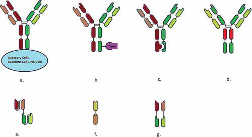
The non-IgG-like BAbs lack the Fc arm and possess only two antigen-binding Fab arms.Citation17 Common non-IgG-like BAbs include BiTE, bispecific nanobody, and diabody. BiTE works by engaging the T-cells of the immune system at the site of infection.Citation18 Bispecific nanobody is a compact molecule containing the variable heavy chains for the two antigens linked by a short peptide called the linker.Citation19 Diabody is a relatively large molecule compared to a nanobody and contains the Fab fragments for the two antigens linked together.Citation20,Citation21 Non-IgG-like BAbs have better tissue penetration, while IgG-like BAbs are more stable and easier to purify in downstream processing.
One of the essential goals of cancer immunotherapy is to redirect immune effector cells toward tumor cells, and BAbs have been designed to achieve this phenomenon.Citation22,Citation23 A more focused approach toward utilizing BAbs in onco-immunotherapy is via the generation of BiTE. BiTE works by engaging T cells at the site of infection. They are responsible for the redirection of cytotoxic T cells against pathogenic target cells. CD3 mediates T-cell activation, and binding of anti-CD3 antibody can mimic the specific antigen recognition by T cells. BAbs capable of recognizing CD3 on the T cell and a second antigen on the surface of a tumor cell are being developed.Citation24 At present, over two dozen BAbs are in clinical development, with BAbs for EpCAM × CD3 and HER2 × CD3 being the most widely studied BAbs.Citation25 Both of these BAbs work on the principle of T cell recruitment and Fc-mediated effector function for EpCAM-positive tumor and Her2/Neu-positive advanced solid tumors, respectively.Citation26,Citation27 Thus, the engagement of T cells for the eradication of tumors using BAbs is presently one of the most compelling concepts for the treatment of cancer. This methodology involves the identification of the biomarkers overexpressed under various cancers and developing antibodies that identify the same.
Apart from engaging T cells to the site of tumor cells, BAbs are being developed for varied applications such as (a) blocking of signaling pathways: HER2/HER3 signaling, IGF-1R/HER3 signaling, and EGFR/HER3 signaling, thus simultaneously neutralizing two targets with a single molecule;Citation28–30 (b) targeting tumor angiogenesis: RG7221 BAb targeting VEGFA and angiopoietin-2 (Ang-2) inhibited angiogenesis and tumor growth strongly as compared to single-pathway inhibitors;Citation31 (c) BAbs have been designed for diagnostic assays to accurately detect bacterial or viral antigens, instead of antibodies generated by immune response, resulting in early-stage diagnosis and treatment of pathogens.Citation32 provides a list of clinically approved BAbs and BAbs under development.
Table 2. List of clinically approved and under development BAbs (https://clinicaltrials.gov)
This review provides an insight into the conventional biomarkers expressed in the two most prevalent cancers in male and female, i.e., prostate and breast cancer, respectively, as mentioned in .
Table 3. Biomarkers expressed in the two most prevalent cancers in male and female, respectively
Common biomarkers in breast cancer
Breast cancer affects women in the middle and old age groups. According to WHO, breast cancer is among the leading causes of female deaths worldwide, with about 2.1 million deaths reported in 2018.Citation52 The various biomarkers that are prevalent in breast carcinoma can be studied for the development of anti-marker sequences.
HER2
HER2 is a transmembrane protein receptor which belongs to Human Epidermal Growth Receptor Family and is a tyrosine kinase.Citation33 The HER family consists of four cell surface receptors (namely HER1, HER2, HER3, and HER4). HER2 acts in a phosphorylation pathway that transmits signals through site-specific phosphorylation and dephosphorylation of various domains of the HER2 receptor,Citation34 as depicted in . Overexpression of the HER2 oncogene is likely to be both a prognostic and a predictive factor in patients with breast cancer.Citation33 The cell surface receptor exists as a homo- or heterodimer and facilitates growth-related signaling of cells. Overexpression on the surface results in enhanced responsiveness to growth factors and malignant growth.Citation35
Figure 3. Activation of various pathways involving HER family of receptors
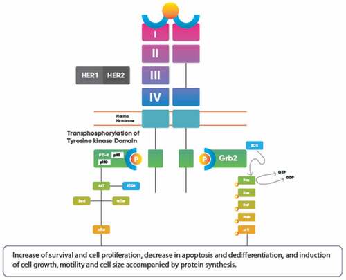
According to the National Cancer Institute (NCI), overexpression of HER2 is the causal agent for breast cancer in about 30% of the patients. Furthermore, HER2 is a potential biomarker related to metastasis, relapse, and decrease in overall survival of the patient. The most recent work done on HER2 included the development of MAbs that attach to the receptor and block its function, thereby preventing malignant growth of tumors. Mainly, two MAbs by the name Transtuzumab (commonly called Herceptin) and Pertuzumab (widely called, Perjeta) are approved.Citation36 The development of BAbs against HER2 can be beneficial since:
HER2 overexpression has the most lethal effect on patients triggering metastasis, relapse, and lower immunity.
It has a high expression rate in ER-positive, PR-positive, and even node-negative breast cancer patients, apart from getting overexpressed in HER2-positive individuals.
Developing a technology as BAbs require a considerable amount of background information about the signaling of the biomarker, its protein structure, and properties. With great effort being put to unveil HER2 dynamics, developing BAbs on the same makes it a potential target molecule, well backed by available data and statistics.
ER/PR
ER/PR represent the progesterone receptor (PR) and estrogen receptor (ER) and are used in the evaluation of breast cancer.
Estrogen receptor
ER is a hormone-dependent receptor that binds to estrogen. It has been one of the most widely studied drug targets in devising treatment methods against breast cancer. It exists as a sequestered multi-protein inhibitory complex in the nucleus, by heat shock proteins (HSP, mainly 50, 70, and 90). Upon interaction with estrogen, the ER undergoes a conformational change and becomes a dimer. This dimerized form then interacts with various estrogen receptor elements (ERE) to control the transcription of several genes. Apart from affecting the genomic mechanisms, ERs also exist in membrane form and bring about rapid, direct action of estrogen on to the cell. They bring about changes in membrane potential by modulating sodium and calcium ion transport and activating nitric oxide pathway and mitogen-activated protein kinase (MAPK) pathway. Two significant classes of ERs are present in a typical mammary cell, namely ERα and ERβ; both of these mediate gene expression with differences in their mode of action:
Complexed ERβ is a weak activator than ERα.
While ERα is mostly involved in cell proliferation, ERβ is linked to suppression of proliferation.
In some instances, ERβ works by modulating/attenuating the action caused by ERα.
Under normal conditions, a breast cell expresses an almost negligible amount of ER, which stimulates cell proliferation. ER is often overexpressed in breast cancer. Hence, during the progression of breast cancer, the production of ERα rises exponentially. The production of ERβ decreases rapidly, given its function of limiting cell growth and division.Citation37 Breast cancer caused by the overexpression of estrogen receptors is regarded as ER-positive breast cancer. The few causes of an increase in expression of ER can be:
Overproduction of estrogen in the body or administration of estrogen through oral contraceptives or hormone replacement therapy.
Binding of mutated forms of estrogen to the preexisting ERs can cause a rapid increase in the production of new, unlinked estrogen receptor molecules.Citation38
According to the American Cancer Society, two out of three breast cancers are hormone dependent and ER positive.Citation39 Furthermore, apart from being overexpressed in the case of ER-positive cancers, ER is a common biomarker showing enhanced production in other types of breast cancer as well as HER2-positive and PR-positive cancers.
Progesterone receptor
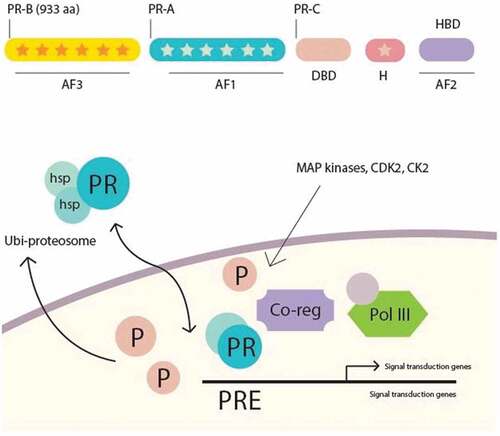
PR is also a hormone-dependent receptor that binds to progesterone, commonly referred to as the pregnancy hormone. Under normal conditions, a mammary cell lacks any PR. PR is expressed in the self-altered cancer cell surface and cytosol. PR functions as a ligand-activated transcription factor molecule acting on both genomic and non-genomic levels. The common isoforms PR-A and PR-B are produced by different post-transcriptional modification of the same gene product. PR-B is a full-length receptor, while PR-A lacks the N-terminal 164 amino acids. Both PR-A and PR-B regulate the same set of target genes. PR-C is another isoform, found in the reproductive tissues, and involved in the induction of labor at the time of pregnancy. At the non-genomic level, the PRs work by activating various signal transduction pathways, many of which are involved in pro-proliferative signaling in the breast as depicted in .
Figure 4. Growth factor-induced signaling by progesterone receptor isoforms
About two-thirds of ER-positive breast cancers are also PR positive and vice-versa.Citation41 Available technologies target ER and PR overexpression in breast cancer using hormone therapy, also known as endocrine therapy, as a mechanism to stop the growth of hormone-sensitive tumors. It involves administering specific drugs that block the ER and PR, thereby preventing any further hormone-induced signaling.Citation42 It also inhibits uncontrolled cell proliferation and differentiation. However, hormone therapy has been found to have mild-to-severe side effects such as nausea, fatigue, blood clots in brain, bone and muscle changes, etc. It also renders the body weak, making it susceptible to secondary infections. Development of BAbs against ER/PR can prove beneficial as follows:
High incidence of double-positive hormone receptor in breast cancer cases. A BAb having dual targeting for ER and PR should be more effective than a MAb against either ER or PR.
Conserved nature of receptor sequences, which makes it relatively easy to design complementary sequences. A BAb having one arm targeting ER/PR because of the conserved sequences and another arm targeting CD3 will result in the recruitment of T-cells and subsequent activation of T-cells at the target site.
Common biomarkers in prostate cancer
Prostate cancer is the second most prevalent cancer affecting males after lung cancer. According to WHO, 1.3 million cases of prostate cancer were reported in 2018, accounted for ~360,000 deaths.Citation52 Two biomarkers expressed during prostate cancer have been the target of BAb development.
PSMA
Prostate-specific membrane antigen (PSMA) is a cell membrane-bound glycoprotein uniquely expressed in large amounts in the vasculature of prostate tumor cells and mostly absent in the vasculature of healthy prostate cells. It consists of a short intracellular domain of 18 amino acids, a transmembrane domain of 24 amino acids, and a glycosylated extracellular domain of 706 amino acids.Citation43 PSMA has two unique enzymatic functions:
Folate hydrolase activity: Cleaves terminal glutamates from folypoly-γ-glutamates to produce folates and their subsequent uptake.
NAALADase activity: Cleaves terminal glutamate from neuro-dipeptide, n-acetyl-aspartyl glutamate (stands for NAAG, it is a chemical that remains localized in neuronal synapse and produces neurotransmitter glutamates upon being metabolized)
Like any other membrane-bound receptor, PSMA also gets recycled through clathrin-coated pit,Citation43 and overexpression of PSMA is a critical feature of prostate cancer. The antigen plays an essential role in carcinogenesis and tumor angiogenesis. However, the mechanism of action of PSMA in cancer cells is unknown. A hypothesis regarding the role of PSMA in metastasis is that the antigen works by undergoing glycosylation, and the alteration in this process is responsible for carcinogenesis. Thus, due to its exciting yet unexplained distribution, it can serve as an essential biomarker for detecting prostate cancer.
PSMA is an excellent diagnostic target because of the following reasons:
It primarily expresses in prostate tumor cells with minimal expression in other tissues; hence, the site-specific action of the anti-PSMA antibody is assured.
Expression in prostate tumor cells is 1000-fold higher than in healthy prostate cells, making the cancer cells to be detected easily.
It is not released into circulation, thereby reducing the chances of side effects or allergy.Citation44
Current immunological interventions for diagnosis and therapeutics that depend on using PSMA target the heavy extracellular domain of the biomolecule as the J591 MAb developed with high binding affinity.Citation45 J591 is in Phase 1/2 study.Citation46 Capromab is another approved anti-PSMA antibody,Citation47 but since it targets the intracellular domain of PSMA, it is not useful on viable cells. While developing MAbs that target the extracellular domain can be advantageous, generating BAbs instead would also bring in the advantages of immune cell-assisted killing of the tumor cells.
HEPSIN
Hepsin is a type-2 transmembrane serine protease frequently overexpressed in prostate cancer cells. It is a 417 amino acid protein composed of a short N-terminal cytoplasmic domain, a transmembrane domain, and a single scavenger receptor cysteine-rich domain that packs tightly against the C-terminal protease domain.Citation48
Several studies have reported high expression levels of hepsin in late-stage prostate cancers. The exact role of hepsin in prostate cancer progression remains unclear. However, some recent insights have been provided by Zhang et al.,Citation49 who reported that hepsin might be acting on the translational machinery to suppress the expression of CDK11p58, a protein responsible for pro-apoptotic signaling in prostate cancer. There is also accumulating evidence that hepsin might be involved in promoting the invasive capacity of the tumor and even metastasis. Upregulation of hepsin in the prostate epithelium of mice is observed to cause disorganization of the basement membrane (BM) of the prostate in probasin (PB) promoter-driven hepsin transgenic mice.Citation50
Immunohistochemical staining using MAbs against hepsin developed in hepsin-knockout mice showed weak hepsin expression in tissues from healthy prostate, benign prostatic hyperplasia, or low grade (2/3) prostate cancer. In contrast, hepsin expression is elevated in advanced prostate tumors (grades 4/5) and bone metastasis, thereby confirming mRNA expression studies.Citation51 Various MAbs against hepsin are being developed. One such antibody is Fab 25,Citation53 which follows noncompetitive inhibition kinetics while binding to hepsin. Thus, BAbs against hepsin can be highly efficient for prostate cancer.
Advantages of BAbs relative to MAbs
(1) They are immunological molecules with dual functionality.
BAbs offer dual functionality since a single antibody molecule can interact with two surface antigens at the same time.
(2) They offer a myriad of features.
BAbs have a highly specialized and wide range of functions such as recognizing two different antigens simultaneously, targeting various disease mediators dually, or delivering payloads to targeted sites.
(3) They can target multiple diseases.
They are next-generation diagnostic options not only for various cancers but also for a myriad of other diseases including genetic disorders, bacterial diseases, and HIV infections.Citation32
(4) They have significant advantages for cancer immunotherapy.
An added advantage is the ability to recognize tumor cells accurately and without using invasive techniques. BAbs allow for direct interaction of immune cells (NK cells, T-cells) with tumor cells. They also enhance the process of killing target cells by blocking more than one signaling pathway simultaneously.Citation54
(5) BiTE is a type of BAb that allows for natural recruitment of T-cells on tumor cells.
BiTEs facilitate direct contact of tumor cells with host T cells, thereby enhancing T-cell potency for acting on tumor cells by creating suitable micro-environments for the release of cytotoxic mediators. BiTEs also can trigger serial killing of tumor cells by a single T cell.Citation54
(6) Usage of BAbs for delivering payloads to target sites.
Given their dual specificity, BAbs can bind to cell surface antigens and molecular payloads simultaneously, thereby serving as excellent delivery systems for transporting cytotoxic entities to malignant cells.Citation55-57
Conclusion
BAbs are an important therapeutic in the field of onco-immunotherapy. Eliminating tumor growth is one of the most important mechanisms that is widely targeted for treating cancer. BAbs can be used in combinatorial therapy with current drugs and can be a cost-effective treatment for cancer. Apart from enhancing T-cell stimulation and tumor cell-binding properties of BAbs, other modifications such as improving the ADCC and Complement-Dependent Cellular Cytotoxicity (CDCC) activity of the molecule also can be explored. Furthermore, developing BAbs against biomarkers for breast and prostate cancer will be beneficial in detection and effective treatment. BAbs should add to the field of immunotherapy as a theragnostic against various cancers.
BAbs also have shortcomings.
The molecules pose difficulty in their downstream processing.
These molecules are mostly short-lived.
They may be immunogenic due to their non-human nature.
Heterodimerization of antibody chains from different sources can be problematic, thus reducing the efficiency of developing these drugs.
MAbs have evolved as site-specific treatments and will continue to have a durable position in the market and in the therapeutic armamentarium.
Disclosure of potential conflicts of interest
No potential conflicts of interest were disclosed.
Abbreviations
| BAbs | = | Bispecific antibodies |
| MAb | = | Monoclonal antibody |
| IgG | = | Immunoglobulin |
| BiTE | = | Bispecific T-cell engager |
| HER2 | = | Human Epidermal Growth Receptor 2 |
| ER/PR | = | Estrogen receptor/progesterone receptor |
| CD3 | = | Cluster of differentiation 3 |
| PSMA | = | Prostate-specific membrane antigen |
| Fc | = | Fragment crystallizable |
| Fab | = | Fragment antibody |
Additional information
Funding
References
- TThe Expresswire. Monoclonal antibodies market demand to grow at a CAGR of 8.5% during 2019-2025 – marketWatch [Internet]. 2019 Sep [ accessed 2019 Nov 22]. https://www.marketwatch.com/press-release/monoclonal-antibodies-market-demand-to-grow-at-a-cagr-of-85-during-2019-2025-2019-09-10.
- Scott AM, Allison JP, Wolchok JD. Monoclonal antibodies in cancer therapy. Cancer Immun. 2012;12:14.
- Minh Ngoc Duong IS. Advances in bispecific antibodies engineering: novel concepts for immunotherapies. J Blood Disord Transfus. 2015;6:1000243.
- Pento JT. Monoclonal antibodies for the treatment of cancer. Anticancer Res [Internet]. 2017;37:5935–39 [accessed 2019 Sep 26]. http://www.ncbi.nlm.nih.gov/pubmed/29061772.
- Staerz UD, Kanagawa O, Bevan MJ. Hybrid antibodies can target sites for attack by T cells. Nature. [Internet]. 1985;314:628–31. doi:10.1038/314628a0.
- Bargou R, Leo E, Zugmaier G, Klinger M, Goebeler M, Knop S, Noppeney R, Viardot A, Hess G, Schuler M, et al. Tumor regression in cancer patients by very low doses of a T cell-engaging antibody. Science [Internet]. 2008;321:974–77 [accessed 2019 Sep 26]. http://www.ncbi.nlm.nih.gov/pubmed/18703743.
- Chames P, Baty D. Bispecific antibodies for cancer therapy: the light at the end of the tunnel? MAbs. [Internet]. 2009;1:539–47. doi:10.4161/mabs.1.6.10015.
- Topp MS, Gökbuget N, Stein AS, Zugmaier G, O’Brien S, Bargou RC, Dombret H, Fielding AK, Heffner L, Larson RA, et al. Safety and activity of blinatumomab for adult patients with relapsed or refractory B-precursor acute lymphoblastic leukaemia: a multicentre, single-arm, phase 2 study. Lancet Oncol [Internet]. 2015;16:57–66. [accessed 2019 Sep 26]. http://www.ncbi.nlm.nih.gov/pubmed/25524800.
- Liu H, Saxena A, Sidhu SS, Wu D. Fc engineering for developing therapeutic bispecific antibodies and novel scaffolds. Front Immunol [Internet]. 2017;8:38 [accessed 2019 Sep 26]. http://www.ncbi.nlm.nih.gov/pubmed/28184223.
- Spiess C, Zhai Q, Carter PJ. Alternative molecular formats and therapeutic applications for bispecific antibodies. Mol Immunol. 2015;67(2):95–106. doi:10.1016/j.molimm.2015.01.003.
- Kontermann RE, Brinkmann U. Bispecific antibodies; different formats. Drug Discov Today. [Internet]. 2015;20(7):838–47. doi:10.1016/j.drudis.2015.02.008.
- Chelius D, Ruf P, Gruber P, Plöscher M, Liedtke R, Gansberger E, Hess J, Wasiliu M, Lindhofer H. Structural and functional characterization of the trifunctional antibody catumaxomab. MAbs. 2010;2(3):309–19. doi:10.4161/mabs.2.3.11791.
- Wang Z, Kim GB, Woo JH, Yuan YL, Mathias A, Stavrou S, Neville DM. Improvement of a recombinant anti-monkey anti-CD3 diphtheria toxin based immunotoxin by yeast display affinity maturation of the scFv. Bioconjug Chem. 2007;18(3):947–55. doi:10.1021/bc0603438.
- Xu Y, Lee J, Tran C, Heibeck TH, Wang WD, Yang J, Stafford RL, Steiner AR, Sato AK, Hallam TJ, et al. Production of bispecific antibodies in “knobs-into-holes” using a cell-free expression system. MAbs. 2015;7(1):231–42. doi:10.4161/19420862.2015.989013.
- Dickopf S, Lauer ME, Ringler P, Spick C, Kern P, Brinkmann U. Highly flexible, IgG-shaped, trivalent antibodies effectively target tumor cells and induce T cell-mediated killing. Biol Chem. 2018;400(3):343–350.
- Ying T, Jung ST, Kontermann RE, Kim Y-S, Ha J-H, Kim J-E. Immunoglobulin Fc heterodimer platform technology: from design to applications in therapeutic antibodies and proteins. Proteins Front Immunol [Internet]. 2016;7:394 [accessed 2020 Jan 16]. www.frontiersin.org.
- Fan G, Wang Z, Hao M, Li J. Bispecific antibodies and their applications. J Hematol Oncol. 2015;8:130.
- Yu L, Wang J. T cell-redirecting bispecific antibodies in cancer immunotherapy: recent advances. J Cancer Res Clin Oncol. 2019;145(4):941–56. doi:10.1007/s00432-019-02867-6.
- Chanier T, Chames P. Nanobody engineering: toward next generation immunotherapies and immunoimaging of cancer. Antibodies. 2019;8(1):13. doi:10.3390/antib8010013.
- Seifert O, Rau A, Beha N, Richter F, Kontermann RE. Diabody-Ig: a novel platform for the generation of multivalent and multispecific antibody molecules. MAbs. 2019;11(5):919–29. doi:10.1080/19420862.2019.1603024.
- Kwon NY, Kim Y, Lee JO. Structural diversity and flexibility of diabodies. Methods. 2019;154:136–42. doi:10.1016/j.ymeth.2018.09.005.
- Satta A, Mezzanzanica D, Turatti F, Canevari S, Figini M. Redirection of T-cell effector functions for cancer therapy: bispecific antibodies and chimeric antigen receptors. Future Oncol. [Internet]. 2013;9(4):527–39. doi:10.2217/fon.12.203.
- Zugmaier G, Klinger M, Schmidt M, Subklewe M. Clinical overview of anti-CD19 BiTE® and ex vivo data from anti-CD33 BiTE® as examples for retargeting T cells in hematologic malignancies. Mol Immunol. 2015;67(2):58–66. doi:10.1016/j.molimm.2015.02.033.
- Baeuerle PA, Reinhardt C. Bispecific T-cell engaging antibodies for cancer therapy. Cancer Res. [Internet]. 2009;69(12):4941–44. doi:10.1158/0008-5472.CAN-09-0547.
- Weidle UH, Kontermann RE, Brinkmann U. Tumor-antigen-binding bispecific antibodies for cancer treatment. Semin Oncol. [Internet]. 2014;41(5):653–60. doi:10.1053/j.seminoncol.2014.08.004.
- Jäger M, Schoberth A, Ruf P, Hess J, Lindhofer H. The trifunctional antibody ertumaxomab destroys tumor cells that express low levels of human epidermal growth factor receptor. Cancer Res. 2009;69(10):4270–76. doi:10.1158/0008-5472.CAN-08-2861.
- Kiewe P, Hasmüller S, Kahlert S, Heinrigs M, Rack B, Marmé A, Korfel A, Jäger M, Lindhofer H, Sommer H, et al. Phase I trial of the trifunctional anti-HER2 x anti-CD3 antibody ertumaxomab in metastatic breast cancer. Clin Cancer Res [Internet]. 2006;12:3085–91. [accessed 2019 Sep 26]. http://www.ncbi.nlm.nih.gov/pubmed/16707606.
- McDonagh CF, Huhalov A, Harms BD, Adams S, Paragas V, Oyama S, Zhang B, Luus L, Overland R, Nguyen S, et al. Antitumor activity of a novel bispecific antibody that targets the ErbB2/ErbB3 oncogenic unit and inhibits heregulin-induced activation of ErbB3. Mol Cancer Ther. [Internet]. 2012;11(3):582–93. doi:10.1158/1535-7163.MCT-11-0820.
- Fitzgerald JB, Johnson BW, Baum J, Adams S, Iadevaia S, Tang J, Rimkunas V, Xu L, Kohli N, Rennard R, et al. MM-141,an IGF-IR-and ErbB3-directed bispecific antibody overcomes network adaptations that limit activity of IGF-IR inhibitors. Mol Cancer Ther. 2014;13(2):410–25. doi:10.1158/1535-7163.MCT-13-0255.
- Duligotuzumab CP. Human anti-EGFR/anti-HER3 MAb, colorectal cancer therapy, head and neck cancer therapy. Drugs Future. 2015;40(3):167. doi:10.1358/dof.2015.040.03.2312450.
- Kienast Y, Klein C, Scheuer W, Raemsch R, Lorenzon E, Bernicke D, Herting F, Yu S, The HH, Martarello L, et al. Ang-2-VEGF-A CrossMab, a novel bispecific human IgG1 antibody blocking VEGF-A and Ang-2 functions simultaneously, mediates potent antitumor, antiangiogenic, and antimetastatic efficacy. Clin Cancer Res. [Internet]. 2013;19(24):6730–40. doi:10.1158/1078-0432.CCR-13-0081.
- Ferrari G, Pollara J, Tomaras GD, Haynes BF. Humoral and innate antiviral immunity as tools to clear persistent HIV infection. J Infect Dis. 2017;215(suppl_3):S152–9. doi:10.1093/infdis/jiw555.
- Viale G. HER2 in breast cancer: ESMO biomarker factsheet | oncologyPRO [Internet] [ accessed 2019 Sep 26]. https://oncologypro.esmo.org/Education-Library/Factsheets-on-Biomarkers/HER2-in-Breast-Cancer.
- Rubin I, Yarden Y. The basic biology of HER2. Ann Oncol. 2001;12(Suppl 1): S3–S8.
- Ferreira PMP, Pessoa C. Molecular biology of human epidermal receptors, signaling pathways and targeted therapy against cancers: new evidences and old challenges. Brazilian J Pharm Sci. 2017;53(2):e16076.
- National Cancer Institute (NIH). HER2 genetic link to breast cancer – National Cancer Institute [Internet] [ accessed 2019 Sep 26]. https://www.cancer.gov/research/progress/discovery/her2.
- Duffy MJ. Estrogen receptors: role in breast cancer. Crit Rev Clin Lab Sci. [Internet]. 2006;43:325–47. doi:10.1080/10408360600739218.
- Fan P, Maximov PY, Curpan RF, Abderrahman B, Jordan VC. The molecular, cellular and clinical consequences of targeting the estrogen receptor following estrogen deprivation therapy. Mol Cell Endocrinol. [Internet]. 2015;418 Pt 3:245–63. doi:10.1016/j.mce.2015.06.004.
- American Cancer Society. Hormone therapy for breast cancer | American Cancer Society [Internet] [ accessed 2019 Sep 27]. https://www.cancer.org/cancer/breast-cancer/treatment/hormone-therapy-for-breast-cancer.html.
- Daniel AR, Hagan CR, Lange CA. Progesterone receptor action: defining a role in breast cancer. Expert Rev Endocrinol Metab. 2011;6:359–69. doi:10.1586/eem.11.25.
- Lim E, Palmieri C, Tilley WD. Renewed interest in the progesterone receptor in breast cancer. Br J Cancer. 2016;115:909–11. doi:10.1038/bjc.2016.303.
- Ratini M. Types of breast cancer: triple negative, ER-positive, HER2-positive [Internet] [ accessed 2019 Sep 27]. https://www.webmd.com/breast-cancer/breast-cancer-types-er-positive-her2-positive#1.
- Ghosh A, Heston WDW. Tumor target prostate specific membrane antigen (PSMA) and its regulation in prostate cancer. J Cell Biochem. 2004;91:528–39. doi:10.1002/jcb.10661.
- Li Y, Cozzi PJ, Russell PJ. Promising tumor-associated antigens for future prostate cancer therapy. Med Res Rev [Internet]. 2010;30:67–101 [accessed 2019 Sep 27]. http://www.ncbi.nlm.nih.gov/pubmed/19536865.
- Liu H, Moy P, Kim S, Xia Y, Rajasekaran A, Navarro V, Knudsen B, Bander NH. Monoclonal antibodies to the extracellular domain of prostate-specific membrane antigen also react with tumor vascular endothelium. Cancer Res [Internet]. 1997;57:3629–34 [accessed 2019 Sep 27]. http://www.ncbi.nlm.nih.gov/pubmed/9288760.
- Tagawa ST, Vallabhajosula S, Christos PJ, Jhanwar YS, Batra JS, Lam L, Osborne J, Beltran H, Molina AM, Goldsmith SJ, et al. Phase 1/2 study of fractionated dose lutetium-177-labeled anti-prostate-specific membrane antigen monoclonal antibody J591 (177 Lu-J591) for metastatic castration-resistant prostate cancer. Cancer [Internet]. 2019;125:2561–69 [accessed 2019 Sep 27]. http://www.ncbi.nlm.nih.gov/pubmed/31012963.
- Kahn D, Williams RD, Manyak MJ, Haseman MK, Seldin DW, Libertino JA, Maguire RT. 111Indium-capromab pendetide in the evaluation of patients with residual or recurrent prostate cancer after radical prostatectomy. The ProstaScint Study Group. J Urol. [Internet]. 1998;159:2041–46. discussion 2046-7. doi:10.1016/S0022-5347(01)63239-7.
- Somoza JR, Ho JD, Luong C, Ghate M, Sprengeler PA, Mortara K, Shrader WD, Sperandio D, Chan H, McGrath ME, et al. The structure of the extracellular region of human hepsin reveals a serine protease domain and a novel scavenger receptor cysteine-rich (SRCR) domain. Structure [Internet]. 2003;11:1123–31 [accessed 2019 Sep 27]. http://www.ncbi.nlm.nih.gov/pubmed/12962630.
- Zhang C, Zhang M, Wu Q, Peng J, Ruan Y, Gu J. Hepsin inhibits CDK11p58 IRES activity by suppressing unr expression and eIF-2α phosphorylation in prostate cancer. Cell Signal. [Internet]. 2015;27:789–97. doi:10.1016/j.cellsig.2014.12.020.
- Klezovitch O, Chevillet J, Mirosevich J, Roberts RL, Matusik RJ, Vasioukhin V. Hepsin promotes prostate cancer progression and metastasis. Cancer Cell. [Internet]. 2004;6:185–95. doi:10.1016/j.ccr.2004.07.008.
- Barwe SP, Maul RS, Christiansen JJ, Anilkumar G, Cooper CR, Kohn DB, Rajasekaran AK. Preferential association of prostate cancer cells expressing prostate specific membrane antigen to bone marrow matrix. Int J Oncol [Internet]. 2007;30:899–904 [accessed 2019 Sep 27]. http://www.ncbi.nlm.nih.gov/pubmed/17332929.
- World Health Organization (WHO). Cancer [Internet]. [ accessed 2019 Sep 26]. https://www.who.int/health-topics/cancer#tab=tab_1.
- Ganesan R, Zhang Y, Landgraf KE, Lin SJ, Moran P, Kirchhofer D. An allosteric anti-hepsin antibody derived from a constrained phage display library. Protein Eng Des Sel. 2012;25:127–33. doi:10.1093/protein/gzr067.
- Spasevska I, Master. An outlook on bispecific antibodies: methods of production and therapeutic benefits. BioSciences Master Reviews; 2014.
- Zhang X, Yang Y, Fan D, Xiong D. The development of bispecific antibodies and their applications in tumor immune escape. Exp Hematol Oncol. [Internet]. 2017;6:12. doi:10.1186/s40164-017-0072-7.
- Yang F, Wen W, Qin W. Bispecific antibodies as a development platform for new concepts and treatment strategies. Int J Mol Sci [Internet]. 2016;18 [accessed 2019 Sep 27]. http://www.ncbi.nlm.nih.gov/pubmed/28036020.
- van Gils MJ, Sanders RW. Opposites attract in bispecific antibody engineering. J Biol Chem. [Internet]. 2017;292:14718–19. doi:10.1074/jbc.H117.793497.