
ABSTRACT
Lower respiratory tract infections (LRTIs) are an important cause of death and bacterial pneumonia is one of the most common causes of mortality in South Korea, but there is little data evaluating the epidemiology of pediatric LRTI in primary care clinics. We evaluated 1,497 pediatric LRTI cases in a primary care clinic over a two-year period from 2015 to 16 for clinical and radiological signs combined with PCR for pathogen detection. In addition, a 1,837 vaccine cohort in the clinic from 2014 to 16 was analyzed separately. Fifty-two percent of cases presented with fever and 15% of 1,423 X-rayed cases had positive pneumonia findings with the grade of fever correlating positively with the proportion of cases with positive chest findings. Bacterial identification was possible for 1,376 cases with Streptococcus pneumoniae, Haemophilus influenzae, and Mycoplasma pneumoniae most common. A higher proportion of 13-valent pneumococcal conjugate vaccinated cases had positive pneumonia findings than 10-valent pneumococcal nontypeable Haemophilus influenzae protein D conjugate vaccine (PHiD-CV) vaccinated cases, although similar proportions for each PCV had confirmed bacterial infections. PHiD-CV vaccinated cases with positive pneumonia findings had proportionally more single S. pneumoniae infections but less co-infections and less cases with H. influenzae infection. The proportions of confirmed bacterial infections in LRTI cases observed in this pediatric primary care setting in South Korea is very high, with co-infections most common. S. pneumoniae and H. influenzae are the most common as expected but this data also highlights M. pneumoniae as an additional important cause of LRTI in primary pediatric care in Korea.
Introduction
Lower respiratory tract infections (LRTIs) are the leading infectious disease cause of death worldwide, with pneumonia being the leading cause of death worldwide for children aged 5 years or less.Citation1 While there are a number of infections involving the lower respiratory tract, in children, the most common acute LRTIs, and those with the greatest clinical relevance, are community-acquired pneumonia (CAP), bronchiolitis, and bronchitis.Citation2 In developed countries, such as South Korea, where mortality rates from pneumonia have reduced over time, pneumonia remains a frequent cause of hospitalization and is one of the most common causes for primary pediatric clinic visits.Citation3
LRTIs may be caused by bacteria, viruses, fungi and even parasites. Prior to antibiotics and vaccines, S. pneumoniae was the primary cause, but currently <15% of cases of CAP are due to this pathogen.Citation4 While viral etiologies are now more common, many patients have bacterial coinfections with viral respiratory pathogens complicating treatment outcomes.Citation4, Citation5 Of particular interest is the impact pneumococcal vaccines have had on the etiology of LRTIs. Pneumococcal conjugate vaccines (PCVs) have been available in South Korea for some time but it was only after 10-valent pneumococcal non-typeable Haemophilus influenzae protein D conjugated vaccine (PHiD-CV) and 13-valent pneumococcal conjugate vaccine (PCV13) replaced 7-valent pneumococcal conjugate vaccine (PCV7) and both PCVs were included into the national immunization program in May 2014, that coverage rates have improved to their current levels of over 95%.Citation6
While there has published a study of the etiologies of acute respiratory infections in children diagnosed in a primary clinic focused on antibiotic resistance of S. pneumonia and H. influenza in Sweden,Citation7 most of the studies of children’s acute respiratory tract infections were focused only on S. pneumonia and its antibiotic resistanceCitation8 and diagnosed from primary clinics and hospitals.Citation9 Bacterial pneumonia is one of the most common causes of mortality in South Korea, but there is no study describing the epidemiology of pediatric LRTI in primary care clinics in the country. The objectives of this study are to understand and analyze the characteristics and patterns of LRTI after pneumococcal NIP introduction in a primary clinic. As both PCVs are used simultaneously in Korea, this also provides an opportunity to compare epidemiological differences between PCV13 and PHiD-CV in clinical practice as a secondary objective.
Patients and methods
Cases of clinically suspected acute lower respiratory tract infections (ALRTI) had been collected prospectively in one primary care center over a two-year period from January 1, 2015 through December 31, 2016. Acute lower respiratory tract infections (ALRTI) are defined in the International Classification of Diseases as those infections that affect airways below the epiglottis and include acute manifestations of laryngitis, tracheitis, bronchitis, bronchiolitis, lung infections, or any combination among them. In this study, patients who had symptoms of respiratory infections such as cough or fever with physical signs of wheezing, rhonchi, or rale as well as positive or negative chest X-ray findings were included. As per Institutional Review Board guidelines in South Korea, preexisting laboratory results and clinical findings could be used in this study without formal IRB approvals required (irb.or.kr). This represented approximately 85% of all ALRTI patients visiting the clinic during the study period (ALRTI cohort). Children aged under 18 years of age who were diagnosed with ALRTI were included in the study conducted at Isaac Pediatric Clinic in Sejong City, Korea and stratified according to prior PCV receipt. The case definition for patients who were clinically diagnosed with ALRTI included those patients having cough, coarse breathing sound with abnormal auscultation findings such as crackles, rhonchi, and rales with or without fever.
Respiratory specimens (one oropharyngeal and one nasopharyngeal swab per case) from all cases included in this study were tested with multiplex bacterial PCR with SeeplexⓇ PneumoBacter ACE Detection assay V 3.0 (Seegene, Seoul, South Korea) which is a qualitative in vitro test for the detection of 6 common pathogens causing lower respiratory tract infections (LRTI): Mycoplasma pneumoniae (MP), Chlamydophila pneumoniae (CP), Legionella pneumophila (LP), Streptococcus pneumoniae (SP), Haemophilus influenzae (HI) and Bordetella pertussis (BP) by applying a dual priming oligonucleotide technology (DUO™).Citation10
Chest X-rays were interpreted by a radiologist and cases were assessed clinically for signs and symptoms and fever was stratified as no fever (<37.5°C), mild fever (37.5-37.9°C), moderate fever (38.0-38.9°C) or high fever (>39.0°C).
As this study started enrolling cases seven months after Korean National Immunization Program had started covering both high valent pneumococcal conjugate vaccines, most of the enrolled cases were not fully vaccinated (3 + 1). That’s why the cases who have received at least one dose of PHiD-CV or PCV13 were included in the PCV evaluations. Patients receiving a mixed schedule of both vaccines (or PCV7) were excluded from the analysis by PCV received. To further evaluate the impact of pneumococcal conjugate vaccine on ALRTI etiology, an additional cohort of children receiving either PHiD-CV or PCV13 at the same clinic was also collected and stratified according to clinical and laboratory findings (PCV cohort).
Statistical analysis
In the analysis of two groups of cases who received PHiD-CV or PCV13 more than one dose, two groups were very different in terms of age distribution, statistical comparisons were not conducted. But in a separate analysis among a cohort of 1,837 cases vaccinated in the clinic between Jan 2014 and Dec 2016, student t-test were used to find a meaningful difference between the two groups.
Results
Demographics
During the study period, 1,497 children aged under 18 years were diagnosed with ALRTI and included in the study (). The numbers of male and female patients were 813 (54.3%) and 684 (45.7%). The median age of the cases (<18 years) was 3.3 years, the mean age was 4.1 years. The median age of cases with positive pneumonia findings in their chest X-ray was 4.3 years and the mean age was 4.5 years.
Table 1. Acute lower respiratory tract infection cohort age and PCV history
Both the median and mean ages were older in cases receiving PCV13 (2.6 and 2.8 years, respectively) compared to those receiving PHiD-CV (1.7 and 2.1 years, respectively). Two hundred cases (13%) were unvaccinated and their median and mean ages were substantially older than vaccinated cases (8.6 and 8.5 years, respectively).
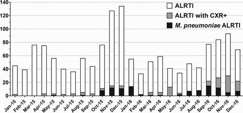
LRTIs occurred all year round during the study period but more frequently during late autumn and winter (). The seasonal distribution of LRTIs with chest X-ray pneumonia findings was more pronounced in the winter months.
Figure 1. Seasonal distribution of ALRTI cases.
Clinical findings
Fever
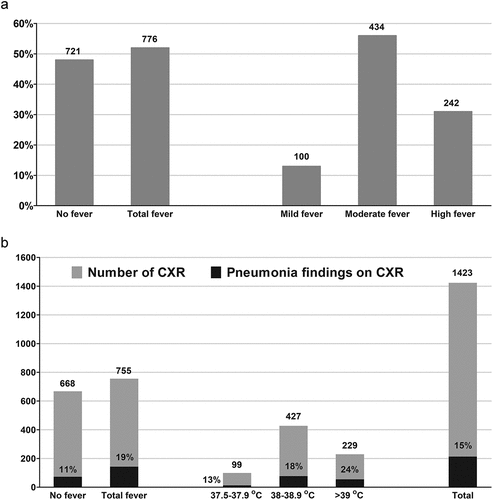
Of the 1,497 cases enrolled, 776 (52%) presented with fever over 37.5°C of those, 434 (56%) had moderate fevers (38–38.9°C) and 242 (31%) had high fevers (over 39°C) ().
Figure 2. Clinical and radiological findings among ALRTI cases.
X-Ray
Of the 1,497 cases enrolled, 1,423 had chest X-ray evaluation with 215 (15%) assessed as having positive pneumonia findings (). Seventy-seven cases (5.1% of all enrolled) were assessed through paranasal sinus X-ray (PNS) with 36 (45%) assesses as having positive sinusitis findings. Overall, there was a higher proportion of pneumonia positive chest findings in cases presenting with a fever (19% vs 11% when no fever present). The grade of fever correlated positively with the proportion of cases with positive chest findings (13% > 18% > 24%). In contrast, there was little difference in the proportion of sinusitis findings between cases with or without fever (44% vs 48%, respectively).
PCR
Positive bacterial identification was defined when one of the patient’s upper respiratory tract specimens (one throat swab plus one nasopharyngeal swab) was positive on multiplex bacterial PCR. Positive bacterial PCR results were obtained from 1,376 cases, of which S. pneumoniae (1,235) was the most frequently identified, followed by H. influenzae (1,060), M. pneumoniae (109), C. pneumoniaee (18) and B. pertussis (3) (). One hundred and twenty-one (8.1%) ALTRI cases were negative by PCR. Less than 30% of the cases presented with a single bacterial pathogen with S. pneumoniae (278) and H. influenzae (124) are the most common single pathogens observed. Co-infection with S. pneumoniae and H. influenzae was observed in 61.4% of cases (845) with another 5.1% detected to have S. pneumoniae, H. influenzae and M. pneumoniae (70).
Table 2. Relationship between fever and bacterial isolations
Overall, there was little difference in fever symptoms between cases with positive bacterial findings compared to those negative for bacteria, or between those presenting with or without fever among confirmed bacterial cases. A trend toward less presentation with fever was apparent with single bacterial infections (H. influenzae 44%, S. pneumoniae 47%), while co-infection with multiple bacteria generally tended to correlate with higher proportions of cases presenting with fever (S. pneumoniae, H. influenzae and M. pneumoniae (69%), H. influenzae and M. pneumoniae (67%), S. pneumoniae and M. pneumoniae (60%), S. pneumoniae and H. influenzae (54%). Further evaluation of M. pneumoniae cases occurring during outbreaks in 2015 and 2016 is described later in this manuscript.
PCV vaccination comparisons
Vaccination histories were available for all 1,497 cases with 889 (59%) receiving PCV13, 205 (14%) receiving PHiD-CV and 205 (7.9%) receiving PCV7, 200 (13%) were unvaccinated (). A small number of cases had received mixed schedules of more than 1 PCV. Only cases receiving at least one dose of only PHiD-CV or PCV13 were included in the analyses related to PCV receipt, all cases receiving PCV7 or mixed PCV schedules were excluded.
Fever
A higher proportion of vaccinated cases presented with fever than unvaccinated cases (53% versus 44%, ) and this difference was primarily apparent via differences in moderate fever (29.9% versus 23.0%). Comparing PCVs, a higher proportion of PHiD-CV vaccinated cases presented with mild and moderate fever than PCV13 vaccinated cases (total fever: 60.5% versus 53.1%, Moderate fever: 35.1% versus 29.4%) although high fever was more frequently associated with PCV13 vaccinated cases (17.3% versus 15.6% PHiD-CV vaccinated) (). PHiD-CV vaccinated cases were generally associated with more fever than PCV13 cases when bacterial infections were confirmed.
Figure 3. Clinical, radiological and PCR findings stratified by PCV received.
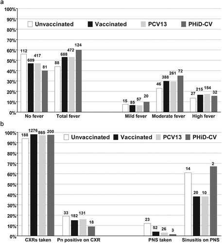
Figure 3. (Continued).
X-Ray
A lower proportion of vaccinated cases than unvaccinated cases had confirmed pneumonia findings (14.5% versus 19.2%) and sinusitis findings (38.5% versus 60.9%), although as mentioned previously, the average age of unvaccinated cases was substantially older than vaccinated cases. Comparing PCVs, a higher proportion of PCV13 vaccinated cases that were X-rayed were confirmed to have positive pneumonia findings than PHiD-CV vaccinated cases (15.1% versus 9.0%). While 26 cases of PCV13 vaccinated had PNS tests and 10 cases had positive sinusitis findings, 3 cases of PHiD-CV vaccinated had PNS tests and 2 cases had positive finding—the limited number (2) of cases vaccinated with PHiD-CV with positive sinusitis findings prevents a valid comparison ().
PCR
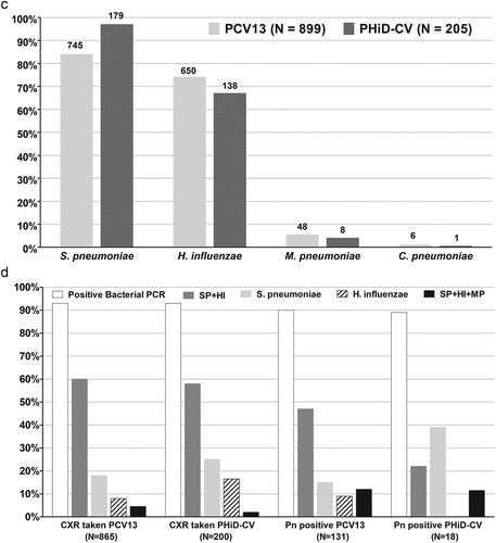
While both groups of vaccinated cases were associated with a similar proportion of bacterial infections, PHiD-CV vaccinated cases had proportionally more S. pneumoniae infections (87.3% versus 83.8%) but less H. influenzae (67.3% versus 73.1%) and M. pneumoniae (3.9% versus 5.4%) infections (). Among cases with positive pneumonia findings on X-ray, similar proportions of PCV13 or PHiD-CV vaccinated cases had confirmed bacterial infections (90.7% versus 88.9%). PHiD-CV vaccinated cases with positive pneumonia findings had proportionally more single S. pneumoniae infections (38.9% versus 15.3%) but less co-infections and no cases with H. influenzae infection were detected, compared to 12.2% of PCV13 vaccinated cases with positive pneumonia findings.
PCV cohort
To reduce confounders between vaccination group comparisons, a separate analysis among a cohort of 1,837 cases vaccinated in the clinic between January 2014 and December 2016 was conducted. For clarity, 275 cases of ALRTI were identified between January 1st, 2015 and December 31st, 2016 within this PCV cohort, which were also included within the 1,497 cases included in the ALRTI cohort ( and ). This separate analysis was included as the number and the age of cases was more comparable between PCV groups than the ALRTI cohort and thus provided a more robust comparison between similar populations.
Figure 4. Number and proportion of pathogens detected via PCR in a separate cohort of 1,837 ALRTI cases vaccinated in the clinic between Jan 2014 and Dec 2016 stratified by PCV received.
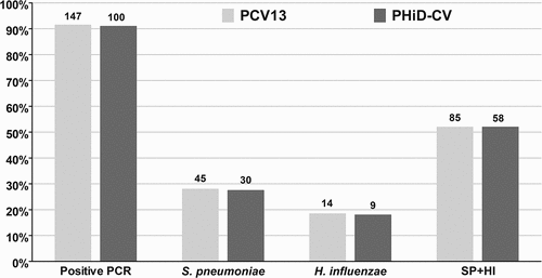
Table 3. PCV cohort demographics
Within this cohort, 990 cases were vaccinated with PHiD-CV versus 847 vaccinated with PCV13. The median ages for PHiD-CV vs PCV13 vaccinated cases were 1.2 versus 1.4 years, respectively, while the mean ages were 1.2 versus 1.6 years. There were 275 cases of LRTI observed of which 110 cases (11.1%) occurred in PHiD-CV vaccinated children, versus 161 cases (19.0%) in PCV13 vaccinated children (p = .039). There were four LRTI cases which had been vaccinated with PHiD-CV and PCV13 alternatively and these were excluded from the analysis.
As for the larger study cohort, slightly more fever was observed in PHiD-CV vaccinated children (64.5% vs 52.8%) although the proportion of high fever was quite similar (17.3% versus 16.1%). Similar proportions of chest X-rays were taken from both groups (96.9% vs 99.1%) although a higher proportion of positive pneumonia findings were observed in PCV13 vaccinated children (9.9% versus 9.1%). Finally, similar proportions of bacteria, either single bacterial infections or co-infections, were observed for both vaccine groups.
M. pneumoniae cases
There was a total of 109 cases with M. pneumoniae PCR positive. Among them, only 4 cases had M. pneumoniae positive only and the other 105 cases had other bacteria co-positive.
Of interest, all four cases with confirmed single infection with M. pneumoniae presented with fever, while all three cases with single C. pneumoniae infection had no fever. The 109 cases with confirmed M. pneumoniae infections were slightly older than the overall group with the median age being 5.3 years and the mean age being 5.9 years and occurred in two well differentiated outbreaks, from October 2015 to January 2016 and from July to December 2016 (). Overall, a higher proportion of cases with M. pneumoniae infections presented with fever (68%) while 58% had positive pneumonia findings in chest X-rays, compared to the overall group averages of 52% and 15%, respectively.
Among the 889 LRTI cases vaccinated with PCV13, 48 (5.4%) had confirmed M. pneumoniae infection compared to 8 (3.9%) of 205 PHiD-CV vaccinated LRTI cases (p > .05). There was little difference observed between vaccinated and unvaccinated M. pneumoniae cases in terms of fever, although greater variations were observed with PHiD-CV vaccinated cases due to the small numbers (eight cases in total) (). Proportionally, almost twice as many unvaccinated cases (7%, n = 14 out of 200 LRTI cases) presented with pneumonia findings compared to vaccinated cases (3.8%, n = 49 of 1,297 LRTI cases). Within those, a slightly higher proportion of PCV13 vaccinated cases had positive pneumonia findings confirmed radiologically than PHiD-CV vaccinated cases (2.8% versus 2.0%).
Figure 5. ALRTI cases with confirmed M. pneumoniae infection stratified by PCV received.
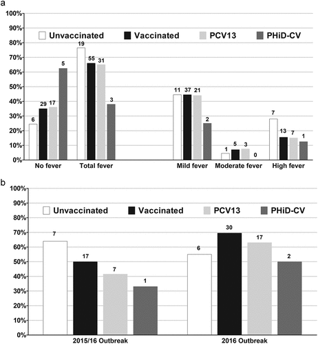
Comparing the 2015/16 M. pneumoniae outbreak with the 2016 outbreak, few differences were seen in the presentation of fever between vaccinated and unvaccinated cases, although slightly higher proportions of confirmed pneumonia findings were observed among vaccinated cases in 2016 ().
Discussion
To date, there has been very limited data available regarding LRTI etiology in South Korea. Importantly, this study not only provides important information regarding the epidemiology of bacterial pneumonia, but enables an evaluation of clinical and radiological findings according to which PCV children received. This is highly relevant to physicians in South Korea as both PCVs are available within a fully reimbursed healthcare environment to enable a more objective evaluation of the benefits of each vaccine.
Our study observed a very high proportion of LRTI cases with confirmed bacterial infection, while a relatively small proportion of LRTI cases presented with X-ray confirmed pneumonia, with an even smaller percentage with sinusitis. Pneumonia confirmation was higher in subjects presenting with fever and the grade of fever correlated positively with pneumonia findings by X-ray. These are logical associations, but there has been little empirical evidence to support this previously.
PCR detection enabled bacterial identification in most cases with the vast majority of cases associated with co-infection by at least two pathogens. As expected, S. pneumoniae and H. influenzae were the most frequently isolated in this study, both well-recognized causative agents of ALRTI. This is consistent with earlier studies validating the development of the current PCR approach.Citation11, Citation12 A small study to validate this novel multiplex PCR assay in 2008 tested 181 nasopharyngeal aspirates collected from pediatric patients with respiratory symptoms at Dankook University Hospital. Of the 44.8% of cases positive by multiplex PCR, 28.7% were identified as S. pneumoniae, 26.0% for H. influenzae and 5.0% for M. pneumoniae.Citation11 A follow-up study at the same institution evaluated the distribution of bacterial pathogens causing respiratory symptoms in different age groups over a 10-year period between January 2008 and September 2017.8 Of 1,861 specimens from 1,664 patients admitted to Dankook University Hospital with respiratory symptoms, bacterial pneumonia pathogens were identified 68.8% of specimens and almost 93% were detected in patients younger than 10 years. S. pneumoniae was the most common (48.6%) followed by H. influenzae (40.1%) while the rate of co-infection was also high among these patients (31.1% overall) peaking in 2015 (54.55%). M. pneumoniae infection also increased in prevalence with age.Citation12 An unrelated nationwide prospective multicenter study in 2009 evaluating the bacterial etiology of CAP in over 600 adults in South Korea also identified an etiology in almost 40% of the patients. The most common pathogen was also S. pneumoniae (21.1%), with M. pneumoniae (16.7%) and K. pneumoniae (10.6%), the next most common.Citation13
In agreement with these earlier studies, M. pneumoniae was the third most frequently identified bacteria in our study, with all but four cases representing coinfections with other pathogens. Compared to the broader ALRTI cohort, M. pneumoniae cases presented as older ages, more frequently with fever, and confirmed the seasonality observed in South Korea from Autumn to winter. When comparing the two M. pneumoniae outbreaks in 2015/16 and 2016, few differences were noted in terms of fever presentations although slightly higher proportions of confirmed pneumonia were noted in the latter outbreak, however a small number of cases prevents robust conclusions.
An important advantage of our study is that we were able to evaluate the effect of PCV vaccination within our ALRTI cohorts. While there were some notable findings when comparing PCV groups, the age differences of the two groups makes a direct comparison challenging. We noted a trend toward more low grade and moderate fever in children vaccinated with PHiD-CV although high fever was not substantially different between the two groups. Anecdotally PCV13 has been associated with slightly higher reactogenicity than PHiD-CV, but this may not be reflected in terms of febrile responses. Interestingly, a higher proportion of PCV13 vaccinated children presented with positive pneumonia findings than PHiD-CV vaccinated children, and this difference increased when considering only cases with confirmed bacterial infections. Interestingly, among these cases with X-ray confirmed pneumonia, less co-infections and no cases of H. influenzae were observed in the PHiD-CV vaccinated children, perhaps suggesting a relationship between H. influenzae (with or without co-infections) and pneumonia severity. PHiD-CV vaccinated children appeared to have more S. pneumoniae infections but less H. influenzae and mycoplasma infections. This is potentially consistent with slightly broader serotype coverage for PCV13 but with additional protection provided by PHiD-CV against H. influenzae. When comparing groups in the PCV cohort study with less differences in ages between groups, differences in bacterial etiology were less apparent, although the same trends regarding higher X-ray confirmed pneumonia in PCV13 vaccinated children despite lower proportions of fever were also observed.
A key strength of this study is the ability to match vaccination status with clinical, laboratory and radiological findings providing robust cohorts for comparative evaluations. It is always challenging to make valid comparisons in observational settings so reducing confounders through data linkage is critical. However, there are also a number of limitations that should be addressed. Firstly, although the majority of ARLTI (over 85%) cases were enrolled in this prospective research, there is still the possibility of selection bias. Additionally, despite the fact that the majority of patients visited the clinic the first time after their symptoms started, there is a possibility that some of the patients could have received antibiotics prescribed from other clinics prior to visiting the Isaac clinic. However, there is no record of previous antibiotics usage for all included subjects. Thirdly, as the focus of this investigation was on bacterial ALRTI pathogens and evaluating the potential PCV impact, this study did not evaluate respiratory viral infections that could be a substantial cause of ARLTI included in these cases. Further, as respiratory samples were collected from the upper airway (one throat swab plus one nasopharyngeal swab per case) there is possibility that the bacteria isolated were not the causing organisms of LRTIs but rather were simply carried. This limitation could be reduced by using control groups and using quantitative PCR methods, but those measures were not available in the plan and execution of the study. This can be a limitation of this study; however, in order to detect the exact bacteria causing LRTIs a bronchial aspirate should be obtained and tested, which is impossible in usual health care settings. Lastly, although the condition of most of the cases enrolled were improved in the clinic, there was the possibility that small numbers of cases were transferred for admission and further treatment. Subsequently, not having a record of treatment and prognosis of these cases can limit the clinical importance of this research.
In conclusion, this study has demonstrated the proportions of confirmed bacterial infections in ALRTI cases are very high, with co-infections most commonly observed. As expected, S. pneumoniae and H. influenzae were the most commonly detected but our data also highlights M. pneumoniae as an additional important cause of ALRTI in South Korea. We have noted some differences in mild to moderate fever presentation between PCVs but less X-ray confirmed pneumonia was also noted in children vaccinated with PHiD-CV. Finally, we observed some differences in bacterial etiology depending on which PCV was used and the current study suggests PHiD-CV may have a greater impact on H. influenzae than PCV13, potentially with implications for the clinical presentation of pneumonia cases.
Acknowledgements
The authors would like to thank Jae Ho Lee, M.D. Ph.D. (Department of Pediatrics, College of Medicine, Chungnam National University, Daejeon, Republic of Korea), Jin Han Kang, M.D. Ph.D. (Department of Pediatrics, Seoul St. Mary’s Hospital, The Catholic University of Korea, Seoul, Republic of Korea), Geun Yong Kwon, M.D. (Sejong Public Health Center, Sejong, Republic of Korea, Bruce A. Mungall Ph.D. (GSK Korea) and Jung Yun Hong, M.D, Ph.D. (GSK Korea) for various aspects of study support, manuscript review or feedback.
Disclosure statement
No potential conflict of interest was reported by the author(s).
Additional information
Funding
References
- Bryce J, Boschi-Pinto C, Shibuya K, Black R, WHO Child Health Epidemiology Reference Group. WHO estimates of the causes of death in children. Lancet. 2005;365(9465):1147–10. doi:10.1016/S0140-6736(05)71877-8.
- Esposito S, Bianchini S, Argentiero A, Neglia C, Principi N. How does one choose the appropriate pharmacotherapy for children with lower respiratory tract infections? Expert Opin Pharmacother. 2020;21(14):1739–47. doi:10.1080/14656566.2020.1781091.
- Wiese A, Grijalva C, Zhu Y, Mitchel E, Griffin M. Changes in childhood pneumonia hospitalizations by race and sex associated with pneumococcal conjugate vaccines. Emerg Infect Dis. 2016;22(6):1109–12. doi:10.3201/eid2206.152023.
- Musher D, Abers M, Bartlett J. Evolving understanding of the causes of pneumonia in adults, with special attention to the role of pneumococcus. Clin Infect Dis. 2017;65(10):1736–44. doi:10.1093/cid/cix549.
- Hendaus M, Jomha F, Alhammadi A. Virus-Induced secondary bacterial infection: A concise review. Ther Clin Risk Manag. 2015;11:1265–71. doi:10.2147/TCRM.S87789.
- Park D, Kim S, Yong D, Suh I, Kim Y, Yi J, Song W, Song S, Moon H, Lee H, et al. Serotype distribution and antimicrobial resistance of invasive and noninvasive Streptococcus pneumoniae isolates in Korea between 2014 and 2016. Ann Lab Med. 2019;39(6):537–44. doi:10.3343/alm.2019.39.6.537.
- Tyrstrup M, Melander E, Hedin K, Beckman A, Mölstad S. Children with respiratory tract infections in Swedish primary care; prevalence of antibiotic resistance in common respiratory tract pathogens and relation to antibiotic consumption. BMC Infect Dis. 2017 Sep 4;17(1):603. 10.1186/s12879-017-2703-3. PMID: 28870173; PMCID: PMC5583975
- Stacevičienė I, Petraitienė S, Vaičiūnienė D, Alasevičius T, Kirslienė J, Usonis V. Antibiotic resistance of Streptococcus pneumoniae, isolated from nasopharynx of preschool children with acute respiratory tract infection in Lithuania. BMC Infect Dis. 2016 May 20;16(1):216. 10.1186/s12879-016-1544-9. PMID: 27206423; PMCID: PMC4875676
- Hjálmarsdóttir MÁ, Haraldsson G, Quirk SJ, Haraldsson Á, Erlendsdóttir H, Kristinsson KG, Melo-Cristino J. Reduction of antimicrobial resistant pneumococci seven years after introduction of pneumococcal vaccine in Iceland. PLoS One. 2020 Mar 17;15(3):e0230332. 10.1371/journal.pone.0230332. PMID: 32182260; PMCID: PMC7077842
- Luznik D, Kosnik M, Tomic V. Comparison of Seeplex PneumoBacter aCE detection assay and in-house multiplex PCR for the identification of Streptococcus pneumoniae. New Microbiol. 2015 ;38(1):51–58. Epub 2015 Jan 1. PMID: 25742147.
- Park J, Kim J, Rheem I, Kim J. [Evaluation of Seeplex Pneumobacter multiplex PCR kit for the detection of respiratory bacterial pathogens in pediatric patients]. Korean J Lab Med. 2009;29(4):307–13. doi:10.3343/kjlm.2009.29.4.307.
- Yook Y, Jeon J, Park J, Kim J. Laboratory investigation of trends in bacterial Pneumonia in Cheonan, Korea, from January 2008 to September 2017. J Microbiol Biotechnol. 2018;28(10):1730–35. doi:10.4014/jmb.1804.04004.
- Chong Y, Jung K, Lee K, Kim M, Moon S, Park S, Hur J, Kim D, Jeon M, Woo J. The bacterial etiology of community-acquired pneumonia in Korea: a nationwide prospective multicenter study. Infect Chemother. 2010;42(6):397–403. doi:10.3947/ic.2010.42.6.397.