
ABSTRACT
Musculoskeletal tissues are diverse and significantly different in their ability to repair upon injury. Current treatments often fail to reproduce the natural functions of the native tissue, leading to an imperfect healing. Gene therapy might improve the repair of tissues by providing a temporarily and spatially defined expression of the therapeutic gene(s) at the site of the injury. Several gene transfer vehicles have been developed to modify various human cells and tissues from musculoskeletal system among which the non-pathogenic, effective, and relatively safe recombinant adeno-associated viral (rAAV) vectors that have emerged as the preferred gene delivery system to treat human disorders. Adapting tissue engineering platforms to gene transfer approaches mediated by rAAV vectors is an attractive tool to circumvent both the limitations of the current therapeutic options to promote an effective healing of the tissue and the natural obstacles from these clinically adapted vectors to achieve an efficient and durable gene expression of the therapeutic sequences within the lesions.
Introduction
Musculoskeletal tissues, including the articular cartilage, bone, tendons, and muscles significantly differ in their ability to repair spontaneously upon injury.Citation1 While the cartilage has a very limited ability to self-repair, most fractures of long bones heal on themselves except for large segmental defects. On the other hand, self-repair of tendons often results in a poor quality tissue.Citation2 In addition, the intrinsic ability of muscles to heal may be compromised under severe trauma conditions, leading to the formation of fibrous scar tissue.Citation3 So far, the limitations associated with the current clinical options and the increment of incidences of musculoskeletal injuries have argued for the necessity of looking for alternative therapeutic options tailored to each tissue type that may lead to its regeneration.
In this scenario, gene transfer has emerged as an alternative technology to directly transfer genes encoding for therapeutic factors in sites of injury, resulting in a temporarily and spatially defined delivery of a candidate agent.Citation4 Diverse nonviralCitation5 and viral gene vehicles (vectors derived from adenoviruses - AdV, retro/lentiviruses, or herpes simplex viruses - HSV)Citation6 have been developed to target human cells that may be affected in a variety of musculoskeletal tissues. Most particularly, vehicles based on the non-pathogenic human adeno-associated virus (AAV) have considerable advantages over other, more classical vectors that currently make them preferred systems to treat human disorders.Citation4
rAAV-mediated gene transfer for musculoskeletal system repair
Features of rAAV vectors
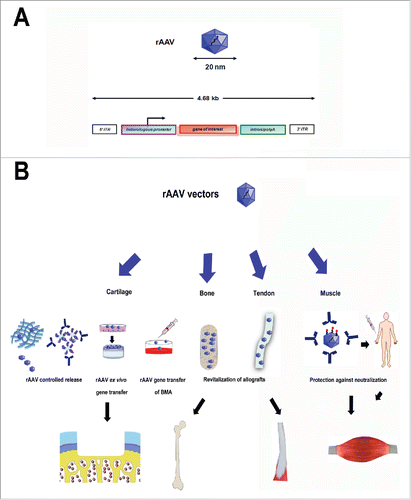
Recombinant AAV (rAAV) vectors are derived from a non-pathogenic, replication-defective human parvovirusCitation7 that can be manipulated to produce recombinant viral constructs by removing all AAV coding sequences and replacing them by a transgene cassette,Citation8,9 making them less immunogenic than AdV and less toxic than HSVCitation10-12 (). Most experimental work has been initiated with the serotype 2 of the virus (AAV-2), including to target tissues of the musculoskeletal system,Citation4,13 yet other natural serotypes have been identified and tested in diverse cells and tissues (AAV-1 to -12, with a focus on AAV-5 in the orthopaedic field).Citation4 One remaining, critical issue when using rAAV is the pre-existence of neutralizing antibodies in the human population, mostly directed against the AAV capsid proteins with a higher prevalence for AAV-1 and -2,Citation14 and possible specific cellular responses for instance by activation of the Toll-like Receptor (TLR) 9/MyD88 and interferon-1 cascade in plasmacytoid dendritic cells by particular serotypes (AAV-1, -2, and -9).Citation14 Active work is ongoing to overcome such hurdles and controlled delivery approaches based on the use of solid scaffolds and hydrogels coated with or encapsulating rAAV vectors to mask potentially immunogenic viral epitopes may allow to produce safer systems of gene transfer.Citation4 rAAV are small (∼20 nm) vectors, capable of transducing both dividing and nondividing cells at relatively high gene transfer efficiencies (up to 100%),Citation4,15 allowing for direct gene transfer approaches in vivo through the dense extracellular matrix of targeted tissues.Citation16 These potential advantages from rAAV make them as the vector of choice to treat human disorders.Citation4
Figure 1. Concepts for rAAV-mediated gene transfer using tissue engineering approaches in the musculoskeletal system. (A) Genomic organization of rAAV vectors. Classical rAAV vector with 2 inverted terminal repeats (ITRs) at either end of a transgene cassette (heterelogous promoter, gene of interest, intron/polyA signal). The arrows show the viral transcription promoter. (B) Principal tissue engineering strategies for rAAV mediated-gene transfer in the musculoskeletal system. rAAV can be encapsulated in different biomaterials such as hydrogels or polymeric micelles to achieve a controlled release profile at the site of injury. The vectors may be delivered ex vivo by genetically modification of cells that are subsequently seeded onto a matrix and implanted in the recipient. Different patient-related materials including bone marrow aspirates (BMA) and allografts can be endowed with biological factors enhancing cell/tissue reparative processes via rAAV-mediated gene transfer. Polymers can be used to overcome rAAV physiological barriers when administered through classical routes to achieve an efficient gene transfer in the target location.
rAAV-mediated gene transfer for tissue regeneration: Implications
rAAV vectors can be directly introduced into the body (in vivo approach) or indirectly by collection, modification and re-implantation of the patient's cells in sites of injury (ex vivo approach).Citation2 Direct administration of rAAV is a simple and cost-effective gene transfer approach but it requires the availability of a considerable cell population in a damaged tissue susceptible to transduction for expression of the transgene being delivered at appropriate therapeutic levels. Also, direct rAAV gene transfer by intra-articular injection as commonly performed in musculoskeletal translational research may lead to the dissemination of the vectors to non-target tissues early on (liver, kidney, lymph nodes)Citation17-20 while vector DNA might be rapidly cleared, becoming detectable only at the site of injection at extended periods of timeCitation17,19-31 and leading to prolonged transgene local expression (at least a year, the longest time points examined).Citation31,32 Another issue is a possible contralateral effect of the gene treatment in nonmodified locations (joints) upon circulation of the therapeutic product and/or by trafficking of vector-modified cells, even though this observation has been mostly reported when using adenoviral and retro-/lentiviral vectorsCitation33-38 and only in rare cases with rAAV.Citation30 Yet, such effects may be prevented when providing the vector treatment by arthrotomy that allows for a more precise delivery of the vectors in the joint.Citation39-42 Also, the existence of patient-associated factors and of physiological barriers (pre-existence of neutralizing antibodies in the host against the viral capsid proteins, inhibition of transduction by particular anticoagulants) may interfere with an effective rAAV delivery, processing, and expression of the transgene in the target cells by blocking vector transduction or by redirecting distribution of rAAV to non-target tissues.Citation4,43 Remarkably, delivery of rAAV via polymeric biomaterials may overcome such limitations by providing a controlled release of the vectors only where necessary.Citation4
Even though ex vivo delivery may obviate these problems by introducing cells instead of gene products in sites of injury, it remains a costly procedure, requiring more complex and laborious steps of cell harvesting and expansion. The identification of alternative, convenient gene delivery procedures is thus under active investigation, such as options based on the supply of tissue biopsy samples (whole bone marrow aspirates, fat, muscle) instead of isolated progenitor cells.
Combination of convenient tissue engineering strategies with clinically adapted rAAV vectors may improve current therapeutic options while increasing the efficiency of rAAV-mediated gene transfer, leading to the elaboration of safe and effective treatments against tissue injuries in patients.
Exploiting the concept of tissue engineering for an effective rAAV-mediated gene transfer
As no single approach is capable of promoting the regeneration of the different musculoskeletal tissues, tailored strategies based on then optimal combination of a therapeutic factor with a biomaterial acting as a vehicle of the gene vector (direct in vivo approach) or as a cell-supportive matrix (indirect ex vivo approach) adequate to the properties of each tissue in question are necessary to promote its regeneration. The most advanced synergic technologies for improving both rAAV-mediated gene transfer and current therapeutic options in the different tissues of the musculoskeletal system are presented in the following sections ().
rAAV gene transfer in cartilage
Articular cartilage is the smooth tissue that covers the ends of bones, allowing for a successful load transmission and mobility within the joints. Due to the lack of access to blood supply, the cartilage has a limited ability to self-healing and full repair of cartilage defects is therefore a major clinical challenge that may progress to osteoarthritis, a critical disorder affecting a large number of patients worldwide.Citation44,45 Despite the availability of several therapeutic options to repair injured cartilage (marrow-stimulating techniques such as microfacture, transplantation of tissue or cells including autologous chondrocytes - ACI - or mesenchymal stem cells - MSCs, replacement surgery),Citation46-48 none of them can reproduce the natural functions of the native, hyaline cartilage (type-II collagen and proteoglycans), rather leading to the formation of a poorly mechanically functional fibrocartilaginous surface (type-I collagen).Citation49
Current approaches for improved rAAV-2-mediated gene transfer in the cartilage focus on the incorporation of the vectors into biomaterials in order to achieve a controlled release profile of rAAV in the site of injury.Citation50-53 These techniques may be combined with bone marrow stimulation for chondral defects and the controlled release of rAAV vectors from the biomaterial could provide a suitable, lasting stimulus to increase the chondrogenic potential of cells that populate the lesion. Hydrogel systems, exhibiting a release pattern via diffusion process, are advantageous materials to achieve this goal as they may be modulated to reduce vector spreading to non-target tissues.Citation4 Different polymers from both naturalCitation50,53 or synthetic originCitation51,53 have been tested to prepare hydrogels as rAAV controlled delivery systems in cartilage regeneration (). Poly(ethylene oxide) (PEO) and poly(propylene oxide) (PPO) tri-block copolymers (poloxamers and poloxamines), described as “smart” or “intelligent” polymers due to their capacity to form polymeric micelles and to undergo sol-to-gel transition upon heating,Citation54 have recently showed to be efficient rAAV-mediated delivery systems.Citation52,53
Table 1. Tissue engineering approaches for rAAV gene transfer in articular cartilage.
Combination of rAAV with tissue engineering approaches has also been exploited to circumvent physiological barriers limiting rAAV-mediated gene transfer such as the neutralization by neutralizing antibodies against the viral capsid proteins present for instance in the synovial fluid from patients with joint diseasesCitation55 or the inhibition of rAAV adsorption at the target cell surface by specific anticoagulants (heparin).Citation4,56 We recently reported that encapsulation of rAAV in poloxamer PF68 and poloxamine T908 polymeric micelles allowed for an effective, durable, and safe modification of human MSCs (hMSCs) to levels similar to or even higher than those noted upon direct vector application (up to 95% of gene transfer efficiency).Citation52 Of further note, these copolymers were capable of restoring the transduction of hMSCs with rAAV in conditions of gene transfer inhibition like in the presence of heparin or of a specific antibody directed against the AAV capsid proteins, enabling effective therapeutic delivery of the chondrogenic sex determining region Y-box 9 (sox9) sequence leading to an enhanced chondrocyte differentiation of the cells.Citation52
Recent gene transfer approaches for cartilage regeneration focused on the use of autologous compounds capable of improving the effectiveness of the microfacture technique. By these methods, a bone marrow aspirate was collected from the patients and transduced ex vivo with viral vectors.Citation57 The resulting clotted bone marrow containing transduced cells and vectors was implanted into cartilage defects.Citation57 While this approach was initially described for the delivery of AdV,Citation57 its use with rAAV may appear more advantageous as these vectors allow for more sustained levels of transgene expression and avoid the undesirable immune responses associated with AdV.Citation58-61 An increase in chondrogenic processes have been described by transfer of transforming growth factor β 1 (TGF-β1),Citation58,60,61 insulin-like growth factor I (IGF-I),Citation59,60 and the transcription factor sox9Citation60 to bone marrow aspirates from both humanCitation58,59,61 or minipig originCitation60 via rAAV-2.
Treatment of large chondral defects is usually performed by ACI using cells from a lesser-weight bearing part of the joint. Although this technique already showed satisfactory long-term results in patients, with the production of hyaline-like repair tissue following transplantation of chondrocytes into chondral defect,Citation62 the ex vivo modification of cells via rAAV may lead to a better quality of the repaired tissue. Fibrin glue (FG) aloneCitation63 or combined with other polymersCitation64,65 has been also used for the ex vivo delivery of rAAV by encapsulation of genetically modified cells such as chondrocytesCitation63 and periosteal cellsCitation64,65 to heal full-thickness chondral defects.Citation63-65 Both IGF-ICitation63-65 and the bone morphogenetic protein 2 (BMP-2)Citation64,65 incorporated in rAAV-5Citation63 or -2Citation64,65) have been reported to be potent factors that increase chondrogenesis of encapsulated cells when implanting in chondral defects from horseCitation63 and minipig models.Citation64,65
rAAV gene transfer in bone
Bone tissue has a highly hierarchical structure based on type-I collagen fibers and nanohydroxyapatite matrix, making it a tissue with unique mechanical properties. The bone has an intrinsic ability to self-repair that may nevertheless be exceeded when the fracture gap is too big or unstable.Citation66 Other critical issues include the rate of morbidity during tissue graft harvesting and the high prevalence of pseudoarthrosis associated with lumbar spine fusion.Citation67 Therefore, complete regeneration of bone tissue remains a challenging issue.
Although autografts are considered the gold standard to treat large bone defects, their use is still restricted by the limited graft availability and by donor site morbidity.Citation68 Even though devitalized cadaveric allograft tissue may help to overcome these issues, its use is hindered by a limited integration with the host bone.Citation68 Revitalization of allografts by rAAV-mediated gene transfer of therapeutic, osteogenic factors is an advantageous approach to increase allograft integration,Citation69-72 based on the administration of morphogens like the BMPsCitation71,72 and of angiogenic factors such as the vascular endothelial growth factor (VEGF)Citation69 ().
Table 2. Tissue engineering approaches for rAAV gene transfer in bone.
An innovative strategy involves the dual immobilization of rAAV carrying VEGF and the receptor activator of nuclear factor kappa-B ligand (RANKL) on the cortical surface of allografts to modulate angiogenesis and bone resorption,Citation69 showing marked remodelization and vascularization that led to a new bone collar around the graft. A limitation for rAAV immobilization on allografts is the limited porosity of the material that compromises the possibility of obtaining a uniform and reproducible coating.Citation73 To overcome this limitation, a demineralization method to increase surface absorbance while retaining the structural integrity of the allograft was further developed.Citation73 Demineralized bone wafers (DBW) obtained by this procedure showed an increased absorbance for uniform rAAV coating, without difference in transduction efficacy when implanted in mice in vivo compared with mineralized allografts.Citation73 The use of self-complementary AAV (scAAV) that bypass the need for second-strand synthesis into the host cells also allowed to increase the transduction efficacy in the hematoma of healing allografts.Citation42,43
An rAAV coating strategy has been also involved for biological activation of bone-related biomaterials.Citation74,75 Nasu et al.Citation74 lyophilized rAAV-lacZ and rAAV-BMP-2 (serotype 2) in hydroxyapatite, β-tricalcium phosphate (β-TCP), and titanium (Ti) alloy. When implanted in rat muscles, a higher β-galactosidase activity and significant induction of bone formation were observed when rAAV-lacZ and rAAV-BMP-2 were immobilized into hydroxyapatite scaffolds.
rAAV-mediated gene transfer in tendons
Tendons are unique connective tissue structures that connect and transmit forces from the muscle to the bone, storing elastic energy and withstanding high tensile forces necessary for locomotion.Citation76,77 Tendon injuries are common pathologies presenting a clinical challenge due to the poor responses of injured tendons to treatments, resulting in a tissue with inappropriate strength or limited mobility.Citation78 Therapeutic options to repair ruptured tendons include suture, autografts, allografts, and synthetic prostheses yet none of them allowed for the successful, long-term healing, resulting instead in incomplete tendon strength and functionality.Citation79 Alternative treatments based on the delivery of morphogens may induce tendon and ligament formation from progenitor cells basede on the use of BMP-12 (or growth/differentiation factor 7 - GDF-7), BMP-13 (GDF-6), and BMP-14 (GDF-5)Citation80,81 for instance (). Of further note, delivery of morphogens via rAAV has been employed to target synovial tenocytes in vitro,Citation80 being more effective that nonviral and adenoviral vectors without eliciting immune responses.Citation82
Table 3. Tissue engineering approaches for rAAV gene transfer in tendons and muscles.
One of the main limitations of using grafts for tendon reconstruction is the appearance of recurrent adhesions that may result in inflammation, fibrosis, and paucity of tendon differentiation signals during healing limiting joint flexion.Citation83 To solve these hurdles, Basile et al.Citation83 loaded rAAV-2/5 vectors expressing GDF-5 in tendon allografts as a means to improve the functional properties and abolish fibrotic adhesions. Coating of freeze-dried allografts with rAAV-GDF-5 resulted in significantly improved metatarsophalangeal joint flexion in a murine model compared with rAAV-lacZ controls. More recently, the same authors optimized rAAV-GDF-5 loading in freeze-dried allografts, showing that lower doses of GDF-5 were more effective to suppress adhesions, without adverse effects on the strength of the repair.Citation84
rAAV-mediated gene transfer in muscles
Skeletal muscles (40–45% of the adult human body mass) generate forces permitting voluntary movement and locomotion.Citation3 Despite a strong ability for self-repair, exposition of muscles to compromised conditions such as severe trauma may impair muscle function, leading to contracture and chronic pain. Current therapeutic approaches to treat these pathologies may not insure the total recover of muscle functionality, often resulting in the formation of dense scar tissue. In this sense, the challenge for muscle repair is to stimulate tissue healing while preventing the fibrosis.
rAAV have been described as potential transfer tools for gene transfer to muscles (), already involved in several clinical trials for the treatment of different pathologies related with this tissue.Citation85 Remarkably, rAAV-mediated gene transfer in muscles has been clearly identified as a safer and more effective methodology than nonviral vectors.Citation86,87 Current routes of administration of rAAV for muscle gene transfer include both localized and systemic gene transfer. One of the challenges limiting rAAV delivery in muscular tissue is the existence of neutralizing antibodies against viral capsid proteins, considerably reducing the efficiency of gene transfer upon intravascular and intravenous injectionCitation88 and in some cases via intramuscular administration.Citation89
The use of polymers to coat rAAV as a means to afford protection against neutralization without compromising transduction efficiency is an attractive strategy to overcome these inconveniences. Lee et al.Citation90 tested the conjugation of rAAV-2 with activated polyethylene glycol chains (PEGylation) to protect gene transfer from neutralizing antibodies. Yet, even though evasion from neutralization was achieved, transduction efficiencies were reduced compared with unmodified vectors.Citation90 Further modification of rAAV-2 using PEG activated by tresyl chloride (TMPEG) allowed to protect AAV against neutralization in vitro and in vivo when administered intravenously in mice.Citation91
Conclusions and perspectives
Adapting tissue engineering platforms to gene transfer approaches mediated by rAAV vectors is an attractive tool to circumvent not only the current limitations from actual therapeutic options but also the natural obstacles from these clinically adapted vectors to achieve an efficient and durable gene expression in the host individual. A variety of systems (hydrogels, solid matrices, microspheres) of both natural and synthetic origin have been exploited to control the delivery of rAAV in a target tissue, showing promising results in different regenerative medicine approaches to treat disorders of the musculoskeletal system. The use of “smart” polymers may also contribute to achieve a productive rAAV-mediated gene transfer by overcoming natural barriers that preclude the effective vector targeting when administered via classical routes. So far, the manipulation of polymeric scaffolds acting as a supportive matrix for rAAV-genetically modified cells is a valuable strategy to increase the healing potential while providing the mechanical strength necessary for the functionality of the tissue. Incorporation of rAAV vectors in autologous materials harvested from the patients may endow them with adapted biological signals to enhance tissue regenerative processes while minimizing the risk of immunological responses and facilitating the integration of the new tissue.
Disclosure of potential conflicts of interest
No potential conflicts of interest were disclosed.
Funding
This work was supported by a grant from Deutsche Forschungsgemeinschaft (DFG RE 328/2-1 to ARR, MC).
References
- Evans CH. Advances in regenerative orthopedics. Mayo Clin Proc 2013;88:1323-39; PMID:24182709; http://dx.doi.org/10.1016/j.mayocp.2013.04.027
- Evans CH, Huard J. Gene therapy approaches to regenerating the musculoskeletal system. Nat Rev Rheumatol 2015;11:234-42; PMID:25776949; http://dx.doi.org/10.1038/nrrheum.2015.28
- Qazi TH, Mooney DJ, Pumberger M, Geissler S, Duda GN. Biomaterials based strategies for skeletal muscle tissue engineering: existing technologies and future trends. Biomaterials 2015;53:502-21; PMID:25890747; http://dx.doi.org/10.1016/j.biomaterials.2015.02.110
- Rey-Rico A, Cucchiarini M. Controlled release strategies for rAAV-mediated gene delivery. Acta Biomater 2016;29:1-10; PMID:26472612; http://dx.doi.org/10.1016/j.actbio.2015.10.015
- Al-Dosari MS, Gao X. Nonviral gene delivery: principle, limitations, and recent progress. AAPS J 2009;11:671-81; PMID:19834816; http://dx.doi.org/10.1208/s12248-009-9143-y
- Giacca M, Zacchigna S. Virus-mediated gene delivery for human gene therapy. J Control Release 2012;161:377-88; PMID:22516095; http://dx.doi.org/10.1016/j.jconrel.2012.04.008
- Rose JA, Berns KI, Hoggan MD, Koczot FJ. Evidence for a single-stranded adenovirus-associated virus genome: formation of a DNA density hybrid on release of viral DNA. Proc Natl Acad Sci USA 1969;64:863-9; PMID:5264145; http://dx.doi.org/10.1073/pnas.64.3.863
- Berns KI, Giraud C. Adenovirus and adeno-associated virus as vectors for gene therapy. Ann N Y Acad Sci 1995;772:95-104; PMID:8546417; http://dx.doi.org/10.1111/j.1749-6632.1995.tb44735.x
- Daya S, Berns KI. Gene therapy using adeno-associated virus vectors. Clin Microbiol Rev 2008;21:583-93; PMID:18854481; http://dx.doi.org/10.1128/CMR.00008-08
- Smith-Arica JR, Bartlett JS. Gene therapy: recombinant adeno-associated virus vectors. Curr Cardiol Rep 2001;3:43-9; PMID:11139798; http://dx.doi.org/10.1007/s11886-001-0009-x
- Grieger JC, Samulski RJ. Adeno-associated virus vectorology, manufacturing, and clinical applications. Methods Enzymol 2012;507:229-54; PMID:22365777; http://dx.doi.org/10.1016/B978-0-12-386509-0.00012-0
- Kotterman MA, Schaffer DV. Engineering adeno-associated viruses for clinical gene therapy. Nat Rev Genet 2014;15:445-51; PMID:24840552; http://dx.doi.org/10.1038/nrg3742
- Cucchiarini M, Henrionnet C, Mainard D, Pinzano A, Madry H. New trends in articular cartilage repair. J Exp Orthop 2015;2:8-15; PMID:26914876; http://dx.doi.org/10.1186/s40634-015-0026-0
- Calcedo R, Vandenberghe LH, Gao G, Lin J, Wilson JM. Worldwide epidemiology of neutralizing antibodies to adeno-associated viruses. J Infect Dis 2009;199:381-90; PMID:19133809; http://dx.doi.org/10.1086/595830
- Arai Y, Kubo T, Fushiki S, Mazda O, Nakai H, Iwaki Y, Imanishi J, Hirasawa Y. Gene delivery to human chondrocytes by an adeno-associated virus vector. J Rheumatol 2000;27:979-82; PMID:10782826
- Venkatesan JK, Rey-Rico A, Schmitt G, Wezel A, Madry H, Cucchiarini M. rAAV-mediated overexpression of TGF-beta stably restructures human osteoarthritic articular cartilage in situ. J Transl Med 2013;11:211-36; PMID:24034904; http://dx.doi.org/10.1186/1479-5876-11-211
- Adriaansen J, Tas SW, Klarenbeek PL, Bakker AC, Apparailly F, Firestein GS, Jorgensen C, Vervoordeldonk MJ, Tak PP. Enhanced gene transfer to arthritic joints using adeno-associated virus type 5: implications for intra-articular gene therapy. Ann Rheum Dis 2005;64:1677-84; PMID:15878906; http://dx.doi.org/10.1136/ard.2004.035063
- Adriaansen J, Khoury M, de Cortie CJ, Fallaux FJ, Bigey P, Scherman D, Gould DJ, Chernajovsky Y, Apparailly F, Jorgensen C, et al. Reduction of arthritis following intra-articular administration of an adeno-associated virus serotype 5 expressing a disease-inducible TNF-blocking agent. Ann Rheum Dis 2007;66:1143-50; PMID:17363402; http://dx.doi.org/10.1136/ard.2006.064519
- Bevaart L, Aalbers CJ, Vierboom MP, Broekstra N, Kondova I, Breedveld E, Hauck B, Wright JF, Tak PP, Vervoordeldonk MJ. Safety, biodistribution, and efficacy of an AAV-5 vector encoding human interferon-beta (ART-I02) delivered via intra-articular injection in Rhesus monkeys with collagen-induced arthritis. Hum Gene Ther Clin Dev 2015;26:103-12; PMID:26086763; http://dx.doi.org/10.1089/humc.2015.009
- Zhou X, Shen L, Liu L, Wang C, Qi W, Zhao A, Wu X, Li B. Preclinical safety evaluation of recombinant adeno-associated virus 2 vector encoding human tumor necrosis factor receptor-immunoglobulin Fc fusion gene. Hum Vaccin Immunother 2016;12:732-9; PMID:26837862; http://dx.doi.org/10.1080/21645515.2015.1090070
- Chan JM, Villarreal G, Jin WW, Stepan T, Burstein H, Wahl SM. Intraarticular gene transfer of TNFR:Fc suppresses experimental arthritis with reduced systemic distribution of the gene product. Mol Ther 2002;6:727-36; PMID:12498769; http://dx.doi.org/10.1006/mthe.2002.0808
- Apparailly F, Khoury M, Vervoordeldonk MJ, Adriaansen J, Gicquel E, Perez N, Riviere C, Louis-Plence P, Noel D, Danos O, et al. Adeno-associated virus pseudotype 5 vector improves gene transfer in arthritic joints. Hum Gene Ther 2005;16:426-34; PMID:15871674; http://dx.doi.org/10.1089/hum.2005.16.426
- Tas SW, Adriaansen J, Hajji N, Bakker AC, Firestein GS, Vervoordeldonk MJ, Tak PP. Amelioration of arthritis by intraarticular dominant negative Ikk beta gene therapy using adeno-associated virus type 5. Hum Gene Ther 2006;17:821-32; PMID:16942442; http://dx.doi.org/10.1089/hum.2006.17.821
- Sun J, Hakobyan N, Valentino LA, Feldman BL, Samulski RJ, Monahan PE. Intraarticular factor IX protein or gene replacement protects against development of hemophilic synovitis in the absence of circulating factor IX. Blood 2008;112:4532-41; PMID:18716130; http://dx.doi.org/10.1182/blood-2008-01-131417
- Zhang T, Yu H, Gong W, Zhang L, Jia T, Wooley PH, Yang SY. The effect of osteoprotegerin gene modification on wear debris-induced osteolysis in a murine model of knee prosthesis failure. Biomaterials 2009;30:6102-8; PMID:19665222; http://dx.doi.org/10.1016/j.biomaterials.2009.07.032
- Khoury M, Courties G, Fabre S, Bouffi C, Seemayer CA, Vervoordeldonk MJ, Tak PP, Jorgensen C, Apparailly F. Adeno-associated virus type 5-mediated intraarticular administration of tumor necrosis factor small interfering RNA improves collagen-induced arthritis. Arthritis Rheum 2010;62:765-70; PMID:20187132; http://dx.doi.org/10.1002/art.27302
- Zhou Q, Guo R, Wood R, Boyce BF, Liang Q, Wang YJ, Schwarz EM, Xing L. Vascular endothelial growth factor C attenuates joint damage in chronic inflammatory arthritis by accelerating local lymphatic drainage in mice. Arthritis Rheum 2011;63:2318-28; PMID:21538325; http://dx.doi.org/10.1002/art.30421
- Shi J, Diao Z, Zhou J, Zhu J, Yuan H, You X, Liu Y, Zheng D. Epirubicin potentiates recombinant adeno-associated virus type 2/5-mediated TRAIL expression in fibroblast-like synoviocytes and augments the anti-arthritic effects of rAAV2/5-TRAIL. Arthritis Rheum 2012;64:1345-54; http://dx.doi.org/10.1002/art.33492
- Zhang W, Wang F, Wang B, Zhang J, Yu JY. Intraarticular gene delivery of CTLA4-FasL suppresses experimental arthritis. Int Immunol 2012;24:379-88; PMID:22354915; http://dx.doi.org/10.1093/intimm/dxs041
- Goodrich LR, Phillips JN, McIlwraith CW, Foti SB, Grieger JC, Gray SJ, Samulski RJ. Optimization of scAAVIL-1ra in vitro and in vivo to deliver high levels of therapeutic protein for treatment of osteoarthritis. Mol Ther Nucleic Acids 2013;2:e70-9; PMID:23385523; http://dx.doi.org/10.1038/mtna.2012.61
- Mason JB, Gurda BL, Engiles JB, Hankenson KD, Wilson JM, Richardson DW. Multiple recombinant adeno-associated viral vector serotypes display persistent in vivo gene expression in vector-transduced rat stifle joints. Hum Gene Ther Methods 2013;24:185-94; PMID:23659250; http://dx.doi.org/10.1089/hgtb.2012.199
- Payne KA, Lee HH, Haleem AM, Martins C, Yuan Z, Qiao C, Xiao X, Chu CR. Single intra-articular injection of adeno-associated virus results in stable and controllable in vivo transgene expression in normal rat knees. Osteoarthritis Cartilage 2011;19:1058-65; PMID:21571082; http://dx.doi.org/10.1016/j.joca.2011.04.009
- Ghivizzani SC, Lechman ER, Kang R, Tio C, Kolls J, Evans CH, Robbins PD. Direct adenovirus-mediated gene transfer of interleukin 1 and tumor necrosis factor alpha soluble receptors to rabbit knees with experimental arthritis has local and distal anti-arthritic effects. Proc Natl Acad Sci USA 1998;95:4613-8; PMID:9539786; http://dx.doi.org/10.1073/pnas.95.8.4613
- Lechman ER, Jaffurs D, Ghivizzani SC, Gambotto A, Kovesdi I, Mi Z, Evans CH, Robbins PD. Direct adenoviral gene transfer of viral IL-10 to rabbit knees with experimental arthritis ameliorates disease in both injected and contralateral control knees. J Immunol 1999;163:2202-8; PMID:10438962
- Boyle DL, Nguyen KH, Zhuang S, Shi Y, McCormack JE, Chada S, Firestein GS. Intra-articular IL-4 gene therapy in arthritis: anti-inflammatory effect and enhanced th2activity. Gene Ther 1999;6:1911-8; PMID:10637442; http://dx.doi.org/10.1038/sj.gt.3301049
- Kim SH, Lechman ER, Kim S, Nash J, Oligino TJ, Robbins PD. Ex vivo gene delivery of IL-1Ra and soluble TNF receptor confers a distal synergistic therapeutic effect in antigen-induced arthritis. Mol Ther 2002;6:591-600; PMID:12409257; http://dx.doi.org/10.1016/S1525-0016(02)90711-2
- Gouze E, Pawliuk R, Gouze JN, Pilapil C, Fleet C, Palmer GD, Evans CH, Leboulch P, Ghivizzani SC. Lentiviral-mediated gene delivery to synovium: potent intra-articular expression with amplification by inflammation. Mol Ther 2003;7:460-6; PMID:12727108; http://dx.doi.org/10.1016/S1525-0016(03)00024-8
- Lechman ER, Keravala A, Nash J, Kim SH, Mi Z, Robbins PD. The contralateral effect conferred by intra-articular adenovirus-mediated gene transfer of viral IL-10 is specific to the immunizing antigen. Gene Ther 2003;10:2029-35; PMID:14566362; http://dx.doi.org/10.1038/sj.gt.3302109
- Cucchiarini M, Madry H, Ma C, Thurn T, Zurakowski D, Menger MD, Kohn D, Trippel SB, Terwilliger EF. Improved tissue repair in articular cartilage defects in vivo by rAAV-mediated overexpression of human fibroblast growth factor 2. Mol Ther 2005;12:229-38; PMID:16043094; http://dx.doi.org/10.1016/j.ymthe.2005.03.012
- Hiraide A, Yokoo N, Xin KQ, Okuda K, Mizukami H, Ozawa K, Saito T. Repair of articular cartilage defect by intraarticular administration of basic fibroblast growth factor gene, using adeno-associated virus vector. Hum Gene Ther 2005;16:1413-21; PMID:16390272; http://dx.doi.org/10.1089/hum.2005.16.1413
- Cucchiarini M, Orth P, Madry H. Direct rAAV SOX9 administration for durable articular cartilage repair with delayed terminal differentiation and hypertrophy in vivo. J Mol Med (Berl) 2013;91:625-36; PMID:23149825; http://dx.doi.org/10.1007/s00109-012-0978-9
- Cucchiarini M, Madry H. Overexpression of human IGF-I via direct rAAV-mediated gene transfer improves the early repair of articular cartilage defects in vivo. Gene Ther 2014;21:811-9; PMID:24989812; http://dx.doi.org/10.1038/gt.2014.58
- Calcedo R, Wilson JM. Humoral immune response to AAV. Front Immunol 2013;4:341. PMID:24151496; http://dx.doi.org/10.3389/fimmu.2013.00341
- Goldring MB, Goldring SR. Osteoarthritis. J Cell Physiol 2007;213:626-34; PMID:17786965; http://dx.doi.org/10.1002/jcp.21258
- Minas T. A primer in cartilage repair. J Bone Joint Surg Br 2012;94:141-6; PMID:23118403; http://dx.doi.org/10.1302/0301-620X.94B11.30679
- Brittberg M, Lindahl A, Nilsson A, Ohlsson C, Isaksson O, Peterson L. Treatment of deep cartilage defects in the knee with autologous chondrocyte transplantation. N Engl J Med 1994;331:889-95; PMID:8078550; http://dx.doi.org/10.1056/NEJM199410063311401
- Horas U, Pelinkovic D, Herr G, Aigner T, Schnettler R. Autologous chondrocyte implantation and osteochondral cylinder transplantation in cartilage repair of the knee joint. A prospective, comparative trial. J Bone Joint Surg Am 2003;85-A:185-92; PMID:12571292
- Knutsen G, Engebretsen L, Ludvigsen TC, Drogset JO, Grontvedt T, Solheim E, Strand T, Roberts S, Isaksen V, Johansen O. Autologous chondrocyte implantation compared with microfracture in the knee. A randomized trial. J Bone Joint Surg Am 2004;86-A:455-64; PMID:14996869
- Johnstone B, Alini M, Cucchiarini M, Dodge GR, Eglin D, Guilak F, Madry H, Mata A, Mauck RL, Semino CE, et al. Tissue engineering for articular cartilage repair–the state of the art. Eur Cell Mater 2013;25:248-67; PMID:23636950
- Lee HH, Haleem AM, Yao V, Li J, Xiao X, Chu CR. Release of bioactive adeno-associated virus from fibrin scaffolds: effects of fibrin glue concentrations. Tissue Eng Part A 2011;17:1969-78; PMID:21449684; http://dx.doi.org/10.1089/ten.tea.2010.0586
- Rey-Rico A, Venkatesan JK, Frisch J, Schmitt G, Monge-Marcet A, Lopez-Chicon P, Mata A, Semino C, Madry H, Cucchiarini M. Effective and durable genetic modification of human mesenchymal stem cells via controlled release of rAAV vectors from self-assembling peptide hydrogels with a maintained differentiation potency. Acta Biomater 2015;18:118-27; PMID:25712390; http://dx.doi.org/10.1016/j.actbio.2015.02.013
- Rey-Rico A, Venkatesan JK, Frisch J, Rial-Hermida I, Schmitt G, Concheiro A, Madry H, Alvarez-Lorenzo C, Cucchiarini M. PEO-PPO-PEO micelles as effective rAAV-mediated gene delivery systems to target human mesenchymal stem cells without altering their differentiation potency. Acta Biomater 2015;27:42-52; PMID:26320543; http://dx.doi.org/10.1016/j.actbio.2015.08.046
- Diaz-Rodriguez P, Rey-Rico A, Madry H, Landin M, Cucchiarini M. Effective genetic modification and differentiation of hMSCs upon controlled release of rAAV vectors using alginate/poloxamer composite systems. Int J Pharm 2015;496:614-26; PMID:26556623; http://dx.doi.org/10.1016/j.ijpharm.2015.11.008
- Alvarez-Lorenzo C, Sosnik A, Concheiro A. PEO-PPO block copolymers for passive micellar targeting and overcoming multidrug resistance in cancer therapy. Curr Drug Targets 2011;12:1112-30; PMID:21443477; http://dx.doi.org/10.2174/138945011795906615
- Cottard V, Valvason C, Falgarone G, Lutomski D, Boissier MC, Bessis N. Immune response against gene therapy vectors: influence of synovial fluid on adeno-associated virus mediated gene transfer to chondrocytes. J Clin Immunol 2004;24:162-9; PMID:15024183; http://dx.doi.org/10.1023/B:JOCI.0000019781.64421.5c
- Rey-Rico A, Frisch J, Venkatesan JK, Schmitt G, Madry H, Cucchiarini M. Determination of effective rAAV-mediated gene transfer conditions to support chondrogenic differentiation processes in human primary bone marrow aspirates. Gene Ther 2015;22:50-7; PMID:25338919; http://dx.doi.org/10.1038/gt.2014.90
- Pascher A, Palmer GD, Steinert A, Oligino T, Gouze E, Gouze JN, Betz O, Spector M, Robbins PD, Evans CH, et al. Gene delivery to cartilage defects using coagulated bone marrow aspirate. Gene Ther 2004;11:133-41; PMID:14712297; http://dx.doi.org/10.1038/sj.gt.3302155
- Frisch J, Venkatesan JK, Rey-Rico A, Schmitt G, Madry H, Cucchiarini M. Determination of the chondrogenic differentiation processes in human bone marrow-derived mesenchymal stem cells genetically modified to overexpress transforming growth factor-beta via recombinant adeno-associated viral vectors. Hum Gene Ther 2014;25:1050-60; PMID:25333854; http://dx.doi.org/10.1089/hum.2014.091
- Frisch J, Rey-Rico A, Venkatesan JK, Schmitt G, Madry H, Cucchiarini M. Chondrogenic differentiation processes in human bone marrow aspirates upon rAAV-mediated gene transfer and overexpression of the insulin-like growth factor I. Tissue Eng Part A 2015;21:2460-71; PMID:26123891; http://dx.doi.org/10.1089/ten.tea.2014.0679
- Frisch J, Rey-Rico A, Venkatesan JK, Schmitt G, Madry H, Cucchiarini M. rAAV-mediated overexpression of sox9, TGF-beta and IGF-I in minipig bone marrow aspirates to enhance the chondrogenic processes for cartilage repair. Gene Ther 2016;23:247-55; PMID:26583804; http://dx.doi.org/10.1038/gt.2015.106
- Frisch J, Rey-Rico A, Venkatesan JK, Schmitt G, Madry H, Cucchiarini M. TGF-beta gene transfer and overexpression via rAAV vectors stimulates chondrogenic events in human bone marrow aspirates. J Cell Mol Med 2016;20:430-40; PMID:26808466; http://dx.doi.org/10.1111/jcmm.12774
- Basad E, Ishaque B, Bachmann G, Sturz H, Steinmeyer J. Matrix-induced autologous chondrocyte implantation versus microfracture in the treatment of cartilage defects of the knee: a 2-year randomised study. Knee Surg Sports Traumatol Arthrosc 2010;18:519-27; PMID:20062969; http://dx.doi.org/10.1007/s00167-009-1028-1
- Ortved KF, Begum L, Mohammed HO, Nixon AJ. Implantation of rAAV5-IGF-I transduced autologous chondrocytes improves cartilage repair in full-thickness defects in the equine model. Mol Ther 2015;23:363-73; PMID:25311491; http://dx.doi.org/10.1038/mt.2014.198
- Gelse K, Muhle C, Franke O, Park J, Jehle M, Durst K, Goken M, Hennig F, von der Mark K, Schneider H. Cell-based resurfacing of large cartilage defects: long-term evaluation of grafts from autologous transgene-activated periosteal cells in a porcine model of osteoarthritis. Arthritis Rheum 2008;58:475-88; PMID:18240212; http://dx.doi.org/10.1002/art.23124
- Gelse K, Muhle C, Knaup K, Swoboda B, Wiesener M, Hennig F, Olk A, Schneider H. Chondrogenic differentiation of growth factor-stimulated precursor cells in cartilage repair tissue is associated with increased HIF-1alpha activity. Osteoarthritis Cartilage 2008;16:1457-65; PMID:18524637; http://dx.doi.org/10.1016/j.joca.2008.04.006
- Balmayor ER, van Griensven M. Gene therapy for bone engineering. Front Bioeng Biotechnol 2015;3:9-15; PMID:25699253; http://dx.doi.org/10.3389/fbioe.2015.00009
- Epstein NE. Iliac crest autograft versus alternative constructs for anterior cervical spine surgery: Pros, cons, and costs. Surg Neurol Int 2012;3:S143-56; PMID:22905321; http://dx.doi.org/10.4103/2152-7806.98575
- Younger EM, Chapman MW. Morbidity at bone graft donor sites. J Orthop Trauma 1989;3:192-5; PMID:2809818; http://dx.doi.org/10.1097/00005131-198909000-00002
- Ito H, Koefoed M, Tiyapatanaputi P, Gromov K, Goater JJ, Carmouche J, Zhang X, Rubery PT, Rabinowitz J, Samulski RJ, et al. Remodeling of cortical bone allografts mediated by adherent rAAV-RANKL and VEGF gene therapy. Nat Med 2005;11:291-7; PMID:15711561; http://dx.doi.org/10.1038/nm1190
- Koefoed M, Ito H, Gromov K, Reynolds DG, Awad HA, Rubery PT, Ulrich-Vinther M, Soballe K, Guldberg RE, Lin AS, et al. Biological effects of rAAV-caAlk2 coating on structural allograft healing. Mol Ther 2005;12:212-8; PMID:16043092; http://dx.doi.org/10.1016/j.ymthe.2005.02.026
- Yazici C, Takahata M, Reynolds DG, Xie C, Samulski RJ, Samulski J, Beecham EJ, Gertzman AA, Spilker M, Zhang X, et al. Self-complementary AAV2.5-BMP2-coated femoral allografts mediated superior bone healing versus live autografts in mice with equivalent biomechanics to unfractured femur. Mol Ther 2011;19:1416-25; PMID:21206485; http://dx.doi.org/10.1038/mt.2010.294
- Ben Arav A, Pelled G, Zilberman Y, Kimelman-Bleich N, Gazit Z, Schwarz EM, Gazit D. Adeno-associated virus-coated allografts: a novel approach for cranioplasty. J Tissue Eng Regen Med 2012;6:e43-50; PMID:22941779; http://dx.doi.org/10.1002/term.1594
- Yazici C, Yanoso L, Xie C, Reynolds DG, Samulski RJ, Samulski J, Yannariello-Brown J, Gertzman AA, Zhang X, Awad HA, et al. The effect of surface demineralization of cortical bone allograft on the properties of recombinant adeno-associated virus coatings. Biomaterials 2008;29:3882-7; PMID:18590929; http://dx.doi.org/10.1016/j.biomaterials.2008.06.007
- Nasu T, Ito H, Tsutsumi R, Kitaori T, Takemoto M, Schwarz EM, Nakamura T. Biological activation of bone-related biomaterials by recombinant adeno-associated virus vector. J Orthop Res 2009;27:1162-8; PMID:19242999; http://dx.doi.org/10.1002/jor.20860
- Dupont KM, Boerckel JD, Stevens HY, Diab T, Kolambkar YM, Takahata M, Schwarz EM, Guldberg RE. Synthetic scaffold coating with adeno-associated virus encoding BMP2 to promote endogenous bone repair. Cell Tissue Res 2012;347:575-88; PMID:21695398; http://dx.doi.org/10.1007/s00441-011-1197-3
- Aslan H, Kimelman-Bleich N, Pelled G, Gazit D. Molecular targets for tendon neoformation. J Clin Invest 2008;118:439-44; PMID:18246194; http://dx.doi.org/10.1172/JCI33944
- Benjamin M, Kaiser E, Milz S. Structure-function relationships in tendons: a review. J Anat 2008;212:211-28; PMID:18304204; http://dx.doi.org/10.1111/j.1469-7580.2008.00864.x
- Nixon AJ, Watts AE, Schnabel LV. Cell- and gene-based approaches to tendon regeneration. J Shoulder Elbow Surg 2012;21:278-94; PMID:22244071; http://dx.doi.org/10.1016/j.jse.2011.11.015
- Docheva D, Muller SA, Majewski M, Evans CH. Biologics for tendon repair. Adv Drug Deliv Rev 2015;84:222-39; PMID:25446135; http://dx.doi.org/10.1016/j.addr.2014.11.015
- Wang XT, Liu PY, Tang JB, Mizukami H, Xin KQ, Ozawa K, Ushijima H. Tendon healing in vitro: adeno-associated virus-2 effectively transduces intrasynovial tenocytes with persistent expression of the transgene, but other serotypes do not. Plast Reconstr Surg 2007;119:227-34; PMID:17255678; http://dx.doi.org/10.1097/01.prs.0000244861.57040.3f
- Wu YF, Mao WF, Zhou YL, Wang XT, Liu PY, Tang JB. Adeno-associated virus-2-mediated TGF-beta1 microRNA transfection inhibits adhesion formation after digital flexor tendon injury. Gene Ther 2016;23:167-75; PMID:26381218; http://dx.doi.org/10.1038/gt.2015.97
- Zhu B, Cao Y, Xin KQ, Wang XT, Summerhayes IC, Liu PY, Tang JB. Tissue reactions of adenoviral, adeno-associated viral, and liposome-plasmid vectors in tendons and comparison with early-stage healing responses of injured flexor tendons. J Hand Surg Am 2006;31:1652-60; PMID:17145387; http://dx.doi.org/10.1016/j.jhsa.2006.09.007
- Basile P, Dadali T, Jacobson J, Hasslund S, Ulrich-Vinther M, Soballe K, Nishio Y, Drissi MH, Langstein HN, Mitten DJ, et al. Freeze-dried tendon allografts as tissue-engineering scaffolds for Gdf5 gene delivery. Mol Ther 2008;16:466-73; PMID:18180771; http://dx.doi.org/10.1038/sj.mt.6300395
- Hasslund S, Dadali T, Ulrich-Vinther M, Soballe K, Schwarz EM, Awad HA. Freeze-dried allograft-mediated gene or protein delivery of growth and differentiation factor 5 reduces reconstructed murine flexor tendon adhesions. J Tissue Eng 2014;5:2041731414528736-43; PMID:24812579; http://dx.doi.org/10.1177/2041731414528736
- Wang D, Zhong L, Nahid MA, Gao G. The potential of adeno-associated viral vectors for gene delivery to muscle tissue. Expert Opin Drug Deliv 2014;11:345-64; PMID:24386892; http://dx.doi.org/10.1517/17425247.2014.871258
- Block TA, Aarsvold JN, Matthews KL 2nd, Mintzer RA, River LP, Capelli-Schellpfeffer M, Wollmann RL, Tripathi S, Chen CT, Lee RC. The 1995 Lindberg Award. Nonthermally mediated muscle injury and necrosis in electrical trauma. J Burn Care Rehabil 1995;16:581-8; PMID:8582934; http://dx.doi.org/10.1097/00004630-199511000-00004
- Djurovic S, Iversen N, Jeansson S, Hoover F, Christensen G. Comparison of nonviral transfection and adeno-associated viral transduction on cardiomyocytes. Mol Biotechnol 2004;28:21-32; PMID:15456960; http://dx.doi.org/10.1385/MB:28:1:21
- High KA. The gene therapy journey for hemophilia: are we there yet? Blood 2012;120:4482-7; PMID:22829631; http://dx.doi.org/10.1182/blood-2012-05-423210
- Manno CS, Chew AJ, Hutchison S, Larson PJ, Herzog RW, Arruda VR, Tai SJ, Ragni MV, Thompson A, Ozelo M, et al. AAV-mediated factor IX gene transfer to skeletal muscle in patients with severe hemophilia B. Blood 2003;101:2963-72; PMID:12515715; http://dx.doi.org/10.1182/blood-2002-10-3296
- Lee GK, Maheshri N, Kaspar B, Schaffer DV. PEG conjugation moderately protects adeno-associated viral vectors against antibody neutralization. Biotechnol Bioeng 2005;92:24-34; PMID:15937953; http://dx.doi.org/10.1002/bit.20562
- Le HT, Yu QC, Wilson JM, Croyle MA. Utility of PEGylated recombinant adeno-associated viruses for gene transfer. J Control Release 2005;108:161-77; PMID:16125817; http://dx.doi.org/10.1016/j.jconrel.2005.07.019
- Moulay G, Boutin S, Masurier C, Scherman D, Kichler A. Polymers for improving the in vivo transduction efficiency of AAV2 vectors. PLoS One 2010;5:e15576-83; PMID:21203395; http://dx.doi.org/10.1371/journal.pone.0015576