 ?Mathematical formulae have been encoded as MathML and are displayed in this HTML version using MathJax in order to improve their display. Uncheck the box to turn MathJax off. This feature requires Javascript. Click on a formula to zoom.
ABSTRACT
This work demonstrated the improvement of the visualization of lesions by modulating the factors of an X-ray mammography imaging system using Taguchi analysis. Optimal combinations of X-ray operating factors in each group of level combination were determined using the Taguchi method, in which all factors were organized into only 18 groups, yielding analytical results with the same confidence as if each factor had been examined independently. The 4 considered operating factors of the X-ray machine were (1) anode material (target), (2) kVp, (3) mAs and (4) field of view (FOV). Each of these factors had 2 or 3 levels. Therefore, 54 (2×3×3×3 = 54) combinations were generated. The optimal settings were Rh as the target, 28 kVp, 80 mAs and 19×23 cm2 FOV. The grade of exposed mammographic phantom image increased from the automatic exposure control (AEC) setting 70.92 to 72.00 under the optimal setting, meeting the minimum standard (70.00) set by Taiwan's Department of Health. The average glandular dose (AGD) of the exposed phantom, 0.182 cGy, was lower than that, 0.203 cGy, under the AEC setting. The Taguchi method was extremely promising for the design of imaging protocols in clinical diagnosis.
Introduction
Mortality and morbidity associated with breast cancer in women have increased steadily. Breast cancer is now the fourth leading cause of death due to cancer in Taiwan.Citation1 In response, the Department of Health (DOH), Executive Yuan, Taiwan provides free annual mammographic screening for females over 40.Citation2,3 Both X-ray and ultrasonic mammography are used alternately for females aged 40–50, whereas annual X-ray mammographic screening alone is used for females aged >50.Citation4 The screening of middle-aged females enables breast cancer to be detected in its early stages. Diagnosis depends on the quality of mammographic images. However, maximizing the quality of a digital X-ray mammographic image depends on many factors that may interact with each other. Complex relationships among factors affect the tuning of each and cannot be accounted for by conventional solutions.Citation5
Unlike Taguchi optimization, many conventional methods for optimizing an imaging system involve modulating a combination of factors and estimating the effect of each on a quality characteristic separately. A revised method based on the robust design yields the optimal result from finite analytical data and identifies significant factors by the analysis of variances (ANOVA). This advanced method, which is applied herein to optimize mammographic phantom imaging quality, is concerned with 4 factors of a digital X-ray machine the target, kVp, mAs, and field of view (FOV, the collimator setting for the X-ray source) and examines the cross interactions among factors. Therefore, the goal of this work is to study the improvement in the visualization of lesions that can be achieved by modulating the factors of an X-ray mammography imaging system for use in clinical diagnosis. The suitability of the ACR-156 mammographic phantom is also discussed.
Materials and methods
Taguchi analysis
The Taguchi method is an efficient and systematic method for optimizing the performance or required quality. Additionally, a Taguchi factor design optimizes performance by optimally setting the operating factors and reducing the fluctuation in system performance that accompanies a variation in any one of them. The Taguchi method uses orthogonal arrays of experimental group to obtain extensive data about factors from only a few experiments. Factor design can be used to maximize the quality of a mammographic phantom image that is captured by a reliable X-ray imaging system. The optimized digital X-ray factor settings should be insensitive to variations in environmental conditions and other factors. Analysis of variance (ANOVA) is used to identify statistically significant factors. Loss function and ANOVA analysis can be combined to find the optimal combination of X-ray image screening factors.
Orthogonal arrays
Unlike other optimal analytical methods, Taguchi's method determines both the optimal combination of values of factors from finite analytical data and identifies dominant factors. It can thus be used to optimize the quality of X-ray mammographic images. This method has been extensively applied in radiological technologyCitation6-8 and other fields.Citation9-13 In this work, the 4 X-ray related factors, target, kVp, mAs and FOV, and 2 or 3 levels of each were considered. Therefore, a total of 54 (2×3×3×3) combinations of factors were considered. However, according to Taguchi, sample setting of the factors can be organized into only 18 groups and still yield results with the same confidence as if they were to be considered separately.Citation14
Analysis of variance; ANVOA
A loss function η is defined to capture any deviation between experimental values and desired values. Dr. Taguchi recommends using a loss function to measure deviations of the performance characteristics from desired values to suppress interference by noise. The value of the loss function is converted into a signal-to-noise ratio. Performance characteristics fall into 3 classes, which are lower-is-better, higher-is-better and nominal-is-best. Each is associated with its own definition of the loss function that is applied in the computation of the optimal combination of values of factors. A larger loss function always corresponds to a better quality characteristic, if considered the factor performance separately. Accordingly, the exposed image of mammographic phantom under various X-ray settings for the ith group is calculated and reorganized as follows:(1)
(1) where ηi is the loss function (S/N unit: dB) of the ith group. A larger η is preferable herein, since the quality of a phantom image is higher-is-better. The value yi,j is the judged grade of phantom image of the ith group in the jth trial, and r is the number of trial in each group, which is 5 herein (the phantom image was graded by 5 radiologists). SSTotal, SSFactor, SSerror and DoF (degree of freedom) are defined as follows,Citation15
(2)
(2)
(3)
(3)
(4)
(4)
(5)
(5) where SSTotal is the sum of squares of all the variance.
is the specific judged grade of phantom image of the ith group in the jth trial, and
is the average of all the judged grade of phantom image. SSFactor is the sum of squares that correlates with the particular operating factor;
is the average judged grade that is associated with the specific factor. L and n is the number of assigned level of the operating factor and all groups, respectively. The factors are target, kVp, mAs and FOV in this work. The corresponding number, L, is 2, 3, 3 and 3, respectively and the number of n is nine. SSerror is the sum of squares of only the random errors. Define Ffactor as the index in the F-test for checking the specific factor and is expressed as,
(6)
(6) where DoFi is the number of degrees of freedom. Its values for target, kVp, mAs, FOV and random error are one [2–1=1], 2, 2, 2 [3–1=2] and 72 [18×(5–1)=72], respectively. The random error is defined herein as the deviation of the grade given by 5 radiologists [Eq. Equation5
(5)
(5) ]. Thus, the confidence level can be easily derived by the program FDIST run in the commercialized software Microsoft Excel 2010.Citation16 The F-test, developed by Dr. Fisher,Citation17 is a test of the assumption that variances of 2 sampled populations are equal. If the variances are equal, there is only a 5% chance that the value of F will exceed F0.05 (the value of F0.05 depends on the number of samples taken from each population). Therefore, if F > F0.05, it is statistically likely that the variance of one population is larger than the variance of the other. Since SSerror is the variance due to random fluctuation, then if factor A is likely to influence η then FA will likely be greater than F0.05.Citation18
X-ray machine
The digital X-ray machine that was used herein was a General Electric Senographe 2000D. The machine's compact geometry, relatively low power output, and dual anode material (target nuclei, Mo/Rh) easily satisfied requirements for mammographic imaging.Citation19,20
Mammographic phantom and grading
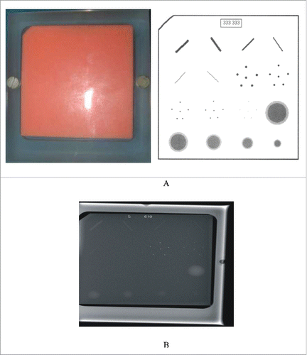
Mammographic phantoms are extensively used in X-ray imaging system for simulating multiple lesions in routine quality-assurance. A well-designed phantom could simulate successfully real lesions and thereby support effective clinical diagnosis during mammography. presents the breast phantom that was used herein. (A) The American College of Radiology (ACR) accreditation mammographic phantom, model 156, had an acrylic (PMMA) block that contained replaceable wax, which approximated a 4.0–4.5 cm-thick compressed breast. The wax contained target structures, comprising (1) 6 fibrils with diameters of 1.56, 1.12, 0.89, 0.75, 0.54 and 0.40 mm; (2) 5 groups of micro-calcifications, each with 6 specks that had diameters of 0.54, 0.40, 0.32, 0.24 and 0.16 mm, and (3) 5 nodules of decreasing diameters and thicknesses of 2.00, 1.00, 0.75, 0.50 and 0.25 mm, all arranged in a 4×4 matrix; and (B) the optimal X-ray image of the phantom. Specifically, the fibrils, micro-calcifications clusters and nodules simulated fibrotic changes, ductal carcinomas in situ (DCIS) (breast cancer), and tumor tissue, respectively, in a real mammographic examination. The contrast and size of the targets in each class of structures declined progressively, and targets were arranged from most visible to least visible.Citation21,22 The grade of each exposed mammographic phantom image was derived using the following equation given various factor set values of the factor of the X-ray machine during optimization,(7)
(7) where Nofi, Nono and NoCa represent the numbers of visible fibrils, nodules, and groups of micro-calcifications, respectively. The group of micro-calcifications was given a partial grade when only some points are visible.Citation18,23 For example, when Nofi, Nono, and NoCa, equal 5, 4 and 4 respectively, the grade was 70, which was the minimum acceptable grade in Taiwan. The highest possible grade of a mammographic image was 80. However, the high accompanying average glandular dose (AGD) constrained the settings of the factors of an X-ray machine, since the principle of “As Low As Reasonably Achievable” (ALARA) must be applied.Citation24
Figure 1. (A) The American College of Radiology (ACR) accreditation mammographic phantom, model 156, had an acrylic (PMMA) block that contained replaceable wax, which approximated a 4.0–4.5 cm-thick compressed breast. The wax contained target structures, comprising (1) 6 fibrils; (2) 5 groups of micro-calcifications and (3) 5 nodules of decreasing diameters and thicknesses, all arranged in a 4×4 matrix. (B) the optimal X-ray image of the phantom.

Orthogonal array of L18 for mammography
presents the 18 groups of settings of the factors for the digital X-ray machine according to Taguchi. The combinations of values of X-ray factors in each group were generated using the Taguchi method. These values clearly defined various setups. The four considered operating factors of the X-ray machine were target, kVp, mAs and FOV. Each operating factor had 2 or 3 levels. Accordingly, 54 (2×3×3×3 = 54) combinations were tested. The values of the factors were organized into only 18 groups, yielding the same degree of confidence as if they were considered separately.Citation14
Table 1. the corresponding 18 groups of orthogonal arrays for optimizing the mammographic phantom image according to the Taguchi. Each factor has 2 or 3 levels to compose the unique combination of X-ray operative setting in this work.
Results
Data analysis
shows the average grade, η (loss function), of the mammographic image and its accompanying AGD for the 18 groups of factor values. The breast phantom was rotated 90° clockwise before each trial to simulate different breast phantoms and to eliminate blind spots. Each mammographic image was labeled and saved. They placed in a random order for grading by all radiologists. The grade of the mammographic image from one radiologist was the mean of 4 independent trials for each group. Each image was, thus, read 4 times by each of the 5 radiologists for a total of 20 times. The AGD was recorded according to the system default feature that was a product from the air kerma (without backscatter), glandularity, and conversion coefficient altogether.Citation25 Additionally, the distance between the target and breast phantom was set to the machine's default value of 100 cm.
Table 2. Average grade, η (loss function), of the mammographic image and its accompanying AGD for the 18 groups of factor values. The average grade was obtained from 5 well-trained radiologists judged data in this work. The automatic glandular dose (AGD) was copied directly from the X-ray machine reading.
The average grade for group 16, 72.00, which was the highest, was converted into the highest η value of 37.13 dB. However, the definition of η involved not only the average grade of each group but also the consistency of the grades given by the professional staff in a real optimization process. Thus, a high loss function need not have a high average grade [Eq. Equation1(1)
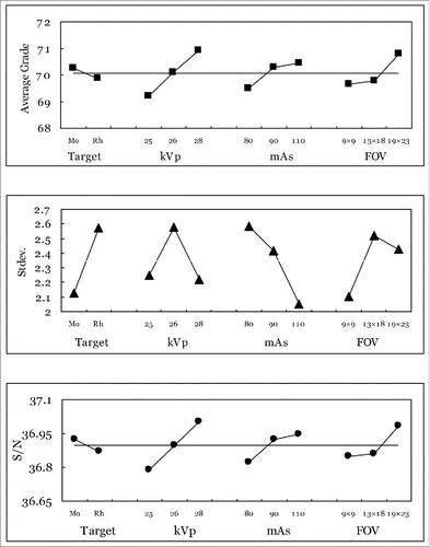
(1) ]. The η values of all groups were rearranged and averaged according to particular factors. For example, averages from groups 1–8 and from groups 9–18 revealed the contributions to image quality made by targets Mo and Rh, respectively. The averages for groups 1, 2, 3, 10, 11 and 12; groups 4, 5, 6, 13, 14 and 15, and groups 7, 8, 9, 16, 17 and 18, revealed the contributions to image quality made by factor B (kVp). The values of kVp were 26 (B1), 25 (B2) and 28 (B3) [].
plots the entire rearranged average grade, standard deviation of the grade and η values against the values of corresponding operating factors of the X-ray machine. It yielded the primary suggestion of each operating factor if considered the loss function, η, of factor individually, which was (A) target, Mo; (B) kVp, 28; (C) mAs, 110, and (D) FOV, 19×23 cm2, since the variable η was of the “higher-is-better” type according to the Taguchi calculations [Eq. Equation1(1)
(1) ]. However, ignoring the cross interactions among factors may mislead the optimization analysis, since over intensifying the loss function of individual factor might reduce the integrated performance in reality. Huda,Citation26 who used the same mammographic phantom to evaluate image quality, obtained a similar result: increasing either kVp or mAs of an X-ray machine effectively increased the visibility of the artificial lesions within the phantom. Notably, he checked image quality by assessing the impact of single factors individually, and so did not discuss interaction among factors.
Figure 2. the average grade of the phantom image from the 5 judged radiologists, standard deviation of the averaged grade and the derived S/N (η) versus various operating factors for X-ray mammography. The highest η values in various factors represent the optimized setting of that specific factor in this work.

Interactions among operating factors
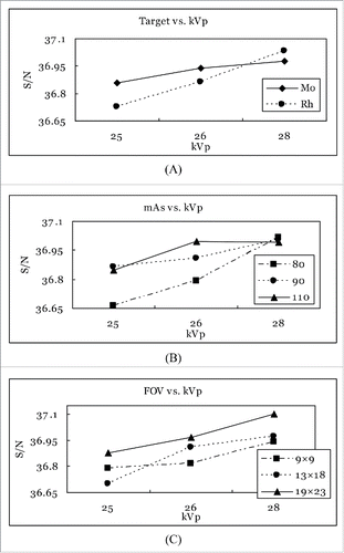
The Taguchi method not only identifies the dominant factor, but also effectively elucidates cross-interactions among factors. In the unique orthogonal arrangement of various values of factors, the frequencies of the levels of the factors are fixed across the 18 groups [], and the data thus obtained are rearranged to elucidate the cross-interactions among the factors. shows 3 cross-interactions between pairs of factors. Parts (A), (B) and (C) plots target vs. kVp, mAs vs. kVp, and FOV vs. kVp, respectively, for the X-ray mammography imaging system.
Figure 3. The cross interaction between factors for various cases. (A) Target vs. kVp, (B) mAs vs. kVp, and (C) FOV vs. kVp. The presence of 2 or 3 lines in the 3 parts reveals weak interactions, indicating that no factors interact with each other in all instances.

The presence of 2 or 3 lines in the 3 parts of reveals weak interactions, indicating that no factors interact with each other in all instances. Part (A) [] plots the S/N values of different targets as functions of kVp. Mo as an X-ray target outperforms Rh when the X-ray imaging is conducted at either 25 or 26 kVp but Rh yields better performance than Mo when the X-ray examination is conducted at 28 kVp. Apparently, the intersection between these 2 factors, kVp and target, complicates the optimization analysis, when any individual factor is adjusted. The first 2 columns in Tab. 1 (target and kVp) are crucial to the analysis of the L18(21×37) orthogonal array, because the associated data arrangement yielded a level V resolution (the highest resolution, with cross-interaction only between factors A and B), as Taguchi noted.
Discussion
Confirming mammographic performance
Confirming the Taguchi recommendation is crucial to any optimization analysis. In practice, any operating factor cannot be identified separately by the highest S/N value, but only by considering factors along with others in determining image quality. Accordingly, the optimal combination of operating factors is ultimately determined to be Rh target, 28 kVp, 80 mAs and 19×23 cm2 FOV. It happens to be the same setting as assigned in the group 16 of , although Mo target, 28 kVp, 110 mAs and 19×23 cm2 FOV yield the highest S/N value if considered separately the highest S/N value of factor []. Notably, that the optimal factor setting may still remain inside or outside the measurement subspace (the original 18 groups of settings according to the Taguchi's suggestion) depends on the optimization analysis and is unique in reality. However, any suggested setting has to be practically verified in order to confirm its suitability; otherwise, emphasizing only on the highest S/N value of any individual factor is inappropriate in the real optimization analysis. compares the AEC settings with the optimal settings of the X-ray machine for mammography. Two conventional settings that are used in most hospitals are also presented for reference. Conventional factor combination II in is exactly the same as that which yields the highest S/N value when factors are considered separately (outside the subspace of the original 18 groups listed in ) []. Additionally, the accompanying AGD was also reduced from 0.203 to 0.182 cGy [].
Table 3. Comparison between AEC setting and optimal recommendation of X-ray device for mammography. Two conventional settings that are used in most hospitals are also presented for reference. Conventional factor combination II is exactly the same as that which yields the highest S/N value when factors are considered separately [].
Dominant factors and ANOVA
Taguchi defines a factor as “dominant” only if it affects decisively to both the average and the standard deviation of average over repeated trials in the optimization process, and “minor” if it affects neither the average nor the standard deviation of average. A “random error reduction” factor is defined as one that affects mainly the standard deviation of average, and a “tune-up” factor is defined as one that affects mainly the average. Therefore, the factor target is “random error reduction” factors because it affects standard deviation of average more than it affects the average. The kVp, mAs and FOV are “dominant” factors because they affect both the average and the standard deviation of average. Additionally, too high the mAs setting may saturate the image contrast then suppress the average of judged grade, although the grade always goes with the high mAs. Thus, a compromised mAs setting is preferable from the practical consideration. The large FOV optimizes the image quality in either increasing the judged grade or minimizing the statistical uncertainty. The preferential interpretation of large FOV may be that it effectively suppresses the ambient heel effect of X-ray exposure and then intensifies the contrast of the phantom image []. Notably, the confidence level of FOV is even higher than that of mAs, although none of them passes the 95% confidence level.
shows the ANOVA of the X-ray mammographic imaging system. Significance is indicated by a percentage of over 95%, so only the factor of kVp passes the F-test and is significant. The deviation of the grades provided by the 5 radiologists is the major sources of variation in SSTotal herein (83% of the variation in SSTotal, ). However, the grades given by 5 radiologists were averaged for each of the 18 groups and so the number of degree of freedom in random error is 72 [18× (5–1) =72]. The factor “target” contributes negligibly to the optimization process (0.6% of the variation in SSTotal, ), indicating that it is the least important in the optimization of X-ray mammographic imaging systems.
Table 4. ANOVA and F-test of factors for X-ray mammographic imaging system. The confidence level was treated as significant if the percentage over 95% and the contribution defines as the ratio of SS of every factor (SSfactor) to their summation (SSTotal).
Disclosure of potential conflicts of interest
No potential conflicts of interest were disclosed.
Funding
The authors would like to thank the Chung Shan Medical University Hospital under contract No. CSH-2011-C-013 and the Ministry of Science and Technology of the Republic of China, under contract No. MOST 103-2221-E-166-003-MY3 for financially supporting this research.
References
- DOH. The establishment of data bank for domestic medicine and its application. Technical Report DoH 82-HP-114-4M16. Department of Health, Taiwan, 1993
- Simonetti D. What's new in mammography. Eur J Radiol 1998; 27s:234-41; http://dx.doi.org/10.1016/S0720-048X(98)00069-2
- DOH. Statistic data for public hygiene in Taiwan. Technical Report DoH 02-PH-2-tab-21. Department of Health, Taiwan, 2002.
- Morimoto T. Effectiveness of mammographic screening for breast cancer in women aged over 50 years in Japan. Japan J Cancer Res 1997; 88:778-884; http://dx.doi.org/10.1111/j.1349-7006.1997.tb00450.x
- Qi J, Moses WW. Instrumentation optimization for positron emission mammography. Nuclear Instruments and Methods in Physics Research A 2004; 527:76-82; http://dx.doi.org/10.1016/j.nima.2004.03.066
- Chen CY, Liu KC, Chen HH, Pan LK. Optimizing the TLD-100 readout system for various radiotherapy beam doses using the Taguchi methodology. Appl Radi and Isot 2010; 68(3):481-8; http://dx.doi.org/10.1016/j.apradiso.2009.12.001
- Cheng TC, Chang CY, Yao KS, Hsieh YH, Hsieh LL, Wang PS. Optimization of preparation of the TiO2 Photocatalytic reactor using the Taguchi method. Adv Mat Res 2008; 47–50:351-4; http://dx.doi.org/10.4028/www.scientific.net/AMR.47-50.351
- Pan LF, Chang MH, Pan LK. Optimization of coronary angiography for spider view via Taguchi methodology. Chin J Radiologic Tech 2012; 34(4):270-7
- Pan LK, Chou DS, Chang BD. Optimization for solidification of low-level-radioactive resin using Taguchi analysis. Waste Management 2001; 21:767-72; PMID:11699633; http://dx.doi.org/10.1016/S0956-053X(01)00012-5
- Pan LK, Wang CC, Shih YC, Sher HF. Optimizing multiple qualities of Nd:YAG laser welding onto magnesium alloy via Grey relational analysis. Sci Technol Welding Joining 2005; 10(4):503-10; http://dx.doi.org/10.1179/174329305X46727
- Pan LK, Wang CC, Wei SL, Sher HF. Optimizing multiple quality characteristics via Taguchi method-based Grey analysis. J Materials Processing Technol 2007; 182:107-16; http://dx.doi.org/10.1016/j.jmatprotec.2006.07.015
- Aggarwal A, Singh H, Kumar P. Optimizing power consumption for CNC turned parts using response surface methodology and Taguchi's technique-A comparative analysis. J Materials Processing Technol 2008; 200(1–3):373-84; http://dx.doi.org/10.1016/j.jmatprotec.2007.09.041
- Yang W, Tarng Y. Optimization of the weld bead geometry in gas tungsten arc welding by the Taguchi method. Int J Adv Manuf Technol 1998; 14:549-54; http://dx.doi.org/10.1007/BF01301698
- Taguchi G. Introduction to quality engineering. Asian productivity organization, Tokyo, 1990
- Phadke MS. Quality engineering using robust design. Prentice Hall, Englewood Cliffs, New Jersey, 1989
- Microsoft Excel. http://www.microsoft.com/taiwan/office2010/excel/default.aspx, 2010.
- Fisher RA. Design and analysis of experiments. Olive and Boyd, London, 1925.
- Lomax RG. Statistical Concepts: A Second Course p. 10, ISBN 0-8058-5850-4, 2007
- Vedantham S, Suryanarayanan S, Karellas A. Physical characteristic of a full-field digital mammography system. Nucl Inst and Meth in Phy Res A 2004; 533:560-70; http://dx.doi.org/10.1016/j.nima.2004.05.128
- Jouan B. Digital mammography performed with computed radiography technology. Eur J Radiol 1999; 31:18-24; PMID:10477094; http://dx.doi.org/10.1016/S0720-048X(99)00065-0
- Dougherty G. Computerized evaluation of mammographic image quality using phantom images. Comput Med Imaging Graph 1998; 22:365-373; PMID:9890181; http://dx.doi.org/10.1016/S0895-6111(98)00043-3
- Gilbert FJ, Undrill PE, O'kane AD. A comparison of digital and screen-film mammography using quality control phantoms. Clin Radiol 2000; 55:782-90; PMID:11052880; http://dx.doi.org/10.1053/crad.2000.0521
- Hillman BJ, Alpert HR. Quality and variability in diagnostic radiology. J Am College Radiol 2004; 1:127-32; http://dx.doi.org/10.1016/j.jacr.2003.11.001
- Moeckli R. Assessment of the image contrast improvement and dose reduction in mammography with synchrotron radiation compared to standard units. Nuclear Instruments and Methods in Physics Research A 2001; 467:1349-52; http://dx.doi.org/10.1016/S0168-9002(01)00662-3
- Geeraert N, Klausz R, Muller S, Bloch I, Bosmans H. Evaluation of exposure in mammography: limitations of average glandular dose and proposal of a new quantity. Radiat Prot Dosimetry 2015; 165(1–4):342-5 1-4
- Huda W, Sajewicz AM, Ogden KM, Scalzetti EM, Dance DR. How good is the ACR accreditation phantom for assessing image quality in digital mammography. Acad Radiol 2002; 9(7):764-72; PMID:12139090; http://dx.doi.org/10.1016/S1076-6332(03)80345-8
