
ABSTRACT
Botulinum toxin type A can temporarily inhibit muscle contraction. Currently, physicians administer this toxin as a bio-drug in treatment of some muscle contraction disorders. TAT-BoNT/A(1–448) is a functional recombinant protein derived from botulinum toxin light chain. Unlike the full length botulinum toxin, TAT-BoNT/A(1–448) is a self-permeable molecule which can pass through bio-surfaces so can be used as a topical therapeutic agent without injection. To maintain the functionality of TAT-BoNT/A(1–448), it is necessary to restore its normal folding upon expression and purification. In this study, we have investigated and optimized expression conditions for this novel recombinant protein. Under denaturing condition (1 mM IPTG, at 37°C), the chimeric protein was produced as inclusion body and required to be purified using denaturing agents (e.g. urea). Yet, lower incubation temperature (18°C) and less IPTG concentration (0.5 mM) induce a protein under native condition. In such condition, about 60% of the chimeric protein was expressed in soluble form.
Introduction
The light chain of botulinum toxin type A (LC-BoNT/A) has a molecular weight of 50kDa and is a zinc (Zn2+)-dependent metalloprotease.Citation1-3 The toxin temporarily prevents muscle contraction due to inhibition of acetylcholine release.Citation4,5 In recent years, this toxin was applied successfully as an injectable biological drug for treatment of some muscle contraction disorders such as blepharospasm, migraine and reducing facial wrinkles.Citation6-11
Despite of medicinal advantages, injection of this drug may cause some undesirable side effects e.g., irritation, inflammation, pain and sometimes bleeding in the site of injection. Hence, it is necessary to find a less/noninvasive means to administer this biological drug. Use of cell penetrating peptides (CPPs) is the most favorable means of transmission of molecules into the cells because of their low toxicity.Citation12,13 CPPs are cationic peptides with less than 30 amino acids. These peptides are able to pass directly through the membrane of many different types of mammalian cells without causing any damage. They can carry various biological cargos (DNA, RNA, peptides and proteins) which are covalently or non-covalently bonded.Citation14 Upon uptake, the cargos maintain their properties. These abilities make CPPs promising candidates for drug delivery applications.Citation15
In our earlier work, we successfully produced TAT-BoNT/A(1–448) recombinant protein through covalent bonding of the light chain of BoNT/A (LC-BoNT/A) to TAT, a cell penetrating peptide.Citation16 TAT-BoNT/A(1–448) is a potent functional recombinant protein that not only does maintain the functionality of the botulinum toxin type A but also has the ability to directly penetrate into the living cells in vitro and in vivo (i.e. mouse skin). Moreover, LC-BoNT/A conjugate has increased potential for proteolytic activity compared to the full-length BoNT/A. Peptide-mediated delivery of BoNT/A is an easy and non-invasive way of administration that may be useful for therapeutic purposes.
In this preliminary study we aimed to optimize expression and purification of TAT-BoNT/A(1–448) under denaturing and native conditions.
Methods
Protein expression under denaturing condition
The recombinant genomic construct containing nucleic acid sequence of TAT-BoNT/A(1–448) was synthesized before.Citation16,17 Briefly, a genomic construct was designed containing tandem nucleic acid sequences of TAT peptide (amino acids 47–57), BoNT/A light chain (amino acids 1–448) and 6 histidine residues at the N-terminus. This construct was based on pET28a expression vector containing T7 promoter. Recombinant vector was then transformed to an expression host, E. coli BL21(DE3). Transformed recombinant bacteria were cultured on LB agar medium containing 20µg/ml kanamycin. Bacterial colonies carrying plasmids containing TAT-BoNT/A(1–448) were cultured in 50ml LB broth plus 20µg/ml kanamycin for 24 h at 37°C. When the culture reached OD600nm of 0.6–0.9, protein production was induced by adding 1mM IPTG at 37°C for 24 h with 150 rpm constant shaking. Bacterial cells were separated from culture supernatant with 5 min centrifugation (5,000 rpm). The cells were lysed for 40 min with lysis buffer (5 mM imidazole, 500 mM NaCl, 10 mM Tris–HCl) containing a denaturing agent (8M urea) and were sonicated on ice (30 cycles/45 sec intervals). After centrifugation (14,000 rpm, 20 min), the supernatant containing soluble proteins was separated from the sediment containing insoluble proteins. Fractions were analyzed on 12% SDS-PAGE. The band density of the proteins related to each bacterial clone were assessed and compared with Quantity One software, ver.. 4.6.5 (Bio-Rad, USA). The bacterial clones possessing high level protein expression were stocked in −80°C until used.
Expression optimization
Several cultures of bacteria containing the recombinant sequence were induced by different IPTG concentrations (0.3, 0.5, 0.8 and 1mM) and were incubated at 37°C to compare the effect of IPTG concentration on protein expression. Bacterial cultures were also incubated at 30°C to compare the effect of temperature on protein expression. Protein expression in each bacterial culture was analyzed by 12% SDS-PAGE.
Protein expression under native condition
In this study, we tried to increase the solubility of recombinant protein by lowering IPTG concentration and reducing the incubation temperature.Citation18 For this purpose, we used 2 different concentrations, 0.3 and 0.5mM, as low IPTG concentrations (data are not shown). Since the protein expression level was very low upon induction with 0.3mM IPTG, we decided to use 0.5mM IPTG for protein expression under native condition. Also, we tested 3 different low temperatures, 25, 18 and 15°C (data are not shown) to check how lower temperatures (compared to 30 and 37°C) may affect protein expression under denaturing condition. Protein expression level at 25 and 18°C was approximately the same, but at 18°C the amount of protein in soluble phase was more than that of 25°C. The protein expression level at 15°C was very low. Hence, we decided to incubate the bacterial culture at 18°C.
Finally, the same protein expression was carried out using 0.5mM IPTG and incubation at 18°C under denaturing condition.
Also, 0.25µM ZnCl2 was added to the culture to maintain the three-dimensional structure of the protein.Citation19 According to some studies, ZnCl2 can help the protein maintain its natural folding and the tertiary structure.Citation20,21
After centrifugation, the bacterial sediment was sonicated and lysed with a lysis buffer lacking urea as a denaturing agent. Fractions obtained from the re-centrifugation containing soluble and insoluble proteins, were separated on a 12% SDS-PAGE.
Protein purification under denaturing condition
The bacterial sediments obtained from 1lit bacterial culture (induced with 1mM IPTG, at 37°C) were collected. The bacterial cells were sonicated and lysed with lysis buffer containing 8M urea and subjected to high speed centrifugation. The resulted supernatant was applied on Ni2+-affinity chromatography column (Ni-NTA, Qiagen, USA)Citation19 and the initial flow were collected. The column was washed twice with 20mM Tris-HCl buffer containing 20 and 100mM imidazole, respectively. 8M urea was added to all washing buffer to avoid protein sedimentation. Subsequently, elution buffer (250mM imidazole), was run through the column. Output flow of the column was collected after each washing step and was analyzed on 12% SDS-PAGE.
Protein purification under native condition
In order to obtain the optimal yield of pure recombinant protein in native structure, we purified the recombinant protein under native condition. For this purpose, the solution containing protein was applied on Ni2+-affinity chromatography column. Washing buffers lacked urea as a denaturing agent which allows the recombinant protein to maintain its native folding. Fractions resulted from each washing step were separated on 12% SDS-PAGE.
Results and discussion
Protein expression under denaturing condition
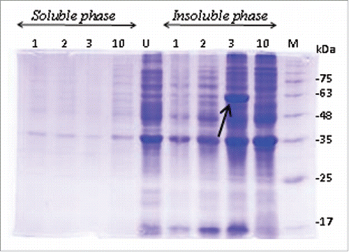
Two clones, among the pool of bacterial clones carrying the recombinant construct showed appropriate protein expression level. The SDS-PAGE pattern of induced bacteria () showed that the recombinant TAT-BoNT/A was produced as inclusion body (insoluble form).
Figure 1. Protein expression evaluation in E. coli Bl21 (DE3). Only one clone (shown with arrow) was able to efficiently express recombinant protein (54kDa). Soluble phase: Supernatant containing soluble proteins; Insoluble phase: Pellets of lysed cells containing insoluble proteins; M: Protein size marker; U: Cell lysate before induction of the expression; 12% SDS-PAGE stained with coomassie brilliant blue G250.
The optimum level of TAT-BoNT/A expression was achieved at 37°C with 1mM IPTG concentration ().
Figure 2. Expression optimization of TAT-BoNT/A recombinant protein under denaturing condition. Protein expression with different concentrations of IPTG was assessed once at (A). 37°C and once at (B). 30°C incubation temperature. Recombinant protein (54kDa band, shown with arrows) had the highest expression level in 1mM IPTG concentration at 37°C. Soluble phase: supernatant containing soluble proteins; Insoluble phase: pellet of cells lysed with 8M urea containing insoluble proteins. M: Protein size marker; U: Cell lysate before induction of the expression; 12% SDS-PAGE stained with coomassie brilliant blue G250.
Protein expression under native condition
Upon reducing IPTG concentration (0.5 mM) and lowering the incubation temperature down to 18°C, the bacterial host, E. coli BL21 could produce more TAT-BoNT/A recombinant protein in soluble form (∼60%) compared to the denaturing conditions ().
Figure 3. Expression of TAT-BoNT/A recombinant protein under native condition. Protein expression was induced in bacterial cells containing recombinant plasmid with 5 mM IPTG. After culture incubation at 18°C, about 60% of recombinant protein was produced in soluble form. Soluble phase: Supernatant containing soluble proteins; Insoluble phase: Pellets of cells lysed with 8M urea containing insoluble proteins. M: Protein size markers; U: Cell lysate before induction of the expression; Protein bands (54kDa) shown with arrows. 12% SDS-PAGE stained with coomassie brilliant blue G250.
One ideal condition in expression of recombinant proteins is to express the protein at high level. Even though the level of protein expression under denaturing condition is much higher than that of native condition, expression under native condition is more favorable for production of functional proteins. Forcing the bacterial host to produce the protein of interest causes aggregation of proteins and formation of inclusion bodies with abnormal folding. Based on a common procedure, denaturing agent (urea) was used to reach maximum protein purification. If the expressed recombinant protein is functional (e.g., enzymes), protein folding should return to normal form to regain its functionality. For this purpose, denaturing agents should be removed from the protein solution. Not all protein molecules can retain the complete 3D native structure. Therefore, during each folding/refolding step a small amount of the produced protein will be lost. It is important for the protein to avoid folding/refolding steps to maintain maximum functionality. The favorable solution is to prevent protein aggregation inside the bacterial host. A solution for this problem, which has been used in many similar studies, is to somehow reduce the speed of protein production.Citation18,19 Under such condition, the host bacteria takes ample time to fold the foreign protein correctly. However, in our study TAT-BoNT/A was highly expressed in soluble form (0.5 mg/ml) and was a major component of the total soluble proteins. Also there was no need of further dialysis after purification. This corresponds with the results of similar studiesCitation22 which produced CPP- BoNT/A fusion protein in soluble form.Citation18,19
In a study performed by Jensen and colleagues, transitional domain sequence of botulinum toxin (amino acids 449–552) was added to that of light chain of botulinum toxin in order to simulate the 3D structure of natural protein. However, no increase was observed in the production of recombinant protein with natural folding. They showed that reduction of incubation temperature is more effective in increasing the solubility of recombinant proteins. Therefore, complete structure of a protein does not necessarily lead to protein production with natural folding. It is likely that with a larger sequence of the recombinant protein, the host bacteria lose the ability of natural folding of proteins.Citation19 In the present study, we showed that reducing the incubation temperature (18°C) makes greater proportion of the proteins to produce in solution form.
In this study, we only expressed the catalytic domain of botulinum toxin type A. Accompany of binding and translocating domains helps light chain domain to maintain its strength and sustainability in natural systems. Yet, according to some studies, immune response to botulinum toxin light chain alone is much less than when it is in association with 2 other domains.Citation19,23 Therefore, the possibility of allergic and inflammatory reactions against the recombinant protein will be very low. In addition, cell-penetrating peptides are one of the non-invasive methods for transferring biological molecules into a variety of cell lines. Studies have shown that these peptides have the least toxicity and minimum damage to their target cells in vivo and in vitro.Citation18
Protein purification
Each fraction of protein purification with resin column containing nickel was run on 12% SDS-PAGE separately (). We could highly purify (>98%) the protein under native condition. While, protein purification in the presence of denaturing agents was not as successful as native condition (data not shown). Also, there was no need for further dialysis. This, on one hand, reduces the purification steps and saves time and on the other hand, eliminates the need for restoring the normal protein folding. To confirm the presence of TAT-BoNT/A recombinant protein, western blotting with HRP-conjugated anti-His-tag rabbit anti-mouse monoclonal IgG (Sigma Aldrich, USA) was performed. We mentioned the result of protein gel blot analysis elsewhere in our previous study.Citation16
Figure 4. Purification of TAT-BoNT/A recombinant protein under native condition using Ni-NTA chromatography column. Column 1: Sample before purification. Column 2: The initial flow. Column 3: Washing sample with a buffer containing 20mM imidazole. Column 4: Washing sample with a buffer containing 100mM imidazole. Columns 5 and 6: Elusion in a buffer containing 250mM imidazole. M: Protein size marker. TAT-BoNT/A protein band shown with arrow. 12% SDS-PAGE stained with Coomassie brilliant blue G250.
In addition, protein purification by affinity chromatography column containing nickel bound resin is very convenient and effective way to purify the recombinant protein with a histidine tail.
Conclusions
Production of recombinant protein requires conditions of high level expression along with inclusion body formation. On the other hand, to preserve the biological activity we should avoid folding/refolding methods during the purification process.
Upon decreasing the concentration of IPTG and incubation temperature about 60% of total volume of TAT-BoNT/A recombinant protein was produced in soluble form. The recombinant protein was purified at a favorable level by nickel affinity chromatography column under native condition.
Disclosure of potential conflicts of interest
No potential conflicts of interest were disclosed.
References
- Johnson EA, Bradshaw M. Clostridium Botulinum and Its Neurotoxins: A metabolic and cellular perspective. Toxicon 2001; 39:1703-22; PMID:11595633; http://dx.doi.org/10.1016/S0041-0101(01)00157-X
- Lacy DB, Tepp W, Cohen AC, Dasgupta BR, Stevens RC. Crystal structure of botulinum neurotoxin type a and implications for toxicity. Nat Struct Biol 1998; 5:898-902; PMID:9783750; http://dx.doi.org/10.1038/2338
- Swaminathan S, Eswaramoorthy S. Structural Analysis of the Catalytic and binding sites of clostridium botulinum Neurotoxin B. Nat Struct Biol 2000; 7:693-9; PMID:10932256; http://dx.doi.org/10.1038/78005
- Schiavo G, Rossetto O, Santucci A, Dasgupta BR, Montecucco C. Botulinum neurotoxins are zinc proteins. J Biol Chem 1992; 267:23479-83; PMID:1429690
- Gul N, Smith LA, Ahmed SA. Light chain separated from the rest of the type a botulinum neurotoxin molecule is the most catalytically active form. PLoS One 2010; 5:e12872; PMID:20877571; http://dx.doi.org/10.1371/journal.pone.0012872
- Mauskop A. The use of Botulinum Toxin in the treatment of headaches. Curr Pain Headache Rep 2002; 6:320-3; PMID:12095468; http://dx.doi.org/10.1007/s11916-002-0054-1
- Mauskop A. Botulinum toxin in headache treatment: The end of the road? Cephalalgia 2007; 27:468; PMID:17448184; http://dx.doi.org/10.1111/j.1468-2982.2007.01290_1.x
- Verheyden J, Blitzer A. Other noncosmetic uses of Botox. Dis Mon 2002; 48:357-66; PMID:12195265; http://dx.doi.org/10.1053/mda.2001.25136
- Dhaked RK, Singh MK, Singh P, Gupta P. Botulinum toxin: Bioweapon & magic drug. Indian J Med Res 2010; 132:489-503; PMID:21149997
- Turton K, Chaddock JA, Acharya KR. Botulinum and tetanus neurotoxins: structure, function and therapeutic utility. Trends Biochem Sci 2002; 27:552-8; PMID:12417130; http://dx.doi.org/10.1016/S0968-0004(02)02177-1
- Masuyer G, Chaddock JA, Foster KA, Acharya KR. Engineered botulinum neurotoxins as new therapeutics. Annu Rev Pharmacol Toxicol 2014; 54:27-51; PMID:24016211; http://dx.doi.org/10.1146/annurev-pharmtox-011613-135935
- Contractor J A, Wadia F, Patel A, Bamania M, Muppidi V. Papillary thyroid carcinoma in a 4-Year-Old Boy. Indian J Otolaryngol Head Neck Surg 2002; 54:148-9; PMID:23119879
- Henriques ST, Melo MN, Castanho MA. Cell-penetrating peptides and antimicrobial peptides: How different are they? Biochem J 2006; 399:1-7; PMID:16956326; http://dx.doi.org/10.1042/BJ20061100
- Mueller J, Kretzschmar I, Volkmer R, Boisguerin P. Comparison of cellular uptake using 22 Cpps in 4 different cell lines. Bioconjug Chem 2008; 19:2363-74; PMID:19053306; http://dx.doi.org/10.1021/bc800194e
- Schmidt N, Mishra A, Lai GH, Wong GC. Arginine-rich cell-penetrating peptides. FEBS Lett 2010; 584:1806-13; PMID:19925791; http://dx.doi.org/10.1016/j.febslet.2009.11.046
- Saffarian P, Peerayeh SN, Amani J, Ebrahimi F, Sedighian H, Halabian R, Fooladi AA. Tat-Bont/a(1-448), a novel fusion protein as a therapeutic agent: analysis of transcutaneous delivery and enzyme activity. Appl Microbiol Biotechnol 2016; 100:2785-95; PMID:26711279; http://dx.doi.org/10.1007/s00253-015-7240-7
- Amani J, Saffarian P, Najar-Peerayeh S, Imani-Fooladi AA. Designing and analyzing the structure of Tat-Bont/a(1–448) fusion protein: an in silico approach. 2014; 3:13
- Kim DW, Kim SY, An JJ, Lee SH, Jang SH, Won MH, Kang TC, Chung KH, Jung HH, Cho SW, et al. Expression, purification and transduction of Pep-1-Botulinum Neurotoxin Type a (Pep-1-Bont/a) into Skin. J Biochem Mol Biol 2006; 39:642-7; PMID:17002886; http://dx.doi.org/10.5483/BMBRep.2006.39.5.642
- Jensen MJ, Smith TJ, Ahmed SA, Smith LA. Expression, purification, and efficacy of the type a botulinum neurotoxin catalytic domain fused to two translocation domain variants. Toxicon 2003; 41:691-701; PMID:12727273; http://dx.doi.org/10.1016/S0041-0101(03)00042-4
- Lacy DB, Stevens RC. Sequence homology and structural analysis of the clostridial neurotoxins. J Mol Biol 1999; 291:1091-104; PMID:10518945; http://dx.doi.org/10.1006/jmbi.1999.2945
- Farasat A, Ebrahimi F, Mousavy J, Salehi M B, Rostamian M. Characterization of antibody titer and immunogenic feature of light chain of botulinum neurotoxin Type A. 2013; 4:6.
- Rawat R, Ashraf Ahmed S, Swaminathan S. High level expression of the light chain of botulinum neurotoxin serotype C1 and an efficient Hplc assay to monitor its proteolytic activity. Protein Expr Purif 2008; 60:165-9; PMID:18482846; http://dx.doi.org/10.1016/j.pep.2008.03.010
- Chaddock JA, Herbert MH, Ling RJ, Alexander FC, Fooks SJ, Revell DF, Quinn CP, Shone CC, Foster KA. Expression and purification of catalytically active, non-toxic endopeptidase derivatives of clostridium botulinum toxin Type A. Protein Expr Purif 2002; 25:219-28; PMID:12135553; http://dx.doi.org/10.1016/S1046-5928(02)00002-5