
ABSTRACT
Fe75Zr25/Cu64Zr36 amorphous multilayers were prepared by magnetron sputtering. Atom probe tomography was employed to analyze the atomic inter-diffusion at the interface of the multilayers before and after annealing (573 K, 60 min). An unexpected enhanced inter-diffusion of the immiscible elements Fe and Cu was detected at the interface of the multilayers. As the inter-diffusion in amorphous multilayers is much faster than that in the crystalline counterparts, this process may open a way to manipulate or create amorphous multilayers with new properties. This idea agrees with the observation of the variation of magnetic properties of Fe75Zr25/Cu64Zr36 amorphous multilayers.
GRAPHICAL ABSTRACT
IMPACT STATEMENT This paper reveals the enhanced atomic inter-diffusion at the interface of amorphous materials, and may open a way to manipulate or create amorphous multilayers with new properties.
In recent years, glasses with tunable nanostructures, called nano-glasses (NGs), have attracted a lot of attention [Citation1–3]. NGs consist of the following two structural components: nanometer-sized amorphous grains and interfacial regions between these grains [Citation2–4]. Since the interfacial regions have an atomic and electronic structure deviating from those of the corresponding melt-quenched glasses, the properties of NGs differ from the properties of the melt-quenched glasses with the same chemical compositions. NGs exhibit remarkable interface-related properties such as the mechanical [Citation5], biological [Citation6], magnetic [Citation7], and catalytic properties [Citation8].
The interfaces in NGs are formed by the atomic inter-diffusion of adjacent amorphous grains. However, the inter-diffusion between the amorphous grains has not yet been studied. In comparison to nano-crystalline materials, the rate of inter-diffusion in NGs is likely to be enhanced because the atoms of the amorphous grains are not forced to remain on lattice sites. In order to study the inter-diffusion in NGs, we propose to use a model system consisting of amorphous multilayers with planar amorphous/amorphous interfaces.
In order to study the variation of the concentration profiles of the planar amorphous/amorphous interfaces, atom probe tomography (APT) [Citation9,Citation10] was applied. APT is based on the controlled field desorption. Atoms are removed one-by-one from the apex of a tip-shaped specimen. Their positions on detector and flight time are recorded. From these data, the chemical nature and the original position of the incoming atom can be deduced. As a result, a 3D reconstruction of the atoms can be achieved with a sub-nanometer resolution.
In this study, we prepared amorphous multilayers by magnetron sputtering with the stoichiometric composition of Fe75Zr25/Cu64Zr36. The reasons for selecting the FeZr and CuZr systems were: (1) Both systems have strong glass forming ability [Citation11,Citation12] and (2) Different types of atomic inter-diffusion at the amorphous/amorphous interface could be investigated in one experiment. The inter-diffusion involves the concentration-driven diffusion of the element Zr as well as of the elements Fe and Cu that are immiscible in the crystalline state [Citation13]. APT was employed to analyze the atomic inter-diffusion at the interface of the FeZr/CuZr multilayers before and after annealing at 573 K for 60 min. After annealing, a pronounced diffusion of Zr between the FeZr and CuZr layers was observed. Moreover, evidence for the enhanced inter-diffusion of Fe and Cu was obtained. As the atomic inter-diffusion at the interface would produce a new layer with different chemical composition, it may open a way to create or manipulate the properties of amorphous multilayers.
Ten layers of Fe75Zr25 (∼17 nm)/Cu64Zr36 (∼38 nm) were deposited by using a JGP650 dual chambers magnetron sputtering system. The detail preparation information was described in Supporting Information. The structure of the multilayers was investigated using XRD (Brucker D8 Advance) with Cu Kα radiation and TEM (Titan G2 60-300) with energy dispersive X-ray spectrometry (EDX). Samples for TEM and APT were prepared using the focused ions beam (FIB) lift-out techniques [Citation14] in a Zeiss Auriga SMT SEM equipped with an Orsay Physics Cobra Z-05 FIB unit with a Ga+ ion source.
APT analysis was performed using a Local Electrode Atom Probe (LEAP 4000X Si) operated in the laser-pulsing mode. During data collection, specimens were cooled to 25 K at a pressure of 6.3 × 10⁻¹¹ Torr. The pulse laser energy was 40 pJ (wavelength 355 nm) with a repetition rate of 200 kHz and a data collection rate of 0.005–0.007 ions per pulse. All concentrations are given in atomic percent. Reconstruction and visualization of the APT data was performed using the Imago Visualization and Analysis Software (IVAS 3.6.8). All APT reconstructions were made by using the shank angle measured from the SEM image of each corresponding tip sample and the dimensions of multilayer structure in the reconstructed volume were calibrated by using the multilayer spacing measured from the SEM image (by adjusting the initial radius of the tip).
A typical XRD pattern of ‘as-prepared’ Fe75Zr25/Cu64Zr36 multilayers is presented in Figure (a). The broad peak corresponds to an amorphous structure. The peak position is around 42°, while for pure amorphous Cu64Zr36 and Fe75Zr25 are 41° and 43°, respectively. This result illustrates that the multilayer thin films are a mixture of amorphous Fe75Zr25 and Cu64Zr34.
Figure 1 (a) A typical XRD pattern of the Fe75Zr25/Cu64Zr36 amorphous multilayers. (b) An HRTEM image of the multilayer sample. The different composition layers were evidenced by their different contrasts. The inset of (b) is the SAED pattern.
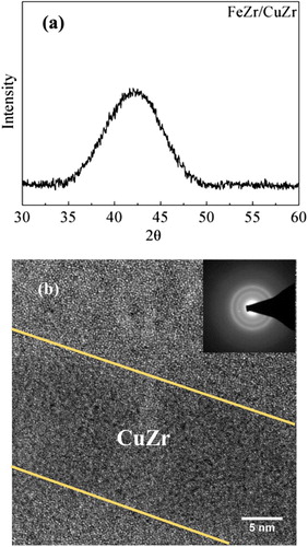
A cross-section of the Fe75Zr25/Cu64Zr36 multilayers sample was prepared for TEM analysis by FIB milling. The initial characterization of the multilayer structure was performed using an aberration-corrected FEI Titan 80-300 operated at 300 kV in the scanning TEM mode (STEM). Figure (b) is an HRTEM image of the multilayer sample. The maze-like pattern is a well-known feature of amorphous materials. The interfaces between two layers were evidenced by their different contrasts. The selected area electron diffraction (SAED) pattern in the inset of Figure (b) displays two halo rings, which may be due to the different compositions in the multilayers [Citation15]. The STEM-EDX mapping (Figure S1) reveals the elemental distribution in the multilayers. In order to find out whether there is atomic inter-diffusion occurring at the interface, APT was performed.
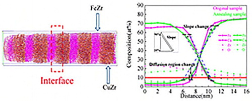
A reconstructed atom map of a FeZr/CuZr multilayers sample in the as-prepared state is presented in Figure (a). A 1D concentration profile across the thickness of the multilayers, as displayed in Figure (b), reveals a significant asymmetry of the interface at both sides of the CuZr layers. The intermixed zone at the interface looks like narrower when CuZr grows on the top of FeZr in comparison to the reversed sequence. Some interfaces with similar asymmetry revealed by APT measurements were also reported in the literature [Citation16–19]. The asymmetric interface region between CuZr and FeZr is likely due to evaporation field difference between Fe and Cu in the two different layers [Citation16,Citation17]. In order to minimize any artifacts that may be introduced when significant roughness exists at the interface, the following procedure was used. The inter-diffusion at the interface was analyzed by positioning 103 analysis cylinders (5 nm diameter) at the interface of CuZr that was sitting on top of FeZr layers. The data obtained from this analysis are plotted in Figure . The inter-diffusion length was deduced by selecting the positions at which the local concentrations were 10 at% Cu to 10 at% Fe [Citation18,Citation20,Citation21]. This approach yielded an interface width of 2.3 ± 0.2 nm. This width is one order of magnitude larger than the width deduced from the data reported for inter-diffusion in crystalline Cu/Fe-based alloy multilayers at room temperature (RT) [Citation16, 21,22]. This result suggests an enhanced inter-diffusion in the amorphous state in comparison to the corresponding crystalline state.
Figure 2 (a) The APT reconstruction of the Fe75Zr25/Cu64Zr36 amorphous multilayers. (b) The concentration profiles of the multilayers.
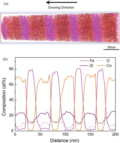
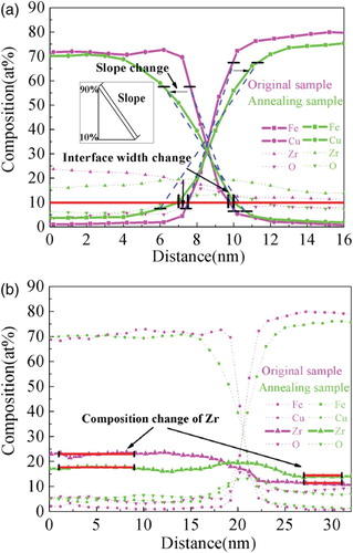
Figure 3 The elements concentration profiles of the Fe75Zr25/Cu64Zr36 amorphous multilayers before and after annealing. The statistical data are acquired from 103 concentration profiles obtained by analysis cylinders with 5 nm in diameter at the CuZr/FeZr interfaces. (a) The inter-diffusion analysis of immiscible elements Fe and Cu. The inter-diffusion length is defined as the distance from concentration 10 at% Cu to 10 at% Fe. The slope was calculated from the concentration profiles of both elements, taking into account the variation between ∼10 and 90 at% of the respective concentration amplitude. The interface width expanded from 2.3 ± 0.2 to 2.6 ± 0.3 nm. The slope of Cu concentration profile was found to decrease from 21.7 ± 1.7 (initially) to 15.4 ± 3.5 (annealed), and the slope of Fe decreased from 23.7 ± 2.0 (initially) to 11.5 ± 2.6 (annealed). (b) The inter-diffusion analysis of Zr. Zr diffused from CuZr (Zr-rich) to FeZr (Zr-poor) and finally the concentration was almost the same everywhere.
In order to further investigate the inter-diffusion at the interfaces, several samples were annealed in a high vacuum system (5 × 10⁻⁵ Pa) for 60 min at 573 K, which is far below Tx of Cu64Zr36 (∼780 K) [Citation23] and Fe75Zr25 (∼880 K) [Citation24] amorphous alloys. The samples were then analyzed by APT.
The concentration profiles of Fe and Cu obtained from these measurements are shown in Figure (a). As may be seen, the interface width expanded from 2.3 ± 0.2 to 2.6 ± 0.3 nm. Considering the positive heat of mixing between Fe and Cu (+13 kJ/mol) [Citation13], the widening of Fe/Cu interface width by about 13% seems remarkable keeping in mind the low annealing temperature (573 K). Further evidence for the inter-diffusion between Fe and Cu is the change of the slope of the concentration profiles. The slope was calculated from the concentration profiles of both elements, taking into account the variation between ∼10 and 90 at% of the respective concentration amplitude. The slope of Cu concentration profile was found to decrease from 21.7 ± 1.7 (initially) to 15.4 ± 3.5 (after annealing), and the slope of Fe decreased from 23.7 ± 2.0 (initially) to 11.5 ± 2.6 (annealing). For Zr, it diffused from CuZr (Zr-rich) to FeZr (Zr-poor) and finally the concentration was almost the same everywhere as shown in Figure (b).
Several groups reported the inter-diffusion in crystalline multilayers consisting of immiscible elements. Ene et al. investigated the atomic inter-diffusion of NiFe/Cu thin films by APT and found the intermixing length at RT to be about 0.5 nm. Even after annealing at 773 K for 40 min, intermixing between Fe and Cu was not observed [Citation21]. Zhou et al. reported the intermixing length of CoFe/Cu multilayers to range from 0.4 to 0.6 nm by APT measurements at ambient temperatures [Citation19]. Kuncser et al. studied the interfacial atomic diffusion of Fe/Cu thin films via 57Fe conversion electron Mössbauer spectroscopy and demonstrated the effective thickness of diffusing Fe atoms is less than 0.35 nm [Citation22]. Our results show a remarkable enhanced interfacial atomic diffusion (2.3 ± 0.2 nm) at ambient temperatures in the amorphous Fe75Zr25/Cu64Zr36 multilayers, which is at least four times larger than the data reported in the literatures. In addition to the extra interfacial energy in amorphous multilayers, there may be two main reasons for the enhancement of the intermixing of the immiscible elements Fe/Cu in our experiments. One reason is the thermodynamic instability in the amorphous state that may enhance the inter-diffusion rate [Citation25]. The other reason is the excess volume existing in the interfaces enhancing the atomic mobility [Citation26]. The enhanced inter-diffusion rate seems consistent with the following published results. At 630 K, the inter-diffusivity of Ge/Si in the amorphous state was noted to be 3.86 × 10⁻²³ m2 s−1, which is almost 4 orders of magnitude larger than that in the crystalline state [Citation27]. The Co isotope diffusion in fcc Co is about 1.0 × 10⁻¹⁸ m2 s−1 at 1100 K, whereas the inter-diffusivity of Co isotopes in amorphous Co76.7Fe2Nb14.3B7 is 2.91 × 10⁻¹⁴ m2 s−1 if extrapolated to this temperature [Citation28].
So far, the atomic inter-diffusion of immiscible elements was only observed in specimens produced by ball-milling, sputtering, or severe plastic deformation. In our case, the enhanced atomic inter-diffusion of immiscible elements was found in vapor deposited amorphous multilayers providing a way to modify the properties of amorphous multilayers. In view of these results, it may be expected that the interfacial regions have a new atomic structure formed by the intermixing of immiscible elements. By controlling the thickness of single layer or the inter-diffusion length at different annealing temperatures, the volume fraction of the interfacial region could be tuned and hence the properties the multilayers could be modified. In fact, if the thickness of the individual layer would be less than the double inter-diffusion length, this approach would open the way to create a new material with unique properties.
In order to support this speculation, the following experiment was carried out. A series of multilayers, with the nominal structure of [a-Fe75Zr25 (10 nm)/a-Cu64Zr36 (10 nm)] × 12, [a-Fe75Zr25(10 nm)/Cu(10) nm] × 12, and a single layer reference sample of a-Fe75Zr25 film with a thickness of 240 nm, were fabricated using magnetron sputtering as described in the supplementary. The magnetic measurements were carried out with the three kinds of thin films before and after annealing at 573 K for 60 min. (The detail of the magnetic measurement is described in the supplementary.) The magnetic moment per Fe atom (In the later part, the ‘magnetic moment’ refer in particular to ‘magnetic moment per Fe atom’.) in those thin films was recorded and it is given in . It is well known that FeZr, CuZr, and Cu are ferromagnetic, paramagnetic, and antiferromagnetic materials, respectively. Here, the 10-nm non-ferromagnetic CuZr or Cu layers are thick enough to suppress any interlayer coupling between the magnetic FeZr layers [Citation29]. The value of the magnetic moment in the original FeZr single layer was 0.8 µB, which is similar to the literature value reported [Citation30]. The magnetic moments in FeZr/CuZr and FeZr/Cu are 0.75 and 0.60 µB, respectively, which is somewhat lower than the magnetic moment in FeZr single layer. This may be due to the effect of introducing non-ferromagnetic layers in magnetic layers [Citation31]. After annealing at 573 K for 60 min, the magnetic moment in all kinds of films increased. If the interface effect is ignored, the ratio of the increase of the magnetic moment in FeZr single layer should be similar to the one of the FeZr/CuZr and FeZr/Cu multilayers. This was indeed observed for the FeZr/Cu multilayers, because the atomic inter-diffusion between the amorphous FeZr and crystalline Cu is quite weak and cause almost no inter-diffusion effect. However, a significant increase of the magnetic moment was noticed in the FeZr/CuZr multilayers after annealing, which is much larger than that the ones noted in the FeZr single layer and FeZr/Cu multilayers. This result indicates a different increasing mechanism for the magnetic moment in FeZr/CuZr multilayers in comparison to FeZr single layer and FeZr/Cu multilayers. The increase of magnetic moment in FeZr single layer and FeZr/Cu multilayers is due to the relaxation in amorphous materials during heating below the Tg [Citation32–34]. Obviously, the dramatic increase of magnetic moment in FeZr/CuZr multilayers could not be explained by the same model. According to the former APT results, the likely reason seems to be the change of the interfacial atomic structure of the FeZr/CuZr multilayers during the annealing treatment. The interfacial regions formed due to the enhanced inter-diffusion ability in the amorphous state contain the immiscible elements Fe and Cu, which may result in higher magnetic moments [Citation35]. This result seems consistent with the idea of manipulating the properties of amorphous multilayers by tuning the interfacial region. The M-H curves of the three kinds of films are shown in Figure S2.
Table 1. The magnetic moment per Fe atom in the three kinds of thin films before and after annealing at 573 K for 60 min.
In conclusion, FeZr/CuZr amorphous multilayers have been prepared by magnetron sputtering. STEM-EDS maps suggest the formation of regions in which Fe and Cu atoms are mixing in the vicinity of the multilayer interfaces. APT concentration profiles confirm this interpretation and suggest a mixing zone width of 2.3 ± 0.2 nm. By studying FeZr/CuZr multilayer samples before and after annealing (573 K, 60 min), it is found that the inter-diffusion of Zr resulted in the same Zr concentration everywhere. The Fe/Cu interface width was found to increase from 2.3 ± 0.2 to 2.6 ± 0.3 nm and the slopes of Cu and Fe concentration profiles decreased for Cu from 21.7 ± 1.7 to 15.4 ± 3.5 and for Fe from 23.7 ± 2.0 to 11.5 ± 2.6. All of these results support the conclusion that enhanced inter-diffusion occurs at the amorphous/amorphous interface in comparison to inter-crystalline interfaces even if the inter-diffusing elements are immiscible. The enhanced inter-diffusion in the amorphous state is important for the understanding of the unique properties of NGs. Such inter-diffusion would form a new nanometer-sized layer with different chemical composition and modify the properties of amorphous multilayers.
In fact, if the original single layers are sufficiently thin (for example, in the case of the system studied here: less than 5 nm), the newly formed layer may take control of the properties of the amorphous multilayers. As the interface diffusion of amorphous multilayers is much faster than in its crystalline counterparts at relatively low temperature, it may open the way to create a new type of amorphous materials with unique properties.
Supplementary_Material
Download MS Word (582.5 KB)Acknowledgements
The authors thank Dr D. Wang and Dr X. K. Mu for the TEM measurement, and also thank Dr M. Ghafari for the useful discussion of the magnetic properties. TF acknowledges the support of the innovation project, Qing Lan project, and the specially appointed professor project of Jiangsu province.
Disclosure statement
No potential conflict of interest was reported by the authors.
Additional information
Funding
Related Research Data
References
- Guo Y, Morozov A, Schneider D, et al. Ultrastable nanostructured polymer glasses. Nat Mater. 2012;11:337–343. doi: 10.1038/nmat3234
- Gleiter H, Schimmel T, Hahn H. Nanostructured solids – from nano-glasses to quantum transistors. Nano Today. 2014;9:17–68. doi: 10.1016/j.nantod.2014.02.008
- Gleiter H. Nanoglasses: a new kind of noncrystalline material and the way to an age of new technologies. Small. 2016;12:2225–2233. doi: 10.1002/smll.201500899
- Fang JX, Vainio U, Puff W, et al. Atomic structure and structural stability of S.75Fe25 nanoglasses. Nano Lett. 2012;12:458–463. doi: 10.1021/nl2038216
- Wang XL, Jiang F, Hahn H, et al. Plasticity of a scandium-based nanoglass. Scripta Mater. 2015;98:40–43. doi: 10.1016/j.scriptamat.2014.11.010
- Chen N, Shi X, Witte R, et al. A novel Ti-based nanoglass composite with submicron–nanometer-sized hierarchical structures to modulate osteoblast behaviors. J Mater Chem B. 2013;1:2568–2574. doi: 10.1039/c3tb20153h
- Witte R, Feng T, Fang JX, Fischer A, Ghafari M, Kruk R, et al. Evidence for enhanced ferromagnetism in an iron-based nanoglass. Appl Phys Lett. 2013;103:073106. doi: 10.1063/1.4818493
- Chen N, Frank R, Asao N, et al. Formation and properties of Au-based nanograined metallic glasses. Acta Mater. 2011;59:6433–6440. doi: 10.1016/j.actamat.2011.07.007
- Miller MK, Kenik EA. Atom probe tomography: a technique for nanoscale characterization. Microsc Microanal. 2004;10:336–341. doi: 10.1017/S1431927604040577
- Seidman DN. Three-dimensional atom-probe tomography: advances and applications. Annu Rev Mater Sci. 2007;37:127–158. doi: 10.1146/annurev.matsci.37.052506.084200
- Yu CY, Liu XJ, Lu J, et al. First-principles prediction and experimental verification of glass-forming ability in Zr-Cu binary metallic glasses. Sci Rep. 2013;3:2124. doi: 10.1038/srep02124
- Arias D, Abriata JP. The Fe−Zr (Iron-Zirconium) system. J Phase Equilib. 1988;9:597–604.
- Ma E. Alloys created between immiscible elements. Prog Mater Sci. 2005;50:413–509. doi: 10.1016/j.pmatsci.2004.07.001
- Miller MK, Russell KF, Thompson GB. Strategies for fabrication atom probe specimens with a dual beam FIB. Ultramicroscopy. 2005;102:287–298. doi: 10.1016/j.ultramic.2004.10.011
- Chen N, Wang D, Feng T, Kruk R, Yao KF, Louzguine-Luzgin DV, et al. A nanoglass alloying immiscible Fe and Cu at the nanoscale. Nanoscale. 2015;7:6607–6611. doi: 10.1039/C5NR01406A
- Larson DJ, Cerezo A, Clifton PH, et al. Atom probe analysis of roughness and chemical intermixing in CoFe/Cu films (invited). J Appl Phys. 2001;89:7517–7521. doi: 10.1063/1.1354593
- Zhou XW, Wadley HNG. Atomistic simulations of the vapor deposition of Ni/Cu/Ni multilayers: The effects of adatom incident energy. J Appl Phys. 1998;84:2301–2315. doi: 10.1063/1.368297
- Larson DJ, Prosa TJ, Geiser BP, et al. Effect of analysis direction on the measurement of interfacial mixing in thin metal layers with atom probe tomography. Ultramicroscopy. 2011;111:506–511. doi: 10.1016/j.ultramic.2010.12.010
- Zhou XW, Wadley HNG, Johnson RA, et al. Atomic scale structure of sputtered metal multilayers. Acta Mater. 2001;49:4005–4015. doi: 10.1016/S1359-6454(01)00287-7
- Stender P, Ene CB, Galinski H, et al. Interface width of immiscible layered elements. Int J Mater Res. 2008;99:480–486. doi: 10.3139/146.101661
- Ene CB, Schmitz G, Kirchheim R, et al. Thermal reaction and stability of NiFe/Cu thin films investigated by atom probe tomography. Surf Interface Anal. 2007;39:227–231. doi: 10.1002/sia.2519
- Kuncser V, Keune W, Hörsten UV, et al. Interfacial atomic diffusion in AF/Fe/Cu/Fe (AF = Fe50Mn50 and Ir50Mn50) multilayer systems. Thin Solid Films. 2010;518:5981–5985. doi: 10.1016/j.tsf.2010.05.100
- Xiao C, Qi DZ, Xiao YL, et al. On crystallization behavior and thermal stability of Cu64Zr36 metallic glass by controlling the melt temperature. J Non-Cryst Solids. 2016;452:336–341. doi: 10.1016/j.jnoncrysol.2016.09.015
- Gorria P, Garitaonandia JS, Pérez MJ, et al. Crystallization of Fe75Zr25 metallic glass: a two-step process involving metastable bcc-Fe and polymorphic transformation. J Phys Status Solidi Rapid Res Lett. 2009;3:28–30. doi: 10.1002/pssr.200802241
- Cahn JW, Hilliard JE. Free energy of nanuniform system I: interfacial free energy. J Chem Phys. 1958;28:258–267. doi: 10.1063/1.1744102
- Rätzke K, Hüppe PW, Faupel F. Transition from single-jump type to highly cooperative diffusion during structural relaxation of a metallic glass. Phys Rev Lett. 1992;68:2347–2349. doi: 10.1103/PhysRevLett.68.2347
- Prokes SM, Spaepen F. Interdiffusion in Si/Ge amorphous multilayer films. Appl Phys Lett. 1985;47:234–236. doi: 10.1063/1.96229
- Faupel F, Hüppe PW, Rätzke K. Pressure dependence and isotope effect of self-diffusion in a metallic glass. Phys Rev Lett. 1990;65:1219–1222. doi: 10.1103/PhysRevLett.65.1219
- Liebig A, Korelis PT, Ahlberg M, et al. Experimental realization of amorphous two-dimensional XY magnets. Phys Rev B. 2011;84:2056–2068. doi: 10.1103/PhysRevB.84.024430
- Ahlberg M, Zamani A, Östman E, et al. Reversed interface effects in amorphous FeZr/AlZr multilayers. Phys Rev B. 2014;90:184403. doi: 10.1103/PhysRevB.90.184403
- Cheng SF, Mansour AN, Teter JP, et al. Structure and magnetic properties of magnetron-sputtered Fe/Cu multilayered thin films. Phys RevB. 1993;47:206–216. doi: 10.1103/PhysRevB.47.206
- Kudryavtsev YV, Oksenenko VA, Lee YP, et al. Evolution of the magnetic properties of Co2MnGa Heusler alloy films: from amorphous to ordered films. Phys Rev B. 2007;76:024430. doi: 10.1103/PhysRevB.76.024430
- Suzuki T, Fujita A, Fukamichi K, Arugakatori H, Goto T, Hashikura M, et al. Changes in the spin-glass state and the atomic structure on annealing the amorphous Y-Fe alloys. J Phys Condens Mat. 1996;8:2219–2231. doi: 10.1088/0953-8984/8/13/013
- Li XL, Wang ZL, Qin XF, et al. Enhancement of magnetic moment of co-doped zno films by postannealing in vacuum. Appl Phys. 2008;103:951–955.
- Gorria P, Martínez-Blanco D, Blanco JA, Hernando A, Garitaonandia JS, Barquín LF, et al. Invar effect in fcc FeCu solid solutions. PhysRev B. 2004;69:1681–1685.